INTRODUCTION
Polymorphous low-grade adenocarcinoma (PLGA) is a low-grade malignant infiltrative tumor of the minor salivary gland. The term polymorphous low-grade adenocarcinoma of the minor salivary glands was coined in 1984 by Evans and Bat- sakis (1), to reflect this tumor’s morphologic appearance and clinical behavior. PLGAs are thought to be indolent tumors that are localized preferentially to the palate and almost exclu- sively affect the minor salivary glands. The local recurrence rates range from 10% to 20%, and metastases to locoregional lymph nodes occur in only 6-10% of the cases (2, 3).
Metastasis of this tumor to the lung occurs very rarely, and there have been only two reported cases, which were both microscopically confirmed (4, 5). Lee and his associates (6) reported the first clinical case of primary lung tumor of this type in 2004. But the patient presented with bilateral lung tumors and low cervical and supraclavicular lymph node involvement, and the possibility of metastases from an un- known primary tumor was not completely ruled out.
CASE REPORT
A 66-yr-old woman was admitted to the department of pulmonology due to her chronic and recently aggravated dys- pnea. She had been treated for three years at another hospi- tal under the diagnosis of asthma. The patient was referred to the department of pulmonology at our hospital. The find- ing of collapse and consolidation of the anterior segment of the right upper lobe was noted on her chest radiography (Fig.
1A). The bronchoscopic examination revealed a cystic fun- gating mass that nearly obstructing the inlet of the right main bronchus (Fig. 1B). A portion of the mass was hyperemic, and the biopsy from the periphery of the mass revealed a few atypical spindle cell proliferations in the stroma. The tenta- tive diagnosis did not exclude the possibility of a spindle cell neoplasm. The patient was referred to the department of tho- racic and cardiovascular surgery for surgical intervention. Pre- operative chest CT scanning revealed about a 1.8-cm sized slightly lobulated and mildly heterogeneously enhancing en- dobronchial mass at the right main bronchus and the upper lobar bronchus with resultant postobstructive pneumonia in
Kyu Do Cho, Ji Han Jung*, Deog Gon Cho, Min Seop Jo, Jinyoung Yoo*, So Hyang Song�, Byoung Yong Shim�, Chi Hong Kim�, Hoon-Kyo Kim�
Departments of Thoracic & Cardiovascular Surgery, Pathology*, and Internal Medicine�, St. Vincent’s Hospital, College of Medicine, The Catholic University of Korea, Suwon, Korea
Address for correspondence Kyu Do Cho, M.D.
Department of Thoracic & Cardiovascular Surgery, St. Vincent’s Hospital, College of Medicine, The Catholic University of Korea, 93-6 Ji-dong, Paldal-gu, Suwon 442-723, Korea
Tel : +82.31-249-7200, Fax : +82.31-251-1755 E-mail : [email protected]
373 J Korean Med Sci 2007; 22: 373-6
ISSN 1011-8934
Copyright � The Korean Academy of Medical Sciences
Primary Polymorphous Low-Grade Adenocarcinoma of Lung Treated by Sleeve Bronchial Resection
: A Case Report
We report a surgical case of primary polymorphous low-grade adenocarcinoma (PLGA) of the minor salivary gland-type of the lung. A PLGA originating from the right upper lobar bronchial inlet was successfully treated by sleeve right upper lobec- tomy. PLGAs are thought to be indolent tumors that are preferentially localized to the palate, and they affect the minor salivary glands almost exclusively. Until now, two cases of distant metastases to the lung have been reported in the English liter- ature. To the best of our knowledge, only one case of PLGA of minor salivary gland- type of the lung without evidence of a previous oropharyngeal primary tumor has been reported in the English literature. But the case was not a single lesion; it was bilateral tumors accompanied by tumors of the cervical lymph nodes. We report here the first case of a single primary PLGA of the minor salivary gland-type of the lung, which was successfully treated by sleeve bronchial resection of right upper lobe.
Key Words : PLGA; Lung; Primary Tumor; Sleeve Lobectomy
Received : 27 October 2005 Accepted : 6 December 2005
374 K.D. Cho, J.H. Jung, D.G. Cho, et al.
the right upper lobe and right middle lobe. The tracheobron- chial lymph nodes and the subcarinal nodes were also noted to be enlarged. The patient was in the high risk group for pneumonectomy and the parameters of the lung function test were as followings; forced vital capacity (FVC) of 1.04 L and 47% of the predicted value, forced expired volume in 1 sec- ond (FEV1) of 0.76 L and 48% of the predicted value, resid-
ual volume (RV) of 2.49 L and 177% of predicted value, and diffusing capacity for carbon monoxide (DLCO) of 7.1 mL/
mmHg/min and 51% of the predicted value. The operation of sleeve right upper lobectomy and mediastinal lymph node dissection was performed. The mass was found to have orig- inated from the right upper lobar bronchial inlet, and it was about 1.0×0.7 cm in size upon fixation with formalin. The mass was lobulated, and the cut surface of the tumor was soft
Fig. 1.(A) Preoperative chest roentgenogram. (B) Preoperative bronchoscopic finding of the lobulated intraluminal mass in the right main bronchus. (C) Bronchoscopic finding on the 5th post- operative day. (D) Postoperative chest roentgenogram on the post- operative 9th day.
A B
C D
Fig. 2.The low-power view shows the various architectural patterns (arrow) including ductal, cystic and trabecular growth (H&E stain,
×40).
Fig. 3.Higher magnification shows the strands of cells with duct- like structures.; the cytoplasm is scanty (H&E stain, ×200).
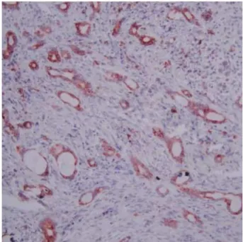
Fig. 4.Most of the tumor cells show diffuse strong staining for epi- thelial membrane antigen (H&E stain, ×200).
and tan-yellow. The microscopic findings revealed a low grade, infiltrating tumor with a mixed growth pattern: there were ductal, tubular, cystic and trabecular patterns. No definite cartilagenous differentiation was observed. Most of the tumor cells had bland-appearing nuclei and finely speckled chro- matin; the cytoplasm was relatively scanty (Fig. 2, 3). Immu- nohistochemically, the tumor cells were positive for cytok- eratin, epithelial membrane antigen (Fig. 4), S-100 protein and smooth muscle actin; they were negative for carcinoem- bryonic antigen (CEA), alpha-1-antitrysin and lysozyme. The bronchial cut margin was free from tumor infiltration, and there was no evidence of invasion to the visceral pleura, the lymphovascular structures and the perineural structures. The dissected lymph nodes were 13 in number and they were as follows; tracheobronchial nodes, hilar nodes, subcarinal nodes and interlobar nodes. The dissected lymph nodes were all negative for tumor invasion.
Her postoperative clinical course was uneventful, and she was discharged on her 9th postoperative day (Fig. 1C, D).
Her head and neck examination was performed after we ob- tained the pathologic report. The study included a clinical examination of the oropharynx and laryngoscopy, and com- puter-assisted tomography (CAT) scans of the head and neck were done. She underwent the whole-body positron emission tomography (PET) scan 2 months after the operation. All the above examinations did not reveal any primary lesion outside the chest. She is still stable without any respiratory symptoms at the time of this report and the parameters of her lung function test, performed 2 months after the opera- tion, were as followings; FVC of 2.37 L and 106% of the pre- dicted value, FEV1of 1.69 L and 107% of the predicted value, RV of 1.11 L and 78% of predicted value, and DLCOof 13.4 mL/mmHg/min and 88% of the predicted value.
DISCUSSION
Although not commonly, it is well recognized that salivary gland-type tumors can also occur in the lung. These are thought to arise from the mucous glands that line the bronchial tree.
The most common salivary gland-type tumors that arise in the lung are adenoid cystic carcinoma and mucoepidermoid carcinoma. Polymorphous low-grade adenocarcinoma (PLGA) is a low-grade malignant infiltrative tumor of the minor salivary gland that displays bland-looking tumor cells and a diverse range of architectural patterns; this tumor was first described by Evans and Batsakis in 1984 (1). Since its origi- nal description and acceptance as a separate entity, more than 200 cases of this tumor have been reported. PLGA comprises 7.4% of the minor salivary gland tumors and 19.6% of those that are malignant (7). Despite its bland morphologic appear- ance, there are also a few reported cases where the tumor has undergone differentiation to a high-grade carcinoma, and often after a considerable period of years since the diagnosis
(8). A large study of 164 cases (9) has shown that 97.6% of all patients were alive or they had died of unrelated disease after a mean follow-up period of 115.4 months after the treat- ment with surgical excision only. Complete surgical excision is the treatment of choice. The macroscopic examination usu- ally displays a circumscribed but nonencapsulated, often light- tan to gray mass. Microscopically, as the name suggests, the tumor has a diverse range of growth patterns that may include tubules, trabeculae, single-cell files, solid nests, and fascicles, or cribriform, papillary or cystic structures. Unifying all these different patterns is the cytologic uniformity of the tumor cells, and these cells exhibit a bland or low-grade cytologic appearance. Immunohistochemically, the tumor cells show diffuse moderate to strong staining for epithelial markers, epithelial membrane antigen (EMA), S-100 and vimetin.
Staining for smooth muscle actin is usually negative or weakly positive and focal. The tumor is generally negative for glial fibrillary acidic protein and monoclonal CEA.
Knowledge concerning the tumor site is rather important.
A tumor arising in a central bronchial site would raise the possibility of a primary salivary gland tumor, as opposed to a conventional adenocarcinoma that is usually peripherally located. The markers CEA and TTF-1 could also be useful targets for antibody testing, as they are quite specific to lung adenocarcinoma. It is also important to distinguish this tumor from other types of salivary gland tumors, especially pleomor- phic adenoma, adenoid cystic carcinoma (ACC), and mucoepi- dermoid carcinoma. The first tumor is benign, whereas the latter two malignant tumors have a more aggressive behav- ior compared to PLGA. Unlike PLGA, pleomorphic adeno- ma is a noninfiltrative, well-circumscribed tumor that con- sists of a biphasic population of epithelial and myoepithelial cells within a chondromyxoid stroma. Pleomorphic adenoma does not demonstrate perineural or stromal invasion. Ade- noid cystic carcinoma shows a greater number of angular, pleomorphic nuclei and increased mitotic activity, as com- pared with the nuclei seen in PLGA, which are slightly larger, rounder and more uniform. The cytoplasm of the tumor cells is usually more eosinophillic in PLGA, whereas it is clearer in ACC. ACC shows EMA staining that is confined to the glandular lumina, and there is weak and patchy S-100 pro- tein staining. In contrast, PLGA shows more diffuse EMA staining and more intense staining for S-100 protein. Mucoe- pidermoid carcinomas usually show definite squamoid or intermediate cells.
Although there are few reports on primary PLGAs of the lung, given the presence of minor salivary glands in the bron- chial glands, this tumorous malady is theoretically possible (6). Primary PLGA of the minor-salivary gland-type of the lung had not been reported until 2004, when Lee and asso- ciates (6) reported a patient who had presented with bilater- al lung tumors and low cervical and supraclavicular lymph nodes involvement without a known history of any previous or concurrent tumor or any oral surgery. The patient was man-
Primary PLGA of Minor Salivary Gland-type of the Lung 375
376 K.D. Cho, J.H. Jung, D.G. Cho, et al.
aged by bilateral wedge resection of the lung. The patient underwent postoperative clinical examination including the laryngoscopy, CAT scans of the head, neck, chest and abdomen, and a whole-body PET scan, which did not reveal any pri- mary lesion. However, the authors discussed a possitility that those lung tumors might have represented metastasis of un- known origin rather than primary lung tumors. In our case, the pathological features were typical of PLGA, where the tumor cells displayed a variety of growth patterns and they had a bland cytologic appearance, which was similar to its counterpart in the minor salivary gland. The diagnosis of PLGA was based on both the typical morphologic appear- ance and the immunohistochemical findings. There was no evidence of primary tumor outside the chest on the detailed head and neck examination including the laryngoscopy, chest CT scanning, and whole body PET-scanning.
The specific operative technique of bronchial sleeve resec- tion was first performed by Price Thomas (10). It avoids the need for pneumonectomy and it spares the healthy lung tissue distal to the pathologic lesion. Benign or malignant tumors confined to the large bronchus can be successfully managed by sleeve lobectomy. Because the surgical risk is not higher than that for pneumonectomy, and a significantly better post- operative quality of life and physical activity is ensured by the preservation of the functioning and intact lung tissue, we regard this type of tumor with its low-grade malignant poten- tial as being well suited for sleeve bronchial resection.
In conclusion, PLGA is a rare minor salivary gland-type tumor that has been thought to occur almost exclusively in the minor salivary glands in the oral cavity and the neck. How- ever, PLGA should be considered in the differential diagnosis of patients presenting with low-grade gland-forming tumors of the lung. When this tumor originates from the large bron- chus, it can be safely resected by the technique of bronchial sleeve resection.
REFERENCE
1. Evans HL, Batsakis JG. Polymorphous low-grade adenocarcinoma of minor salivary glands. A study of 14 cases of a distinctive neoplasm.
Cancer 1984; 53: 935-42.
2. Vincent SD, Hammond HL, Finkelstein MW. Clinical and thera- peutic features of polymorphous low-grade adenocarcinoma. Oral Surg Oral Med Oral Pathol 1994; 77: 41-7.
3. Gnepp DR, Chen JC, Warren C. Polymorphous low-grade adenocar- cinoma of minor salivary gland. An immunohistochemical and clin- icopathologic study. Am J Surg Pathol 1988; 12: 461-8.
4. Tanaka F, Wada H, Inui K, Mizuno H, Ike O, Yokomise H, Fukuse T, Hitomi S, Shoji K, Nakashima Y. Pulmonary metastasis of poly- morphous low-grade adenocarcinoma of the minor salivary gland.
Thorac Cardiovasc Surg 1995; 43: 178-80.
5. Hannen EJ, Bulten J, Festen J, Wienk SM, de Wilde PC. Polymor- phous low grade adenocarcinoma with distant metastases and dele- tions on chromosome 6q23-qter and 11q23-qter: a case report. J Clin Pathol 2000; 53: 942-5.
6. Lee VK, McCaughan BC, Scolyer RA. Polymorphous low-grade adenocarcinoma in the lung: a case report. Int J Surg Pathol 2004;
12: 287-94.
7. Ellis LG, Auclair Paul L. Tumors of the Salivary Glands. In atlas of tumor pathology. Third series. Fascicle no.17. Armed Forces Institute of Pathology, Washington, DC. 1996
8. Simpson RH, Reis-Filho JS, Pereira EM, Ribeiro AC, Abdulkadir A. Polymorphous low-grade adenocarcinoma of the salivary glands with transformation to high-grade carcinoma. Histopathology 2002;
41: 250-9.
9. Castle JT, Thompson LD, Frommelt RA, Wenig BM, Kessler HP.
Polymorphous low-grade adenocarcinoma: a clinicopathologic study of 164 cases. Cancer 1999; 86: 207-19.
10. Thomas PC. Conservative resection of the bronchial tree. J R Coll Surg Edinb 1956; 1: 169-86.