INTRODUCTION
Fractures of the clivus are very rare, occurring in only 0.55
% of head injury patients. Clival fractures are classified as lon- gitudinal, transverse, or oblique. Longitudinal fractures are associated with a 67-80% mortality rate (1, 2), apparently due to vertebrobasilar artery occlusion (3-9) and direct brain stem trauma (10, 11). There are only 11 reported cases of vertebro- basilar artery entrapment or prolapse associated with longi- tudinal clivus fractures. Eight of those cases were only iden- tified at autopsy, while the other 3 cases (12-14) survived but had various neurological deficits.
The present report describes a rare case of traumatic ver- tebrobasilar junction entrapment due to a longitudinal cli- val fracture. The patient was treated using surgical decom- pression of the left cerebellar infarction caused by the left ver- tebral artery occlusion. The patient survived and showed an improved Glasgow coma scale (GCS) score.
CASE REPORT
A 54-yr-old man sustained major craniofacial injuries fol- lowing a traffic collision. The patient was riding bicycle and landed face-down on a hard road surface. He presented with
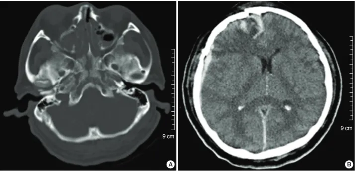
severe right facial and ocular swelling, and profuse cerebrospinal fluid (CSF) rhinorrhea. Neurological findings included a stu- porous mental state (GCS=8) and left hemiparesis (Grade 3). Brain and facial computed tomography (CT) revealed a comminuted fracture of the right frontal bone and anterior skull base extending to the clivus posteriorly, inferior and medial orbital wall fractures with extension to the maxillary sinus, diffuse but predominantly right-sided subdural hema- toma, and a right-sided parenchymal frontotemporal hem- orrhagic contusion with a 6.65 mm midline shift to the left side (Fig. 1). The initial CT scan demonstrated no cerebellar or brainstem lesion.
The patient underwent surgical decompression and removal of the hematoma. After surgery, the patient showed decreased consciousness (GCS=5), aggravated left hemiparesis (Grade 2), and worse right arm and leg response (Grade 3) under noxious stimuli. A brain CT scan on the second postoperative day demonstrated low-density lesions in the left cerebellum and lateral pons (Fig. 2), indicating a left posterior inferior cerebellar artery (PICA) territory infarction. A CT angiogram (Fig. 3A) and its source images (B) revealed the right verte- bral artery (a, b), and the entrapped vertebrobasilar junction (c, d) and basilar artery (e).
A transfemoral conventional angiogram showed obstruc- tion of the left vertebral artery in the cervical region (Fig. 4).
747
Joon Cho, Chang Taek Moon, Hyun Seung Kang, Woo Jin Choe, Sang Keun Chang, Young Cho Koh, and Hong Gee Roh*
Departments of Neurosurgery and Radiology*, Konkuk University Hospital, Seoul, Korea
Address for correspondence Chang Taek Moon, M.D.
Department of Neurosurgery, Konkuk University Hospital, 4-12 Hwayang-dong, Gwangjin-gu, Seoul 143-729, Korea
Tel : +82.2-2030-7623, Fax : +82.2-2030-7469 E-mail : [email protected]
*This paper was supported by Konkuk University in 2007.
DOI: 10.3346/jkms.2008.23.4.747
Traumatic Entrapment of the Vertebrobasilar Junction Due to a Longitudinal Clival Fracture: A Case Report
Vertebrobasilar junction entrapment due to a clivus fracture is a rare clinical obser- vation. The present case report describes a 54-yr-old man who sustained a major craniofacial injury. The patient displayed a stuporous mental state (Glasgow Coma Scale [GCS]=8) and left hemiparesis (Grade 3). The initial computed tomography (CT) scan revealed a right subdural hemorrhage in the frontotemporal region, with a midline shift and longitudinal clival fracture. A decompressive craniectomy with removal of the hematoma was performed. Two days after surgery, a follow-up CT scan showed cerebellar and brain stem infarction, and a CT angiogram revealed occlusion of the left vertebral artery and entrapment of vertebrobasilar junction by the clival fracture. A decompressive suboccipital craniectomy was performed and the patient gradually recovered. This appears to be a rare case of traumatic verte- brobasilar junction entrapment due to a longitudinal clival fracture, including a cere- bellar infarction caused by a left vertebral artery occlusion. A literature review is provided.
Key Words : Cranial Fossa, Posterior; Skull Fracture, Basilar; Vertebral artery; Brain Infarction
Received : 25 August 2007 Accepted : 23 April 2008
The patient underwent a suboccipital decompressive craniec- tomy and a C1 laminectomy in order to widen the foramen magnum (Fig. 5). The patient initially made a fair recovery in terms of consciousness and quadriparesis. At one month after surgery, the patient had normal cognitive function. A tracheostomy had to be maintained. The patient remained dependent in terms of daily living activities, and required a wheelchair for movement.
DISCUSSION
Fractures in the clivus are usually associated with high-impact blunt head trauma. While longitudinal clivus fractures are associated with a 67-80% mortality, transverse or oblique fractures have a better prognosis but are still associated with cranial nerve deficits. They are sometimes associated with carotid artery injury (1, 2).
Menku et al. identified nine cases involving clival fractures from the CT scans of 2,500 head injury patients. Five of those patients had longitudinal fractures, and three of them died (15). Corradino et al. reported 17 cases of clival fracture from the CT scans of 3,000 head injury patients. Six patients had longitudinal fractures. There were four deaths among them, including a case involving a trapped vertebral artery accord- ing to the vertebral angiogram (2). Joslyn et al. identified 11 cases of clival fracture from another series of 2,000 head injury patients. Five patients had longitudinal fractures, and four of those five died (1). All of the three aforementioned reports suggested that vascular injury affecting the posterior circu- lation was a key reason for such a high mortality rate.
To date, there have only been 11 confirmed cases (includ- ing Corradino’s case) of longitudinal clival fractures associ- ated with prolapse or entrapment of the vertebrobasilar arter- ies (Table 1). Most of those cases were fatal. Autopsy results in several cases showed complete occlusion of the prolapsed artery within the fracture, or thrombosis of the proximal basi- lar artery (2-7, 10-13). In the present case, even though there was occlusion of the left vertebral artery and narrowing of the vertebrobasilar junction according to the CT angiogram, there was good distal basilar arterial flow. Thus, the degree of brain
Fig. 1. Initial CT scan showing a longitudinal clivus fracture extending from the frontal basal skull fracture (A), and an acute subdural hem- orrhage with a partial midline shift in the right frontotemporal region (B).
A B
9 cm
9 cm
Fig. 2. Follow-up CT scan showing a left cerebellar and brain stem infarction.
10 cm
stem and cerebellar infarction was manageable. In addition, the decompressive suboccipital craniectomy was probably helpful in reducing brain stem compression caused by the infarction.
In the current study, CT angiography was used to confirm the diagnosis of vascular entrapment. However, in most other cases, the patients died almost instantaneously as a result of the injury, and further imaging studies were not possible. Mag- netic resonance (MR) angiography may reveal vascular occlu-
sion and other additional findings in some cases (13, 14).
Clival fractures are created as a result of three potential direc- tional forces: frontal, occipital, or axial impact (14). Sights et al. suggested (3) that frontal impact contributes to multi- ple facial fractures, and temporary deformation and an increased coronal dimension create a longitudinal clival fracture, such as that seen in the present patient. Occipital impact may have a similar mechanism in the formation of a longitudinal frac- ture (1, 8, 9). A direct blunt injury to the vertex may cause
Fig. 3. CT angiogram (A) and its source images (B). White arrows indicate the right vertebral artery (a), entrapped vertebrobasilar junction (b, c) and basilar artery (d, e).
A B-a
B-b B-c
B-d B-e
enough axial force to form a fracture (3, 6, 9).
Clivus fractures may be under-diagnosed due to the diffi- culty of demonstrating them on plain head radiographs. How- ever, current bony window CT imaging has clearly shown the condition is not as infrequent as previously believed (9). The longitudinal subgroup of clival fractures is considered vulner- able. A CT angiogram is an essential diagnostic work-up in terms of associated vascular injury. In selected cases, surgical decompression may be helpful to reduce brain stem and cere- bellar compression.
REFERENCES
1. Joslyn JN, Mirvis SE, Markowitz B. Complex fractures of the clivus:
Diagnosis with CT and clinical outcome in 11 patients. Radiology 1988; 166: 817-21.
2. Corradino G, Wolf AL, Mirvis S, Joslyn J. Fractures of the clivus:
Classification and clinical features. Neurosurgery 1990; 27: 592-6.
3. Sights WP Jr. Incarceration of the basilar artery in a fracture of the clivus. case report. J Neurosurg 1968; 28: 588-91.
4. Lindenberg R. Incarceration of a vertebral artery in the cleft of a lon- gitudinal fracture of the skull. case report. J Neurosurg 1966; 24: 908- 10.
5. Loop JW, White LE Jr, Shaw CM. Traumatic occlusion of the basi- lar artery within a clivus fracture. Radiology 1964; 83: 36-40.
6. Anthony DC, Atwater SK, Rozear MP, Burger PC. Occlusion of the basilar artery within a fracture of the clivus. case report. J Neurosurg 1987; 66: 929-31.
7. Shaw CM, Alvord EC Jr. Injury of the basilar artery associated with closed head trauma. J Neurol Neurosurg Psychiatry 1972; 35: 247-57.
8. Sato H, Sakai T, Uemura K. A case of incarceration of the vertebral and basilar arteries in a longitudinal fracture of the clivus. No Shinkei Geka 1990; 18: 1147-50.
Fig. 5. Post-surgical CT scan showing the decompressive suboc- cipital craniectomy state.
Fig. 4. Transfemoral conventional left vertebral angiogram antero- posterior (A) and lateral (B) views. Arrows show the vertebral artery obstruction in the cervical region.
A B
VA, vertebral angiogram; ND, not described; GCS, Glasgow coma scale.
Author (yr) (Ref) Age Sex Mechanism of Injury GCS Trapped artery Outcome Method of
confirmation
Loop et al. (1964) (5) 59 M Hit by a falling beam 3 Basilar Death day 14 Autopsy
Lindenberg (1966) (4) 42 M Fall 15 Left vertebral Death day 14 Autopsy
Sight et al. (1968) (3) 23 M Motor vehicle accident 3 Basilar Death day 35 Autopsy
Shaw et al. (1972) (7) 59 M Hit by a tree onto forehead 3 Basilar Death day 13 Autopsy
Anthony et al. (1987) (6) 70 M Fall 3 Basilar Death day 5 Autopsy
Guha et al. (1989) (12) 27 M Fall 3 Basilar Vegetative VA
Corradino et al. (1990) (2) ND ND ND ND Vertebral Death VA
Sato et al. (1990) (8) 80 M Motor vehicle accident 3 Right vertebral Death day 1 Autopsy and basilar
Taguchi et al. (2000) (13) 52 M Fall 3 Basilar Quadriparesis CT angiogram
Sato et al. (2001) (9) 56 M Fall 3 Basilar Death day 4 VA, autopsy
Bala et al. (2003) (14) 46 M Fall 15 Basilar Mild left hemi- CT angiogram,
paresthesia MR angiogram Table 1. Reported cases of vertebrobasilar artery trapping in a longitudinal clivus fracture
9. Sato S, Iida H, Hirayama H, Endo M, Ohwada T, Fujii K. Traumatic basilar artery occlusion caused by a fracture of the clivus-case report.
Neurol Med Chir (Tokyo) 2001; 41: 541-4.
10. Zhu BL, Quan L, Ishida K, Taniguchi M, Oritani S, Fujita MQ, Maeda H. Longitudinal brainstem laceration associated with complex basilar skull fractures due to a fall: an autopsy case. Forensic Sci Int 2002;
126: 40-2.
11. Sato M, Kodama N, Yamaguchi K. Post-traumatic brain stem distor- tion: a case report. Surg Neurol 1999; 51: 613-6.
12. Guha A, Fazl M, Cooper PW. Isolated basilar artery occlusion asso-
ciated with a clivus fracture. Can J Neurol Sci 1989; 16: 81-3.
13. Taguchi Y, Matsuzawa M, Morishima H, Ono H, Oshima K, Hayakawa M. Incarceration of the basilar artery in a longitudinal fracture of the clivus: case report and literature review. J Trauma 2000; 48: 1148- 52.
14. Bala A, Knuckey N, Wong G, Lee GY. Longitudinal clivus fracture associated with trapped basilar artery: unusual survival with good neurological recovery. J Clin Neurosci 2004; 11: 660-3.
15. Menku A, Koc RK, Tucer B, Durak AC, Akdemir H. Clivus fractures:
clinical presentations and courses. Neurosurg Rev 2004; 27: 194-8.