INTRODUCTION
Pulmonary epithelioid hemangioendothelioma (PEH) is the currently preferred term for the neoplastic process, originally described in the lung as intravascular bronchioloalveolar tumor (1-3). The term ‘‘epithelioid hemangioendothelioma (EH)’’
was first applied by Weiss and Enzinger to a soft tissue vascular tumor of borderline malignancy (2). PEH typically occurs as bilateral multiple nodules among young women. Only rare PEH develops as a solitary lung nodule (3-7). Moreover cavi- tary nodule is exceptional. We describe an unusual case of PEH presented as a single cavitary nodule.
CASE REPORT
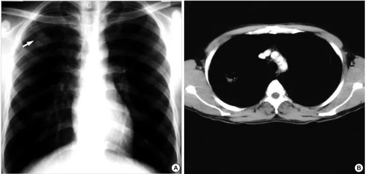
A 33-yr-old asymptomatic man presented for a routine chest radiograph in May 1998. A plain chest radiograph and com- puterized tomograph revealed a 2 cm sized, well-defined cav- itary mass in the apical segment of right upper lobe (Fig. 1).
The physical examination and laboratory findings were not significant. Percutaneous transthoracic needle biopsy was per- formed with unremarkable results. Only a small amount of inflamed lung tissue was seen. Open lung biopsy was recom- mended to confirm the diagnosis. However, the patient de- clined. Thereafter he was irregularly followed up with plain
chest radiograph.
In January 2001, the patient began to complain of chest dis- comfort. Follow-up films showed slight growth of tumor. Bron- choscopic examination revealed narrowing of the lumen with protruded intraluminal mass. With microscopic examina- tion of the bronchoscopic biopsy specimen, the diagnosis of PEH was rendered. Thoracotomy was recommended, but was declined by the patient. Four months later, the patient com- plained more developed chest discomfort. A simple chest radi- ograph showed marked increased size and atelectatic change in the right upper lobe (Fig. 2).
In August 2001, the patient underwent a lobectomy of the right upper lobe. Grossly, the cut surface showed a 5.5×3.3 cm sized and ill-defined grayish white mass expanding to vis- ceral pleura. The mass protruded into the lumen of the bron- chus. Histologically, the tumor showed typical PEH appear- ance. At the periphery of the tumor, tumor cells extended to adjacent alvoli, in a contiguous, micropolypoid fashion (Fig.
3A). The tumor cells were either round or oval. Mitotic activity averaging 2 mitoses per 10 high-power fields was identified (Fig. 3B). Cytoplasmic vacuoles were seen and sometimes, ery- throcytes or fibrin were identified within these intracytoplas- mic lumina (Fig. 3C). Foci of spindle-shaped tumor cells were occasionally seen (Fig. 4A). The tumor showed vascular and lymphatic invasion, pleural invasion, and endobronchial spread (Fig. 4B-D). Immunohistochemically, the tumor cells were
Kyu Yun Jang, Gong Yong Jin*, Yong Chul Lee�, Hung Bum Lee�, Myoung Jae Kang�, Ho Yeul Choi�, Myoung Ja Chung��
5th Criminal Division, Daegu District Public Prosecutor’s Office, Daegu; Department of Radiology*, Internal Medicine�, and Pathology�, Chonbuk National University Medical School, Institute for Medical Sciences�, Jeonju, Korea
Address for correspondence Myoung Ja Chung, M.D.
Department of Pathology, Chonbuk National University Medical School, San 2-20 Keumam-dong, Dukjin-gu, Jeonju 560-182, Korea
Tel : +82.63-270-3071, Fax : +82.63-270-3135 E-mail : [email protected]
599 J Korean Med Sci 2003; 18: 599-602
ISSN 1011-8934
Copyright � The Korean Academy of Medical Sciences
Pulmonary Epithelioid Hemangioendothelioma: A Tumor Presented as a Single Cavitary Mass
Pulmonary epithelioid hemangioendothelioma (PEH) is a rare tumor that occurs among young women and typically presents as bilateral multiple nodules. In the present report, we describe an uncommon case of PEH presented as a single cavitary nodule in a 33-yr-old asymptomatic man. This is the first case of PEH presented as a single cavitary nodule in the English literature. Three years of the follow-up without treatment was performed. Overall histologic findings were accord with conventional PEH, but some atypical features such as, increased mitotic activi- ty (mean; two per ten high power fields), necrosis, spindling, and pleural and vas- cular invasion were recognized. Immunohistochemically, the tumor cells were pos- itive for CD34. This report may contribute to the data on clinical findings and natural history of this rare tumor.
Key Words : Lung; Hemangioendothelioma, Epithelioid
Received : 29 July 2002 Accepted : 26 September 2002
600 K.Y. Jang, G.Y. Jin, Y.C. Lee, et al.
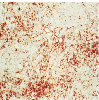
positive for CD34 (Fig. 5) and epithelial membrane antigen, while cytokeratins and S-100 protein were negative. The pa- tient was not followed up after operation.
DISCUSSION
PEH is a rare pulmonary tumor and it shows distinctive cli-
nical features. PEH occurs among young women and typically presents multiple, bilateral nodules up to 2 cm in diameter (1-6). However, PEH rarely develops as a solitary lung nodule (3, 5-7). The frequency of this presentation lies between 10%
and 19% of PEH cases (3, 5-7). Our case revealed a single cav- itary nodule. Although a few cases of solitary nodules with ex- tensive necrosis were reported, there was no case that presented as a cavitary lesion (3, 5-7). This unusual radiographic finding and inadequate biopsy specimen, due to the patient’s refusal of further evaluation, obstructed correct diagnosis at first ad- mission. Additionally, the high incidence of tuberculosis in Korea brought difficulty in diagnosis.
Usually, PEH shows a slowly progressive clinical course. How- ever, the biologic behavior can be influenced by its origin, clinical extend, and histologic features (3-5). The reported mor- tality associated with EH is 13% in soft tissue, 35% in liver, and 65% in lung after a minimum of 4 yrs of follow-up (8).
Metastatic disease occurs in approximately 20% of the patients with soft tissue disease, 15% of those with lung disease, and 25% of those with liver disease (8). Respiratory symptoms at presentation, pleural effusion on chest radiography, peripheral lymphadenopathy, pleural invasion, extensive intravascular and endobronchial tumor spread, hepatic metastasis, and spindle tumor cells at histology were reported as unfavorable prognos- tic factors (3, 5). Our case showed poor prognostic factors, such as spindling of tumor cells, pleural invasion, lymphatic and vascular invasion, and endobronchial spread. Therefore, it was predicted that the prognosis of our patient would be poor. It is not certain that the patient with solitary nodule has a more favorable clinical course than the patient with multiple nod- ules. Weiss and Enzinger used the term malignant epithelioid
Fig. 1.On first admission, simple chest radiography shows about 2 cm sized, well-defined cavitary mass with thickened wall in the right upper lobe (A). Contrast-enhanced CT shows mild heterogeneous enhancement on the thickened wall of the cavitary nodule (B).
A B
Fig. 2.On the 32-month follow-up, the cavitary nodule in the right upper lobe marks increased size and extension to right hilum. Also, atelectasis in the right upper lobe occurred.
Pulmonary Epithelioid Hemangioendothelioma 601
hemangioendothelioma of soft tissue for the tumor showing cellular atypia, mitotic activity (>1 mitosis per 10 high power field), necrosis, or extensive spindling (4). Histologically, our case showed atypical features, such as increased mitotic activity (mean; 2 per 10 high power fields), spindling of tumor cells and necrosis, but there was no definition for malignant PEH and necrosis was not a prognostic factor for PEH.
Histologically, our case should be distinguished from epithe- lioid angiosarcoma and carcinoma. However, angiosarcoma
and carcinomas display far more nuclear atypia and mitotic activity than EH (2, 4, 5). Immunohistochemical and ultra- structural studies may provide the clues that the proliferating cells of PEH are endothelial cells (2, 4, 5).
In the present report, we described an uncommon case of PEH presented as a single cavitary nodule. This report may contribute to the data on clinical findings and natural history of this rare tumor.
Fig. 4.Spindling (A), vascular invasion (B), pleural invasion (C), and endobronchial spread of tumor cells (D) are noted (H&E, ×100).
A
Fig. 3.Tumor cells show micropolypoid growth (A), occasional mitosis (arrow) (B), and frequent cytoplasmic vacuolization (arrowheads) (C) (H&E, ×100 and ×200).
B C
A B C D
602 K.Y. Jang, G.Y. Jin, Y.C. Lee, et al.
REFERENCES
1. Dail DH, Liebow AA. Intravascular bronchioloalveolar tumor (Abstr).
Am J Pathol 1975; 78: 6a.
2. Weiss SW, Enzinger FM. Epithelioid hemangioendothelioma: a vas- cular tumor often mistaken for a carcinoma. Cancer 1982; 50: 970-81.
3. Kitaichi M, Nagai S, Nishimura K, Itoh H, Asamoto H, Izumi T, Dail DH. Pulmonary epithelioid hemangioendothelioma in 21 patients, in- cluding three with partial spontaneous regression. Eur Respir J 1988;
12: 89-96.
4. Weiss SW, Goldblum JR. Hemangioendothelioma: vascular tumors of intermediate malignancy. In: Strauss M, editor, Soft tissue tumors.
4 th ed., St. Louis: Mosby Yearbook, 2001; 891-915.
5. Dail DH, Liebow AA, Gmelich JT, Friedman PJ, Miyai K, Myer W, Patterson SD. Intravascular, bronchiolar, and alveolar tumor of the lung (IVBAT): an analysis of twenty cases of a peculiar sclerosing endothe- lial tumor. Cancer 1983; 51: 452-64.
6. Yousem SA, Hochholzer L. Unusual thoracic manifestation of epithe- lioid hemangioendothelioma. Arch Pathol Lab Med 1987; 111: 459-63.
7. Buchenroth M, Beus J, Bittinger F, Kelbel C, Kersjes W, Mayer E, Ferlinz R. Solitary epithelioid hemangioendothelioma, a rare vascular tumor of the lung. Pneumologie 1995; 49: 239-42.
8. Weiss SW, Ishak KG, Dail DH, Sweet DE, Enzinger FM. Epithelioid hemangioendothelioma and related lesions. Semin Diagn Pathol 1986;
3: 259-87.
Fig. 5.Intracytoplasmic vacuoles are characteristically accentu- ated by the CD34 immunostaining (×200).