ISSN: 2233-601X (Print) ISSN: 2093-6516 (Online)
Received: October 25, 2016, Revised: April 3, 2017, Accepted: April 7, 2017, Published online: October 5, 2017
Corresponding author: Joung Hun Byun, Department of Thoracic and Cardiovascular Surgery, Gyeongsang National University Changwon Hospital, 11 Samjeongja-ro, Seongsan-gu, Changwon 51472, Korea
(Tel) 82-55-214-3849 (Fax) 82-55-214-3260 (E-mail) [email protected]
© The Korean Society for Thoracic and Cardiovascular Surgery. 2017. All right reserved.
This is an open access article distributed under the terms of the Creative Commons Attribution Non-Commercial License (http://creativecommons.org/
licenses/by-nc/4.0) which permits unrestricted non-commercial use, distribution, and reproduction in any medium, provided the original work is properly cited.
Risk Factors for Pneumonia in Ventilated Trauma Patients with Multiple Rib Fractures
Hyun Oh Park, M.D. 1 , Dong Hoon Kang M.D. 2 , Seong Ho Moon, M.D. 1 , Jun Ho Yang, M.D. 1 , Sung Hwan Kim, M.D. 1 , Joung Hun Byun, M.D. 1
1
Department of Thoracic and Cardiovascular Surgery, Gyeongsang National University Changwon Hospital, Gyeongsang National University School of Medicine,
2Department of Thoracic and Cardiovascular Surgery,
Gyeongsang National University Hospital, Gyeongsang National University School of Medicine
Background: Ventilator-associated pneumonia (VAP) is a common disease that may contribute to morbidity and mortality among trauma patients in the intensive care unit (ICU). This study evaluated the associations between trauma factors and the development of VAP in ventilated patients with multiple rib fractures.
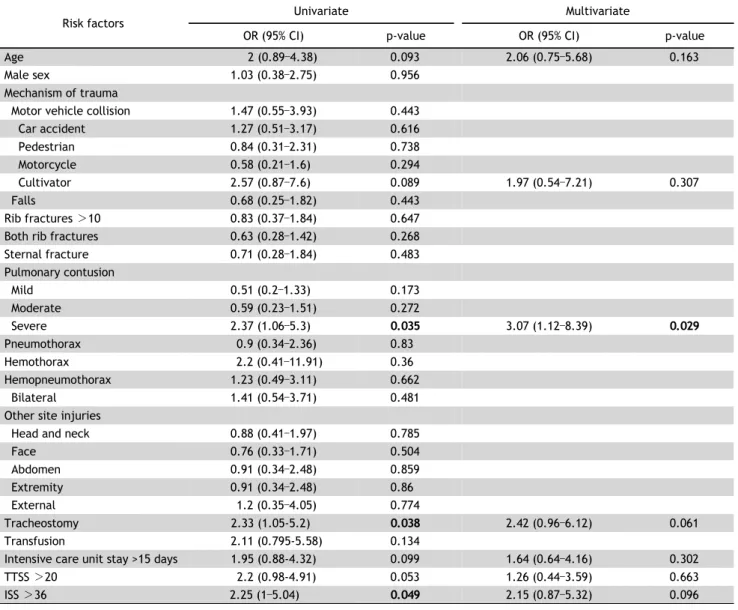
Methods: We retrospectively and consecutively evaluated 101 patients with multiple rib fractures who were ventilated and managed at our hospital between January 2010 and December 2015, analyzing the associa- tions between VAP and trauma factors in these patients. Trauma factors included sternal fracture, flail chest, diaphragm injury, traumatic aortic dissection, combined cardiac injury, pulmonary contusion, pneumothorax, hemothorax, hemopneumothorax, abbreviated injury scale score, thoracic trauma severity score, and injury se- verity score. Results: Forty-six patients (45.5%) had at least 1 episode of VAP, 10 (21.7%) of whom died in the ICU. Of the 55 (54.5%) patients who did not have pneumonia, 9 (16.4%) died in the ICU. Using logistic regression analysis, we found that VAP was associated with severe lung contusion (odds ratio, 3.07; 95%
confidence interval, 1.12 to 8.39; p=0.029). Conclusion: Severe pulmonary contusion (pulmonary lung con- tusion score 6–12) is an independent risk factor for VAP in ventilated trauma patients with multiple rib fractures.
Key words: 1. Ventilator-associated pneumonia 2. Thoracic injuries
3. Wounds and injuries 4. Injuries
Introduction
Hospital-acquired infections (HAI) are infections acquired in hospitals and other healthcare facilities.
HAIs are common complications among patients ad- mitted to intensive care units (ICUs), especially those requiring endotracheal intubation with mechanical ventilation [1]. Ventilator-associated pneumonia (VAP)
results from an invasion of the lower respiratory tract and lung parenchyma by microorganisms. It is defined as pneumonia occurring more than 48 hours after intubation and mechanical ventilation with ra- diologic evidence of new or progressive infiltrates, laboratory detection of the causative agent, and symptomatic evidence of systemic infection [2].
Green et al. [3] reported a high incidence of pul-
https://doi.org/10.5090/kjtcs.2017.50.5.346
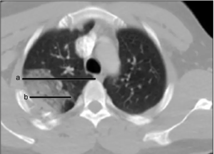
Fig. 1. The PCS. The PCS is determined by summing each of the lengths (a and b). a: The length of the transverse axis of the lobe with the largest pulmonary contusion visualized by computed to- mography of the chest. b: The length of the longest pulmonary contusion. PCS of 1 lobe=(b/a)×3. Fractions of 0.5 and higher were rounded up to the nearest whole number. PCS, pulmonary contusion score.
monary infection in patients with multiple trauma that was attributable to a decrease in the efficacy of immune defense mechanisms. Upper airway colo- nization, trauma requiring surgical treatment, im- munosuppression by sedative drugs, or excessive bleeding due to multiple trauma can reduce the im- mune defense functions [3]. Previous studies found that scoring systems reflecting the severity of trau- ma, such as the injury severity score (ISS), the thora- cic trauma severity score (TTSS), and the rib score, were independent risk factors for the development of pneumonia. In addition, multiple studies have shown that pulmonary contusion and sternal fracture in- crease the risk of developing pneumonia [4-6].
However, the incidence of pneumonia and the related risk factors in cases of multiple trauma are still un- der debate, especially for patients with multiple rib fractures who require mechanical ventilation. There- fore, the present study evaluated the association be- tween trauma factors and the development of VAP in ventilated patients with multiple rib fractures.
Methods
1) Study population
This retrospective study was approved by the in- stitutional review board of the Gyeongsang National University Hospital (GNUH 2016-10-028-003). We retrospectively and consecutively evaluated all pa- tients with multiple rib fractures who required me- chanical ventilation in the ICU; all patients were ad- mitted at a single center between January 2010 and December 2015. We identified 110 trauma patients who received mechanical ventilation. We excluded patients who died within 48 hours of injury, those who were discharged from the ICU within 24 hours, those who had single rib fractures, and those who had stab injuries. Based on these criteria, 9 patients were excluded, and 101 ventilated patients were fi- nally included in our study. Data extracted from the medical records included demographic characteristics, mechanism of injury, number and laterality of rib fractures, associated injuries, rib score, TTSS, ISS, clinical course, and outcomes.
2) Definitions
We defined multiple rib fractures as more than 3 fractures of the ribs. Pulmonary contusion was cate-
gorized into 3 levels of severity based on a scoring system. We calculated 2 lengths (a and b) after di- viding both lungs into the following 4 areas: right upper plus right middle lobe, right lower lobe, left upper lobe, and left lower lobe. ‘a’ was the length of the transverse axis of the lung, with the largest area of pulmonary contusion visualized and measured by means of computed tomography (CT) of the chest. ‘b’
was the length of the longest pulmonary contusion.
The pulmonary contusion score (PCS) for a lobe was calculated as (b/a)×3. We rounded up fractions of 0.5 or greater to the next whole-number unit (Fig. 1) and summed the scores of the lobes. The total PC S was then defined as mild (0–2), moderate (3–5), or severe (6–12) [7].
The ISS is an anatomic scoring system that pro- vides an overall score for patients with multiple injuries. Each injury is assigned an abbreviated injury scale (AIS) score and is associated with 1 of the fol- lowing 6 body regions: head, face, chest, abdomen, extremities (including the pelvis), and external. Only the highest AIS score in each body region is used.
The sum of the squares of the 3 most severely in-
jured body regions is then used to calculate the ISS
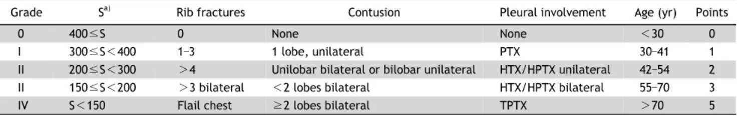
[8]. The TTSS combines the patient’s age, resuscita-
tion parameters, and radiologic assessment of thora-
cic trauma (Table 1) [6].
Table 1. The thoracic trauma severity score
Grade S
a)Rib fractures Contusion Pleural involvement Age (yr) Points
0 400 ≤S 0 None None <30 0
I 300 ≤S<400 1 –3 1 lobe, unilateral PTX 30 –41 1
II 200 ≤S<300 >4 Unilobar bilateral or bilobar unilateral HTX/HPTX unilateral 42 –54 2
II 150 ≤S<200 >3 bilateral <2 lobes bilateral HTX/HPTX bilateral 55 –70 3
IV S <150 Flail chest ≥2 lobes bilateral TPTX >70 5
For calculation of the total score, all categories are summed. The score can range from 0 to 25.
PTX, pneumothorax; HTX, hemothorax; HPTX, hemopneumothorax; TPTX, tension pneumothorax.
a)
S is PaO
2/FiO
2.
The patients with pneumonia satisfied all of the following criteria: (1) core temperature >38.3
oC; (2) a white blood cell count >10.0×10
9/L; (3) purulent tracheobronchial secretions; (4) worsening of pulmo- nary gas exchange levels; and (5) persistence of pul- monary infiltrates on radiographic images (>24 hours) [9]. VAP was defined as pneumonia occurring more than 48 hours after intubation and initiation of mechanical ventilation with radiologic evidence of new or progressive infiltrates, laboratory detection of a causative agent, and symptomatic evidence of sys- temic infection [2,4].
3) Treatment
Endotracheal intubation was performed in patients with at least 1 of the following 5 indications: (1) in- ability to maintain airway patency; (2) inability to protect the airway against aspiration; (3) ventilator compromise; (4) failure to adequately oxygenate pul- monary capillary blood; and (5) anticipation of a de- teriorating course that would eventually lead to the inability to maintain airway patency or protection [10]. All patients received prophylactic antibiotics.
The most commonly used antibiotics were second- generation cephalosporins, such as cefotiam. We used additional antibiotics for other conditions, such open wounds, and as a surgical prophylaxis.
4) Data analysis
Missing data were not replaced or imputed. We calculated p-values using the Fisher exact test or the Pearson chi-square test for categorical variables and used the Mann-Whitney U-test for continuous variables.
Significance was set at p<0.05. To evaluate the risk factors for a poor outcome, we used logistic re- gression analysis. In the multivariate model of effi- cacy, we included relevant variables with p<0.10 in
the univariate analysis. We calculated associations between the variables included in the multivariate analysis, with significance set at p<0.05. All stat- istical analyses were performed using IBM SPSS soft- ware ver. 24.0 (IBM Corp., Armonk, NY, USA).
Results
Between January 2010 and December 2015, Gyeongsang National University Hospital provided mechanical ventilation to 101 patients with multiple rib fractures. The median age of the patients was 66 years, and the majority of patients were male (80.2%). Fifty-three patients (52.5%) were smokers or ex-smokers (the latter group had not smoked within the last 1 year), and 13 patients (12.9%) had a history of lung disease: 4 (4%) had chronic ob- structive lung disease, 8 (7.9%) had asthma, and 1 (1.0%) had pulmonary tuberculosis. Motor vehicle collisions were the main cause of trauma (79.2%), and included car (23.8%), motorcycle (19.8%), pedes- trian (18.8%), and cultivator (16.8%) accidents.
Overall, 63 patients (62.4%) had bilateral rib frac- tures, and the median number of rib fractures was 10. Of the 101 patients in the study, 46 (45.5%) de- veloped pneumonia (the VAP group) and 55 (54.5%) did not (the non-VAP group). There were no sig- nificant differences in age or sex between the groups.
Additionally, the mechanisms of trauma and the mean number of rib fractures was not significantly different between the groups (Table 2).
The differences in individual trauma factors be-
tween the VAP and control groups were studied. We
assessed sternal fracture, flail chest, diaphragm in-
jury, traumatic aortic dissection, combined cardiac in-
jury, pulmonary contusion, pneumothorax, hemothor-
ax, hemopneumothorax, AIS score, TTSS, and ISS.
Table 2. Demographic and clinical characteristics of patients with multiple rib fractures receiving mechanical ventilation
Characteristic Total (n=101) No pneumonia (n=55) Pneumonia (n=46) p-value
Age (yr) 66 (50 –74) 61 (43 –75) 68 (45 –74) 0.179
Male sex 81 (80.2) 44 (80) 37 (80.4) 0.956
Smoking 53 (52.5) 28 (50.9) 25 (54.3) 0.842
Comorbidity
Chronic obstructive pulmonary disease 4 (4) 1 (1.8) 3 (6.5) 0.328
Asthma 8 (7.9) 4 (7.3) 4 (8.7) 1
Tuberculosis 1 (1) 0 1 (2.2) 0.455
Mechanism of trauma
Motor vehicle collision 80 (79.2) 42 (76.4) 38 (82.6) 0.472
Car accident 24 (23.8) 12 (21.8) 12 (26.1) 0.645
Pedestrian 19 (18.8) 11 (20) 8 (17.4) 0.471
Motorcycle 20 (19.8) 13 (23.6) 7 (15.2) 0.211
Cultivator 17 (16.8) 6 (10.9) 11 (23.9) 0.11
Falls 21 (20.8) 13 (23.6) 8 (17.4) 0.472
No. of fractures 10 (5 –14) 10 (6 –14) 9 (5 –13) 0.547
Bilateral rib fractures 63 (62.4) 37 (67.3) 26 (56.5) 0.306
Values are presented as median (interquartile range) or number (%). All p-values were calculated using the Fisher exact test or the Pearson chi-square test for categorical variables and the Mann-Whitney U-test, as appropriate.
Sternal fractures were noted in 18 patients (17.8%), flail chest in 75 patients (74.3%), and pulmonary contusion in 101 patients (100%). Traumatic dia- phragm injury was noted in 5 patients (5%), all of whom underwent surgical repair. Seven patients (6.9%) had traumatic aortic dissection; 3 of these pa- tients underwent thoracic endovascular aortic repair once their condition was suitable (1–2 weeks post-in- jury). Three patients (3%) sustained traumatic heart injuries: 1 had a traumatic tricuspid injury, 1 had a traumatic aortic valve injury, and 1 had a ruptured left atrium. Pneumothorax was noted in 80 patients (79.2%), hemothorax in 94 patients (93.1%), and he- mopneumothorax in 77 patients (76.2%). The most commonly combined injuries were external, extremity, abdomen, face, and head and neck injuries. Emergency neurosurgery due to intracranial hemorrhage was performed in 24 patients (23.8%). Thirteen patients (12.9%) underwent emergency abdominal surgery.
Emergency embolization was performed in 25 patients (24.8%) due to hepatic (n=10 [9.9%]), splenic (n=7 [6.9%]), or retroperitoneal (n=9 [8.9%]) injury. Severe pulmonary contusion (p=0.046) and the ISS (p<0.001) were the only trauma factors that differed signifi- cantly between the VAP and control groups (Table 3).
Patients with suspected VAP were initially treated with ceftriaxone or an antimicrobial agent such as a respiratory fluoroquinolone, ampicillin/sulbactam, or
meropenem. Bronchoalveolar lavage fluid and blood culture specimens were obtained from patients with suspected VAP. The causative organisms identified from these specimens were Staphylococcus aureus (45.7%), Acinetobacter baumanii (21.7%), Klebsiella pneumoniae (13%), Pseudomonas aeruginosa (13%), Candida albicans (4.3%), and Staphylococcus epi- dermidis (2.2%) (Table 4). When a causative organ- ism was identified, the administered antibiotic treat- ment was appropriately modified upon consultation with specialist physicians.
Generally, patients in the VAP group had longer hospital stays than those in the control group.
Additionally, the duration of the ICU stay (p=0.002) and the duration of mechanical ventilation (p<
0.001) were significantly longer in the VAP group than in the control group. We assessed complications, such as acute respiratory distress syndrome, acute renal failure, gastrointestinal bleeding, atrial fi- brillation, and disseminated intravascular coagulation.
Overall, the rate of complications was higher in the VAP group than in the control group; however, the difference was not significant. The mortality rate was defined as the rate of all-cause mortality, and it was comparable between the 2 groups (Table 5).
Univariate logistic regression analysis revealed that
the development of VAP was significantly associated
with severe pulmonary contusion and the ISS. The
Table 3. Intrathoracic injuries, combined injuries, AIS score, TTSS, and ISS of patients with multiple rib fractures receiving mechanical ventilation
Characteristic Total (n=101) No pneumonia (n=55) Pneumonia (n=46) p-value
Sternal fracture 18 (17.8) 14 (25.5) 9 (19.6) 0.634
Flail chest 75 (74.3) 40 (72.7) 35 (76.1) 0.82
Bilateral 51 (50.5) 31 (56.4) 20 (43.5) 0.233
Diaphragm injury 5 (5) 4 (7.3) 1 (2.2) 0.373
Traumatic aortic dissection 7 (6.9) 4 (7.3) 3 (6.5) 1
Combined cardiac injury 10 (9.9) 4 (7.3) 6 (13) 0.506
Pulmonary contusion
a)101 (100) 55 (100) 46 (100) 1
Mild 24 (23.8) 16 (29.1) 8 (17.4) 0.241
Moderate 25 (24.8) 16 (29.1) 9 (19.6) 0.356
Severe 52 (51.5) 23 (41.8) 29 (63) 0.046
Pneumothorax+ 80 (79.2) 44 (80) 36 (78.3) 1
Hemothorax 94 (93.1) 50 (90.9) 44 (95.7) 0.45
Hemopneumothorax 77 (76.2) 41 (74.5) 36 (78.3) 0.815
Both hemopneumothorax 21 (20.8) 10 (18.2) 11 (23.9) 0.623
Other site injuries
Head and neck 52 (51.5) 29 (52.7) 23 (50) 0.843
Face 65 (64.4) 37 (67.3) 28 (60.9) 0.537
Abdomen 82 (71.3) 45 (81.8) 37 (80.4) 0.859
Extremity 82 (81.2) 45 (81.8) 37 (80.4) 1
External 89 (88.1) 48 (87.3) 41 (89.1) 1
AIS score
Head and neck 1 (0 –3) 2 (0 –3) 1 (0 –4) 0.746
Face 1 (0 –1) 1 (0 –1) 1 (0 –1) 0.635
Thorax 4 (3 –5) 4 (3 –5) 4 (3 –5) 0.539
Abdomen 3 (1 –4) 2 (1 –3) 3 (2 –4) 0.067
Extremity 2 (1 –3) 2 (2 –3) 2 (1 –3) 0.795
External 1 (1 –2) 1 (1 –2) 1 (1 –2) 0.775
TTSS 20 (18 –23) 21 (17 –23) 20 (19 –24) 0.056
ISS 36 (29 –42) 34 (29 –38) 38 (33 –43) <0.001
Values are presented as median (interquartile range) or number (%).
AIS, abbreviated injury scale; TTSS, thoracic trauma severity score; ISS, injury severity score.
a)