ISSN: 2233-601X (Print) ISSN: 2093-6516 (Online)
Received: November 27, 2018, Revised: December 27, 2018, Accepted: December 31, 2018, Published online: June 5, 2019
Corresponding author: Youngkyu Moon, Department of Thoracic and Cardiovascular Surgery, Seoul St. Mary’s Hospital, College of Medicine, The Catholic University of Korea, 222 Banpo-daero, Seocho-gu, Seoul 06591, Korea
(Tel) 82-2-2030-4507 (Fax) 82-2-2030-2617 (E-mail) [email protected]
*Current affiliation: Department of Thoracic & Cardiovascular Surgery, Eunpyeong St. Mary’s Hospital, College of Medicine, The Catholic University of Korea, Seoul, Korea
© The Korean Society for Thoracic and Cardiovascular Surgery. 2019. All right reserved.
This is an open access article distributed under the terms of the Creative Commons Attribution Non-Commercial License (http://creativecommons.org/
licenses/by-nc/4.0) which permits unrestricted non-commercial use, distribution, and reproduction in any medium, provided the original work is properly cited.
Prognostic Factors in Stage IIB Non-Small Cell Lung Cancer according to the 8th Edition of TNM Staging System
Jin Won Shin, M.D. 1 , Deog Gon Cho, M.D., Ph.D. 1 , Si Young Choi, M.D., Ph.D. 2, *, Jae Kil Park, M.D., Ph.D. 3 , Kyo Young Lee, M.D., Ph.D. 4 , Youngkyu Moon, M.D., Ph.D. 3, *
1
Department of Thoracic and Cardiovascular Surgery, St. Vincent’s Hospital, College of Medicine, The Catholic University of Korea, Suwon;
2Department of Thoracic and Cardiovascular Surgery, St. Paul’s Hospital, College of Medicine, The
Catholic University of Korea; Departments of
3Thoracic and Cardiovascular Surgery and
4Hospital Pathology, Seoul St. Mary’s Hospital, College of Medicine, The Catholic University of Korea, Seoul, Korea
Background: The purposes of this study were to evaluate the appropriateness of the stage migration of stage IIA non-small cell lung cancer (NSCLC) in the seventh edition of the tumor, node, and metastasis classi- fication for lung cancer to stage IIB lung cancer in the eighth edition, and to identify prognostic factors in patients with eighth-edition stage IIB disease. Methods: Patients with eighth-edition stage IIB disease were subclassified into those with seventh-edition stage IIA disease and those with seventh-edition stage IIB dis- ease, and their recurrence-free survival and disease-specific survival rates were compared. Risk factors for re- currence after curative resection were identified in all included patients. Results: Of 122 patients with eighth-edition stage IIB NSCLC, 101 (82.8%) had seventh-edition stage IIA disease and 21 (17.2%) had sev- enth-edition stage IIB disease. Nonsignificant differences were observed in the 5-year recurrence-free survival rate and the 5-year disease-specific survival rate between the patients with seventh-edition stage IIA disease and those with seventh-edition stage IIB disease. Visceral pleural invasion was a significant risk factor for re- currence in patients with eighth-edition stage IIB NSCLC. Conclusion: The stage migration from seventh-edi- tion stage IIA NSCLC to eighth-edition stage IIB NSCLC was appropriate in terms of oncological outcomes.
Visceral pleural invasion was the only prognostic factor in patients with eighth-edition stage IIB NSCLC.
Key words: 1. Prognosis
2. Non-small-cell lung carcinoma 3. Stage IIB
4. 8th edition
Introduction
Lung cancer is the leading cause of cancer death worldwide [1]. The eighth edition of the tumor, node, and metastasis (TNM) classification of lung cancer was proposed by the International Association for
the Study of Lung Cancer (IASLC) in 2015 and enact- ed on January 1, 2017 [2]. The changes in the new TNM staging system consist of adjustments of the T descriptors, emphasizing the prognostic impact of tu- mor size and redefining the classification of addi- tional tumor nodules; a redefinition of malignant
https://doi.org/10.5090/kjtcs.2019.52.3.131
pleural effusion and the subclassification of M1; and a rearrangement of the stage groups; however, the N descriptors were left unchanged [2-4].
The eighth edition of the TNM classification pres- ents many changes in the T categories, which are based on tumor size, as follows: T1a ≤1 cm, T1b ≥ 1 to 2 cm, T1c ≥2 to 3 cm, T2a ≥3 to 4 cm, T2b
≥ 4 to 5 cm, T3 ≥5 to 7 cm, and T4 ≥7 cm [5].
The previous T3 classification (>7 cm) was up- graded to T4 in the eighth edition. The seventh-edi- tion T2b (>5 cm) classification was also changed in the eighth edition to T3. However, other T3 factors such as lung-to-lung metastasis (same lobe) and pari- etal pleural invasion were not changed. Therefore, previous stage IIA (T2bN0M0) disease was changed in the eighth edition to stage IIB (T3N0M0) disease.
As a result, stage IIB lung cancer in the eighth edi- tion of the TNM classification is a combination of seventh-edition stage IIA (T2bN0M0) and sev- enth-edition stage IIB (T3N0M0) non-small cell lung cancer (NSCLC).
Even though the N descriptors remained un- changed, because almost all N1 disease was upgraded from stage IIA to stage IIB [5], eighth-edition stage IIB comprises many heterogeneous TNM groups, as follows: T1aN1M0, T1bN1M0, T1cN1M0, T2aN1M0, T2bN1M0, and T3N0M0. In contrast, seventh-edition stage IIB only consisted of T2bN1M0 and T3N0M0 disease. To the best of our knowledge, however, studies have not yet evaluated the appropriateness of combining seventh-edition IIA and seventh-edition stage IIB disease and reclassifying the combination in the eighth edition as stage IIB disease. The eighth-ed- ition stage IIB classification subdivides the T category into 6 groups (T1a to T3) and incorporates several factors that are associated with outcomes (visceral pleural invasion [T2], parietal pleural invasion [T3], lung to lung metastasis [T3], and lymph node meta- stasis [N1]). Therefore, identifying predictive factors of outcomes in patients with eighth-edition stage IIB disease would potentially enable the subclassification of stage IIB disease into additional subgroups in a future staging system.
The purposes of this study were to evaluate the appropriateness of the stage migration of stage IIA NSCLC in the seventh edition of the TNM classi- fication for lung cancer to stage IIB lung cancer in the eighth edition, and to identify prognostic factors
in patients with eighth-edition stage IIB disease.
Methods
1) Patients
From January 2006 to July 2016, 1,551 consecutive patients were diagnosed with and surgically treated for NSCLC at a tertiary hospital in South Korea. Of those patients, 1,126 underwent standard anatomical pulmonary resection (more than lobectomy) with systematic nodal dissection. Patients were reclassified according to the eighth edition of the TNM staging system. Among the reclassified patients, 122 patients were diagnosed with eighth-edition stage IIB NSCLC.
None of the study patients received incomplete resection. These consecutive patients were reviewed retrospectively. The clinicopathological characteristics of the tumors and oncological outcomes were analyzed. Patients with eighth-edition stage IIB dis- ease were subclassified into those with seventh-edi- tion stage IIA disease and those with seventh-edition stage IIB disease, and their recurrence-free survival (RFS) and disease-specific survival (DSS) rates were compared. Risk factors for recurrence after curative resection were identified. This study was approved by the Institutional Review Board of Seoul St. Mary’s Hospital, the Catholic University of Korea (IRB appro- val no., KC19RESI0173).
2) Surgical procedures and systematic nodal dissection
Patients with stage II NSCLC are considered candi-
dates for curative surgery at Seoul St. Mary’s
Hospital. Every patient underwent surgery, which in-
cluded more than only lobectomy, and they all un-
derwent mediastinal lymph node dissection of more
than 3 mediastinal lymph node stations (operations
on the right lobe included paratracheal and sub-
carinal lymph node dissection, and operations on the
left lobe included subaortic and subcarinal lymph
node dissection). They all underwent dissection of
every visible hilar, peribronchial, and perivascular N1
lymph node. The en bloc resection technique was
used for lymph node dissection, including adjacent
fat tissue.
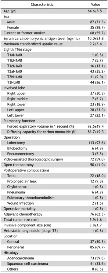
Table 1. Clinicopathological characteristics of patients with stage IIB non-small cell lung cancer after curative surgery
Characteristic Value
Age (yr) 64.6±8.5
Sex
Male 87 (71.3)
Female 35 (28.7)
Current or former smoker 68 (55.7)
Serum carcinoembryonic antigen level (ng/mL) 10.0±21.8 Maximum standardized uptake value 9.2±5.4 Eighth TNM stage
T1aN1M0 1 (0.8)
T1bN1M0 7 (5.7)
T1cN1M0 16 (13.1)
T2aN1M0 43 (35.2)
T2bN1M0 11 (9.0)
T3N0M0 44 (36.1)
Involved lobe
Right upper 37 (30.3)
Right middle 7 (5.7)
Right lower 23 (18.9)
Left upper 28 (23.0)
Left lower 27 (22.1)
Pulmonary function
Forced expiratory volume in 1 second (%) 92.6±19.4 Diffusing capacity for carbon monoxide (%) 86.7±19.3 Operation
Lobectomy 113 (92.6)
Bilobectomy 6 (4.9)
Pneumonectomy 3 (2.5)
Video-assisted thoracoscopic surgery 72 (59.0)
Open thoracotomy 50 (41.0)
Postoperative complications
Total 22 (18.0)
Prolonged air leak 12 (9.8)
Chylothorax 1 (0.8)
Pneumonia 6 (4.9)
Pulmonary thromboembolism 1 (0.8)
Wound infection 2 (1.6)
Operative mortality 1 (0.8)
Adjuvant chemotherapy 76 (62.3)
Total tumor size (cm) 3.9±1.6
Invasive component size (cm) 3.8±1.7
Metastatic lung nodule (stage T3) 1 (0.8) Location
Central 37 (30.3)
Peripheral 85 (69.7)
Histology
Adenocarcinoma 73 (59.8)
Squamous cell carcinoma 41 (33.6)
Others 8 (6.6)
(Continued to the next page)
Table 1. Continued
Characteristic Value
No. of dissected lymph nodes 14.7±7.6
Pleural invasion
Visceral pleural invasion 33 (27.0)
Parietal pleural invasion 16 (13.1)
Lymphovascular invasion 104 (85.2)
Values are presented as mean±standard deviation or number (%).
TNM, tumor, node, and metastasis.
3) Histological evaluation and restaging to the eighth-edition staging system
All clinical specimens were examined by patholo- gists, whose observations were recorded. Each pa- tient report was reviewed for tumor size, location, lymph node status, pleural invasion, and lymphovas- cular invasion. The presence of visceral pleural in- vasion was defined as tumor extension beyond the elastic layer of the visceral pleura. Lymphovascular invasion was defined by the presence of tumor cells in the lymphatic or vascular lumen. Histologic fea- tures, as well as the presence of pleural invasion, lymphatic invasion, or vascular invasion, were de- termined by examining slides of tissue sections stained by hematoxylin and eosin. If the findings could not be determined on hematoxylin-and-eosin stained tissue alone, special staining such as the Verhoeff-Van Gieson elastic stain was performed as necessary. In particular, Verhoeff-Van Gieson elastic staining was performed for a detailed evaluation of visceral pleural invasion. TNM staging was based on the eighth IASLC edition of the TNM staging system [2]. To reclassify the T category according to the eighth edition, tumor size was measured again by the pathologist as the greatest dimension of the invasive component on the histopathologic study [6]. In the eighth edition, the N category remained unchanged from the seventh edition, and was thus not re- classified [4].
4) Follow-up evaluations
All patients were evaluated from the day of
surgery. For the first 2 years, the patients were eval-
uated via physical examinations and chest radiog-
raphy every 3 months, and chest computed tomog-
raphy (CT) that included cervical and abdominal
views every 6 months. Thereafter, they were as-
sessed with physical examinations and chest CT ev-
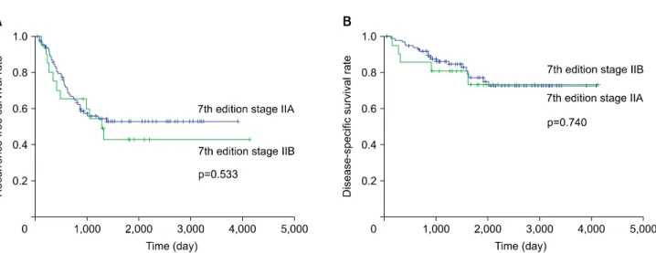
Fig. 1. Comparisons of recurrence-free survival (A) and disease-specific survival (B) rates in patients with eighth-edition stage IIB non-small cell lung cancer after surgery who were subdivided into seventh-edition stage IIA and seventh-edition stage IIB groups.
ery 6 months for 5 years, after which they were evaluated annually.
5) Statistical analysis
The clinicopathological characteristics of the study patients were compared by the Student t-test or the Wilcoxon rank-sum test for continuous variables, and the chi-square test or Fisher exact test for categorical variables. The Kaplan-Meier method was used to ana- lyze data collected from the interval between the time of surgical resection and the time of the final follow-up visit. RFS and DSS were estimated by the Kaplan-Meier method from the collected data on con- firmed cases of recurrence and cancer-related deaths.
The Cox proportional hazards model was used in a univariate analysis to determine the risk of re- currence and cancer-related death for all the study patients. A p-value <0.05 was considered to indicate statistical significance. Statistical analysis was per- formed using IBM SPSS ver. 24.0 software (IBM Corp, Armonk, NY, USA).
Results
The clinicopathological characteristics of the 122 study patients are shown in Table 1. The mean±
standard deviation age was 64.6±8.5 years, and there were more male (71.3%) than female patients. The number of patients with stage T1aN1M0, T1bN1M0, T1cN1M0, T2aN1M0, T2bN1M0, and T3N0M0 NSCLC were as follows: 1 (0.8%), 7 (5.7%), 16 (13.1%), 43
(35.2%), 11 (9.0%), and 44 (36.1%), respectively.
There were 22 complications (18.0 %) among the patients, as follows: 12 patients with prolonged air leakage, 1 with chylothorax, 6 with pneumonia, 1 with pulmonary thromboembolism, and 2 with wound infection. All complications were managed successfully during the period of hospitalization for curative surgery. There was 1 (0.8%) postoperative mortality.
Adjuvant chemotherapy was recommended for all patients, and 76 (62.3%) of the study patients did receive adjuvant chemotherapy. The remaining 37.7%
of patients did not undergo adjuvant chemotherapy for the following reasons: fear of chemotoxicity, un- derlying comorbidities, postoperative complications, and advanced age.
The mean size of the total tumor and mean size of
the invasive component were 3.9 cm and 3.8 cm,
respectively. One patient was diagnosed with stage
T3 lung cancer because of unilateral lung metastasis,
and 37 patients (30.3%) had central lesions. Most of
the patients had adenocarcinoma (59.8%), and the
others had squamous cell carcinoma (33.6%) and
other histologic types (6.6%). The mean number of
dissected lymph nodes was 14.7±7.6. The incidences
of visceral pleural invasion, parietal pleural invasion,
and lymphovascular invasion were 33 (27.0%), 16
(13.1%), and 104 (85.2%), respectively.
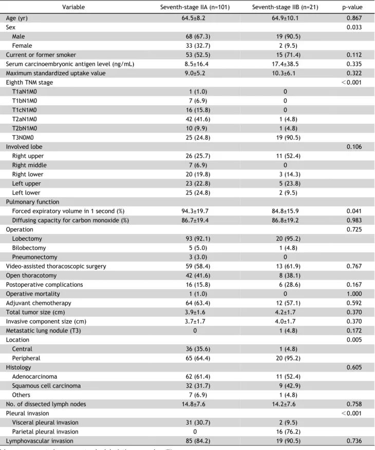
Table 2. Comparison of clinicopathological characteristics between the seventh-edition TNM classifications of IIA and IIB non-small cell lung cancer in patients with eighth-edition stage IIB disease
Variable Seventh-stage IIA (n=101) Seventh-stage IIB (n=21) p-value
Age (yr) 64.5±8.2 64.9±10.1 0.867
Sex 0.033
Male 68 (67.3) 19 (90.5)
Female 33 (32.7) 2 (9.5)
Current or former smoker 53 (52.5) 15 (71.4) 0.112
Serum carcinoembryonic antigen level (ng/mL) 8.5±16.4 17.4±38.5 0.335
Maximum standardized uptake value 9.0±5.2 10.3±6.1 0.322
Eighth TNM stage <0.001
T1aN1M0 1 (1.0) 0
T1bN1M0 7 (6.9) 0
T1cN1M0 16 (15.8) 0
T2aN1M0 42 (41.6) 1 (4.8)
T2bN1M0 10 (9.9) 1 (4.8)
T3N0M0 25 (24.8) 19 (90.5)
Involved lobe 0.106
Right upper 26 (25.7) 11 (52.4)
Right middle 7 (6.9) 0
Right lower 20 (19.8) 3 (14.3)
Left upper 23 (22.8) 5 (23.8)
Left lower 25 (24.8) 2 (9.5)
Pulmonary function
Forced expiratory volume in 1 second (%) 94.3±19.7 84.8±15.9 0.041
Diffusing capacity for carbon monoxide (%) 86.7±19.4 86.8±19.2 0.983
Operation 0.725
Lobectomy 93 (92.1) 20 (95.2)
Bilobectomy 5 (5.0) 1 (4.8)
Pneumonectomy 3 (3.0) 0
Video-assisted thoracoscopic surgery 59 (58.4) 13 (61.9) 0.767
Open thoracotomy 42 (41.6) 8 (38.1)
Postoperative complications 16 (15.8) 6 (28.6) 0.167
Operative mortality 1 (1.0) 0 1.000
Adjuvant chemotherapy 64 (63.4) 12 (57.1) 0.592
Total tumor size (cm) 3.9±1.6 4.2±1.7 0.370
Invasive component size (cm) 3.7±1.7 4.0±1.7 0.370
Metastatic lung nodule (T3) 0 1 (4.8) 0.172
Location 0.005
Central 36 (35.6) 1 (4.8)
Peripheral 65 (64.4) 20 (95.2)
Histology 0.605
Adenocarcinoma 62 (61.4) 11 (52.4)
Squamous cell carcinoma 32 (31.7) 9 (42.9)
Others 7 (6.9) 1 (4.8)
No. of dissected lymph nodes 14.8±7.6 14.2±7.6 0.758
Pleural invasion <0.001
Visceral pleural invasion 31 (30.7) 2 (9.5)
Parietal pleural invasion 0 16 (76.2)
Lymphovascular invasion 85 (84.2) 19 (90.5) 0.736
Values are presented as mean±standard deviation or number (%).
TNM, tumor, node, and metastasis.
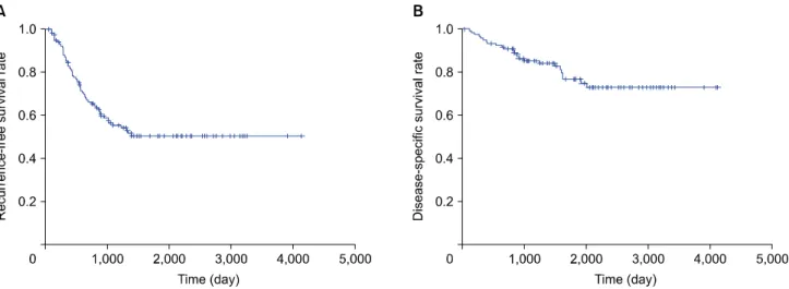
Fig. 2. Recurrence-free survival (A) and disease-specific survival (B) rates of patients with eighth-edition stage IIB non-small cell lung cancer after surgery.
Table 3. Summary of cases of recurrence in study patients
Variable No. (%)
Overall recurrence 54 (100.0)
Locoregional recurrence
a)19 (35.2)
Distant recurrence 15 (27.8)
Both
b)20 (37.0)
a)
Recurrence within the ipsilateral hemithorax, including the pleura and mediastinal lymph nodes.
b)Locoregional recurrence+
distant recurrence.
1) Comparison of the survival rate between the seventh-edition stage IIA group and the seventh-edition stage IIB group in patients with eighth-edition IIB NSCLC
Among the 122 patients with eighth-edition stage IIB NSCLC, 101 (82.8%) were classified as having seventh-edition stage IIA disease and 21 (17.2%) were classified as having seventh-edition stage IIB disease. We compared the survival rates of these pa- tients (Fig. 1) and their clinicopathological character- istics (Table 2). The differences in most of the clin- icopathological characteristics between the 2 groups were not significant. There were differences in T fac- tors between the 2 groups (metastatic lung nodule [T3], parietal pleural invasion [T3]). The patients with seventh-edition stage IIA disease had more cen- tral tumors than the patients with seventh-edition stage IIB disease.
The difference in the RFS rate between patients with seventh-edition stage IIA and seventh-edition stage IIB disease was not significant (52.8% versus 42.8%, respectively; p=0.533) (Fig. 1A). The differ- ence in the DSS rate between patients in the 2 groups was also not significant (77.4% versus 73.6%, respectively; p=0.740) (Fig. 1B).
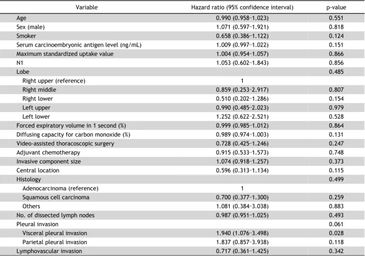
2) Survival analysis and risk factors for recurrence in patients with eighth-edition stage IIB NSCLC after curative surgery
The median follow-up time for patients with
eighth-edition stage IIB NSCLC was 1,463 days (range, 37–4,127 days), with recurrence identified in 54 patients (Table 3). Among those 54 patients, lo- coregional recurrence occurred in 19 patients (35.2%), locoregional recurrence with distant re- currence occurred in 20 (37.0%), and distant re- currence only occurred in 15 (27.8%). The 5-year RFS and DSS rates were 50.6% and 76.8%, re- spectively (Fig. 2). The results of the univariate anal- ysis are shown in Table 4. The single variable identi- fied as significant (p<0.05) in the univariate analysis was visceral pleural invasion. Visceral pleural in- vasion (hazard ratio, 1.940; p=0.028) was a sig- nificant risk factor for recurrence. Table 5 shows the recurrence pattern of patients with eighth-edition stage IIB disease with visceral pleural invasion.
Distant recurrence without locoregional recurrence
occurred in 50.0% of the patients who developed
recurrence. The 2 major sites of distant recurrence
were the contralateral lung and brain.
Table 4. Univariate analysis of risk factors for recurrence in patients with eighth-edition stage IIB non-small cell lung cancer after cura- tive surgery
Variable Hazard ratio (95% confidence interval) p-value
Age 0.990 (0.958–1.023) 0.551
Sex (male) 1.071 (0.597–1.921) 0.818
Smoker 0.658 (0.386–1.122) 0.124
Serum carcinoembryonic antigen level (ng/mL) 1.009 (0.997–1.022) 0.151
Maximum standardized uptake value 1.004 (0.954–1.057) 0.866
N1 1.053 (0.602–1.843) 0.856
Lobe 0.485
Right upper (reference) 1
Right middle 0.859 (0.253–2.917) 0.807
Right lower 0.510 (0.202–1.286) 0.154
Left upper 0.990 (0.485–2.023) 0.979
Left lower 1.252 (0.622–2.521) 0.528
Forced expiratory volume in 1 second (%) 0.999 (0.985–1.012) 0.864
Diffusing capacity for carbon monoxide (%) 0.989 (0.974–1.003) 0.131
Video-assisted thoracoscopic surgery 0.728 (0.425–1.246) 0.247
Adjuvant chemotherapy 0.915 (0.533–1.573) 0.748
Invasive component size 1.074 (0.918–1.257) 0.373
Central location 0.596 (0.313–1.134) 0.115
Histology 0.499
Adenocarcinoma (reference) 1
Squamous cell carcinoma 0.700 (0.377–1.300) 0.259
Others 1.081 (0.384–3.038) 0.883
No. of dissected lymph nodes 0.987 (0.951–1.025) 0.493
Pleural invasion 0.061
Visceral pleural invasion 1.940 (1.076–3.498) 0.028
Parietal pleural invasion 1.837 (0.857–3.938) 0.118
Lymphovascular invasion 0.717 (0.361–1.425) 0.342
TNM, tumor, node, and metastasis.
Table 5. Summary of cases of recurrence in patients with vis- ceral pleural invasion
Variable No. (%)
Overall recurrence 20 (100)
Locoregional recurrence
a)4 (20)
Distant recurrence
b)10 (50)
Both
c)6 (30)
a)