• 검색 결과가 없습니다.

Palatal Rugae Pattern in Korean Children and Adolescents JOMP

N/A
N/A
Protected

Academic year: 2021

Share "Palatal Rugae Pattern in Korean Children and Adolescents JOMP"

Copied!
5
0
0

로드 중.... (전체 텍스트 보기)

전체 글

(1)

pISSN 2288-9272 eISSN 2383-8493 J Oral Med Pain 2019;44(4):169-173 https://doi.org/10.14476/jomp.2019.44.4.169

Palatal Rugae Pattern in Korean Children and Adolescents

Na-Hee Kim 1 , Yeong-Gwan Im 1 , Ji-Yeon Kim 2 , Byung-Gook Kim 1

1

Department of Oral Medicine, School of Dentistry, Chonnam National University, Gwangju, Korea

2

Department of Oral Medicine, Seoul Veterans Hospital, Seoul, Korea

Received November 26, 2019 Revised December 19, 2019 Accepted December 20, 2019

Purpose: To determine whether the morphological features of the palatal rugae are associ- ated with sex and age in children and adolescents.

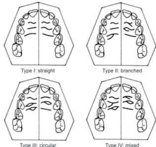
Methods: A total of 300 diagnostic models of the palatal rugae of children and adolescents were collected. The models were classified into male and female and<13- and ≥13-year-old groups. The palatal rugae pattern, and the number and length of palatal rugae plicae, were analyzed.

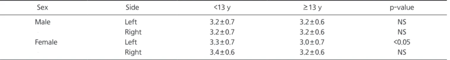
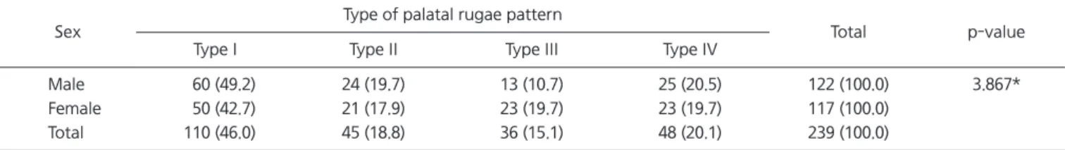
Results: The number of palatal rugae plicae was higher in females than in males, however, the difference was negligible. In the group aged 13 years or more, the number was higher in the male group on the left side. There was no association between the number of palatal rugae plicae and age group. The type I pattern was the most common in both males and females. The length of palatal rugae plicae was greater in males than in females. There was no association between the length of palatal rugae plicae and age group.

Conclusions: The number and length of palatal rugae were associated with sex, but the morphological features of the palatal rugae could not distinguish between children and ad- olescents. These findings suggest that the palatal rugae have limited value for identification of individuals.

Key Words: Forensic dentistry; Individual identification; Palatal rugae; Palate

Correspondence to:

Byung-Gook Kim

Department of Oral Medicine, School of Dentistry, Chonnam National University, 33 Yongbong-ro, Buk-gu, Gwangju 61186, Korea

Tel: +82-62-530-5574 Fax: +82-62-530-5679 E-mail: [email protected]

https://orcid.org/0000-0002-3602-4720

JOMP Journal of Oral Medicine and Pain

Copyright Ⓒ 2019 Korean Academy of Orofacial Pain and Oral Medicine. All rights reserved.

CC

This is an open-access article distributed under the terms of the Creative Commons Attribution Non-Commercial License (http://creativecommons.org/licenses/by-nc/4.0/), which permits unrestricted non-commercial use, distribution, and reproduction in any medium, provided the original work is properly cited.

INTRODUCTION

Various types of disasters, including structural collapses, fires, and explosions, as well as accidents involving trains, ships, and aircraft, can result in large numbers of casual- ties. Forensic identification of individuals is required when such disasters occur and cause many deaths. The identifica- tion of individuals in this context involves the examination of both living and dead bodies. Many methods for human identification have been developed. In 1932, Hermosa et al. introduced palatoscopy based on palatal rugae patterns for human identification, which can be applied even if the teeth are severely damaged [1].

The palatal rugae are anatomical ridges with asymmetric and irregular shapes located in the anterior one-third of the

palate. They are formed between 12 and 14 weeks of birth, and extend outward on both sides from the palatal closure [2,3]. Thomas [4] studied the histologic features of rugae cores from human embryos aged over 20 weeks and found that the reticulin fiber content was miniscule; moreover, the fibroblasts differed from those in adjacent palatal tissues only in size and amount. The palatal rugae can be used to identify a dead body because of their specificity and stabil- ity [5]. Moreover, the palatal rugae can be used as an alter- native method when individual identification based on fin- gerprints or dental records is difficult, or when postmortem dental identification is impossible, as in cases with edentu- lous mouths [6].

Many studies have attempted to use the palatal rugae for

human identification [7]. Thomas and Kotze [8] studied the

(2)

pattern of palatal rugae plicae of six South African popula- tions and found that the palatal rugae are unique and can be used for genetic research. Chae and Shin [9] reported in- dividuality of palatal rugae pattern in twin children and ad- olescents. However, although prior studies used palatal ru- gae data for individuals of all ages, they did not specifically evaluate the effect of age on the palatal rugae [8].

We investigated the morphological features of the palatal rugae of children and adolescents, and examined the as- sociation between those features and age. Additionally, we explored the possibility of using palatal rugoscopy for iden- tification of individual children and adolescents.

MATERIALS AND METHODS

1. Subjects

Diagnostic models of adolescent male and female sub- jects aged 5 to 18 years were obtained from the Department of Pediatric Dentistry of Chonnam National University Dental Hospital. Diagnostic models with intact palatal rugae patterns were selected and divided into male and female groups. The models were further divided into children (<13 years old) and adolescents (≥13 years old) (Table 1).

2. Analysis of Palatal Rugae

The plicae of the palatal rugae of each diagnostic model were sketched using a sharp mechanical pencil. Photographs were obtained with a standard ruler placed adjacent to the models at a standardized distance [10]. The patterns, num- bers, and lengths of palatal rugae plicae were analyzed.

Palatal rugoscopy was based on the methods developed by Thomas and Kotze [8] and Lysell [11]. The palatal rugae pattern was classified into the following four types: type I, straight; type II, branched; type III, circular; and type IV, mixed (Fig. 1) [10,12].

3. Statistical Analysis

The Pearson chi-squared test was used to compare the palatal rugae pattern distributions between the male and fe- male groups. The independent t-test was used to compare the number and length of the palatal rugae plicae, and to examine differences between the two age groups. Statistical analyses were performed using IBM SPSS Statistics for Windows, Version 21.0 (IBM Co., Armonk, NY, USA). Ethics approval was obtained from the Institutional Review Board of Chonnam National University Dental Hospital (IRB no.

CNUDH-2019-002).

RESULTS

1. Number of Palatal Rugae Plicae

The mean number of palatal rugae plicae on the left and right sides of all age groups ranged from 3.2 to 3.4.

The difference in number of palatal rugae plicae between males and females was significant on both the left (p<0.05) and the right (p<0.05) side. Although the mean number was higher in the female group on both sides, the differ- ence was only between 0.1 and 0.2 (Table 2). In the group aged less than 13 years, the difference was significant both on the left (p<0.05) and the right (p<0.05) side (Table 3).

In the group aged 13 years or more, however, the number

Type I: straight

Type III: circular

Type II: branched

Type IV: mixed

Fig. 1. Classification of the types of palatal rugae pattern based on the methods developed by the article of Surekha et al. (J Forensic Dent Sci 2012;4:93-96) [10] and Hauser et al. (J Anat 1989;165:237- 249) [12].

Table 1. Distribution of the subjects according to age group and sex

Age group Male Female Total

<13 y 122 117 239 (79.7)

≥13 y 38 23 61 (20.3)

Total 160 (53.3) 140 (46.7) 300 (100.0)

Values are presented as number only, number (%).

(3)

was significantly higher in the male group on the left side (p<0.05) (Table 3). A comparison of the number of palatal rugae plicae between the two age groups showed no signif- icant difference, except in the female group on the left side (p<0.05) (Table 4).

2. Distribution of the four types of palatal rugae pattern The most frequent type of palatal rugae pattern among all subjects (n=300) was type I, which was found in 135 of

300 subjects (45.0%), followed by type IV (20.3%), type II (19.0%), and type III (15.7%). For the male subjects, type I was the most common pattern, followed by type II, type IV, and type III, in order. For the female subjects, the order was type I, followed by types III, IV, and II; however, the differ- ence in proportions of the four types of palatal rugae pattern were not significantly different (p=5.465) (Table 5, Fig. 2).

The proportions of the four types of palatal rugae pattern were similar in the group aged less than 13 years (p=3.867) (Table 6). Similar results were obtained in the group aged 13 years old or higher.

Table 2. Number of palatal rugae plicae according to sex Side Male (n=160) Female (n=140) p-value

Left 3.2±0.7 3.4±0.7 <0.05

Right 3.2±0.7 3.3±0.6 <0.05

Values are presented as mean±standard deviation.

p-values were determined by independent t-test.

Table 3. Number of palatal rugae plicae according to sex and age group

Age group Side Male Female p-value

<13 y Left 3.2±0.7 3.3±0.7 <0.05 Right 3.2±0.7 3.4±0.6 <0.05

≥13 y Left 3.2±0.6 3.0±0.7 <0.05

Right 3.2±0.6 3.2±0.6 NS

NS, not significant.

Values are presented as mean±standard deviation.

p-values were determined by independent t-test.

Table 4. Number of palatal rugae plicae in the two age groups

Sex Side <13 y ≥13 y p-value

Male Left 3.2±0.7 3.2±0.6 NS

  Right 3.2±0.7 3.2±0.6 NS

Female Left 3.3±0.7 3.0±0.7 <0.05

  Right 3.4±0.6 3.2±0.6 NS

NS, not significant.

Values are presented as mean±standard deviation.

p-values were determined by independent t-test.

Table 5. Distribution of the four types of palatal rugae pattern (n=300)

Sex Type of palatal rugae pattern

Total p-value

Type I Type II Type III Type IV

Male 75 (46.9) 34 (21.3) 18 (11.3) 33 (20.6) 160 (100.0) 5.465*

Female 60 (42.9) 23 (16.4) 29 (20.7) 28 (20.0) 140 (100.0)

Total 135 (45.0) 57 (19.0) 47 (15.7) 61 (20.3) 300 (100.0)

Type I, straight; type II, branched; type III, circular; type IV, mixed.

Values are presented as number (%).

*p-values were determined by the χ

2

test.

Type I 160

140 120 100 80 60 40 20

Type IV 0

Type II Type III

Male Female Total

Number of subjects

Fig. 2. Distribution of the four types of palatal rugae pattern.

(4)

3. Length of the Palatal Rugae Plicae According to Sex The mean length of the palatal rugae plicae ranged from 7.4 to 8.6 mm. The length was greater in the male group on both the left and right sides. The difference in length be- tween the two sex groups was significant on the left (p<0.05) and the right (p<0.05) (Table 7) side. The mean length was greater in the male group on both sides among those aged less than 13 years, and among those aged 13 years or more (Table 8). There was no significant difference in the length of the palatal rugae plicae between the two age groups (Table 9).

DISCUSSION

The aim of this study was to determine whether the mor- phological features of the palatal rugae are associated with sex and age in children and adolescents.

Analysis of the palatal rugae has been used to aid

identification of individuals in the field of forensics. Most previous studies focused on the individual uniqueness of the palatal rugae. Hauser et al. [12] found that the shape and length of the palatal rugae of the palate were un- changed between birth and 9 years of age. Moreover, van der Linden [13] reported that the length of the palatal rugae did not change after 10 years of age, and their shape, ori- entation, and other qualitative characteristics also remained stable throughout the lifetime. According to Carrea [14], the palatal rugae remain unchanged for life, and orthodontic treatments and tooth loss do not influence their form. These findings illustrate the stability of the palatal rugae during developmental growth.

Indira et al. [15], Bing et al. [16], and Hermosilla et al.

[17] found that males have a larger number of palatal rugae than females. In this study, the number of palatal rugae pli- cae was significantly higher in female than male subjects.

However, the difference in absolute measurements between the two sexes was negligible; mean values were similar. We also found no difference in the number of palatal rugae pli- cae between children and adolescent subjects. It is thought that the number of palatal rugae plicae does not change throughout childhood and adolescence.

Hauser et al. [12] found a clear difference between Swazi

Table 6. Distribution of the four types of palatal rugae pattern in the group aged less than 13 years (n=239)

Sex Type of palatal rugae pattern

Total p-value

Type I Type II Type III Type IV

Male 60 (49.2) 24 (19.7) 13 (10.7) 25 (20.5) 122 (100.0) 3.867*

Female 50 (42.7) 21 (17.9) 23 (19.7) 23 (19.7) 117 (100.0)

Total 110 (46.0) 45 (18.8) 36 (15.1) 48 (20.1) 239 (100.0)

Type I, straight; type II, branched; type III, circular; type IV, mixed.

Values are presented as number (%).

*p-values were determined by the χ

2

test.

Table 7. Length of the palatal rugae plicae according to sex Side Male (n=160) Female (n=140) p-value

Left 8.0±1.5 7.4±1.4 <0.05

Right 8.6±1.6 8.0±1.4 <0.05

Values are presented as mean±standard deviation.

Unit of length, mm.

p-values were determined by independent t-test.

Table 8. Length of the palatal rugae plicae according to sex and age group

Age group Side Male Female p-value

<13 y Left 7.8±1.5 7.5±1.4 <0.05

Right 8.5±1.5 8.1±1.4 <0.05

≥13 y Left 8.3±1.5 7.0±1.6 <0.05

Right 8.8±1.8 7.8±1.6 <0.05

Values are presented as mean±standard deviation.

Unit of length, mm.

p-values were determined by independent t-test.

Table 9. Length of the palatal rugae plicae in the two age groups

Sex Side <13 y ≥13 y p-value

Male Left 7.8±1.5 8.3±1.5 NS

  Right 8.5±1.5 8.8±1.8 NS

Female Left 7.5±1.4 7.0±1.6 NS

  Right 8.1±1.4 7.8±1.6 NS

NS, not significant.

Values are presented as mean±standard deviation.

Unit of length, mm.

p-values were determined by independent t-test.

(5)

and Greek populations in the pattern of the plicae palatine.

In our study, type I was the most common pattern in both male and female subjects, and the distribution of the types was not significantly different between the sexes. This sug- gests that the type of palatal rugae pattern cannot be used for distinguishing between the sexes.

Lysell [11] reported that between the ages of 5 and 10 years, the length of the palatal rugae in the palate increases by 11% in males and 9% in females. In the present study, male subjects had longer palatal rugae plicae than female subjects. There was no difference in the length of the palatal rugae plicae between the children and adolescent subjects.

One major shortcoming of this study was that it involved only children and adolescents. Future studies should in- clude a broader age range and larger numbers of children and adolescents, to verify our findings.

In conclusion, in general, palatal rugoscopy has limited value and cannot be considered a useful method for identi- fying individuals. However, some characteristics of the pal- atal rugae, such as their number and length of plicae, may be useful for distinguishing between the sexes.

CONFLICT OF INTEREST

No potential conflict of interest relevant to this article was reported.

ORCID

Na-Hee Kim

https://orcid.org/0000-0002-1254-2240 Yeong-Gwan Im

https://orcid.org/0000-0003-2703-1475 Ji-Yeon Kim

https://orcid.org/0000-0001-9127-096X Byung-Gook Kim

https://orcid.org/0000-0002-3602-4720

REFERENCES

1. Paliwal A, Wanjari S, Parwani R. Palatal rugoscopy: establishing identity. J Forensic Dent Sci 2010;2:27-31.

2. Caldas IM, Magalhães T, Afonso A. Establishing identity using cheiloscopy and palatoscopy. Forensic Sci Int 2007;165:1-9.

3. Patil MS, Patil SB, Acharya AB. Palatine rugae and their signifi- cance in clinical dentistry: a review of the literature. J Am Dent Assoc 2008;139:1471-1478.

4. Thomas CJ. The prenatal developmental microscopic anatomy of the palatal rugae. J Dent Assoc S Afr 1984;39:527-533.

5. Jain A, Chowdhary R. Palatal rugae and their role in forensic odontology. J Investig Clin Dent 2014;5:171-178.

6. Thomas CJ. The role of the denture in identification: a review. J Forensic Odontostomatol 1984;2:13-16.

7. Ahn Y, Ko C, Huh K, et al. Consideration to the role of forensic odontology in determination of unidentified human body. J Oral Med Pain 2010;35:111-117.

8. Thomas CJ, Kotze TJ. The palatal ruga pattern: a new classifica- tion. J Dent Assoc S Afr 1983;38:153-157.

9. Chae G, Shin K. Individuality and stability of the palatal rugae pattern. J Oral Med Pain 2002;27:285-307.

10. Surekha R, Anila K, Reddy VS, Hunasgi S, Ravikumar S, Ramesh N. Assessment of palatal rugae patterns in Manipuri and Kerala population. J Forensic Dent Sci 2012;4:93-96.

11. Lysell L. Plicae palatinae transversae and papilla incisiva in man; a morphologic and genetic study. Acta Odontol Scand 1955;13(Suppl. 18):5-137.

12. Hauser G, Daponte A, Roberts MJ. Palatal rugae. J Anat 1989;

165:237-249.

13. van der Linden FP. Changes in the position of posterior teeth in relation to ruga points. Am J Orthod 1978;74:142-161.

14. Carrea JU. La Identificacion humana por las rugosidades palati- nas. Rev Orthodont (Buenos Aires) 1937;1:3-23.

15. Indira A, Gupta M, David MP. Usefullness of palatal rugae pat- terns in establishing identity: preliminary results from Bengaluru city, India. J Forensic Dent Sci 2012;4:2-5.

16. Bing L, Wu XP, Feng Y, Wang YJ, Liu HC. Palatal rugae for the construction of forensic identification. Int J Morphol 2014;32:

546-550.

17. Hermosilla Venegas V, San Pedro Valenzuela J, Cantín López

M, Suazo Galdames IC. Palatal rugae: systematic analysis of its

shape and dimensions for use in human identification. Int J Mor-

phol 2009;27:819-825.

수치

Fig. 1. Classification of the types of palatal rugae pattern based on  the methods developed by the article of Surekha et al
Table 5. Distribution of the four types of palatal rugae pattern (n=300)
Table 6. Distribution of the four types of palatal rugae pattern in the group aged less than 13 years (n=239)

참조

관련 문서

The above-mentioned findings suggested that the children with normal weight fared better with others and were more confident of their own physical ability

However, in that a group fine art program may result in truthful interactions between teachers and children and among children and, consequently, the

Methods: I conducted a retrospective analysis of clinical records of children aged between 4 and 19 years who were treated with flunarizine for headache at

By emphasizing sorrowful and unfair features of Meiji children and women, she wanted to expose the social situation of the under-represented Meiji class and

Background: 8,000 children died in North Korea in 2013 before they could reach 1 year of age. Consequently, the health of children has been at risk. The

Conclusion: The mean CGRP level in overweight and obese children with headache was not significantly higher than in overweight and obese children without headache. There

According to process analysis ‘the process of establishing peer-relationships between disabled children and non-disabled children in inclusive classeses of

In doing this, the films reflect policies of the colonial education and the children which embodied a dominant ideology representing the reconciliation