Introduction
Pheochromocytoma is a rare tumor derived from chromaffin cells of the adrenal gland. It can secrete excessive catecholamine and cause clinical problems.1)2) Stimulation of alpha-adrenergic receptors results in elevated blood pressure, increased cardiac contractility, glycogenolysis, gluconeogenesis, and intestinal re- laxation. Stimulation of beta-adrenergic receptors results in an increase in heart rate and contractility. Excessive catecholamine
release can cause transient reversible cardiomyopathy associated with pheochromocytoma.3)4) There are many sporadic case re- ports about transient episode of heart failure associated with pheochromocytoma. However, there is no known article about the incidence of cardiomyopathy in the patients with pheo- chromocytoma. Thus we evaluated the patterns of clinical pre- sentation and the incidence of catecholamine cardiomyopathy in Korean patients with pheochromocytoma.
ORIGINAL ARTICLE J Cardiovasc Ultrasound 2011;19(2):76-82
Prevalence and Patterns
of Left Ventricular Dysfunction in Patients with Pheochromocytoma
Jae-Hyeong Park, MD, PhD1, Kyu Seop Kim, MD1, Ji-Young Sul, MD, PhD2, Sung Kyun Shin, MD1, Jun Hyung Kim, MD1, Jae-Hwan Lee, MD, PhD1,
Si Wan Choi, MD, PhD1, Jin-Ok Jeong, MD, PhD1 and In-Whan Seong, MD, PhD1
1Division of Cardiology, Departments of Internal Medicine, 2Surgery, School of Medicine, Chungnam National University, Regional Cardiocerebrovascular Center, Chungnam National University Hospital, Daejeon, Korea
Background: Excessive catecholamine release in pheochromocytoma is known to cause transient reversible left ventricular (LV) dysfunction, such as in the case of pheochromocytoma-associated catecholamine cardiomyopathy. We investigated patterns of clinical presentation and incidence of LV dysfunction in patients with pheochromocytoma.
Methods: From January 2004 to April 2011, consecutive patients with pheochromocytoma were retrospectively studied with clinical symptoms, serum catecholamine profiles, and radiologic findings. Patterns of electrocardiography and echocardiography were also analyzed.
Results: During the study period, a total of 36 patients (21 males, 49.8 ± 15.8 years, range 14-81 years) with pheochromocytoma were included. In the electrocardiographic examinations, normal findings were the most common findings (19, 52.8%). LV hypertrophy in 12 cases (33.3%), sinus tachycardia in 3 (8.3%), ischemic pattern in 1 (2.8%) and supraventricular tachycardia in 1 (2.8%). Echocardiographic exam was done in 29 patients (80.6%). Eighteen patients (62.1%) showed normal finding, 8 (27.6%) revealed concentric LV hypertrophy with normal LV systolic function, and 3 (10.3%) demonstrate LV systolic dysfunction (LV ejection fraction < 50%). Three showed transient LV dysfunction (2 with inverted Takotsubo-type cardiomyopathy and 1 with a diffuse hypokinesia pattern). Common presenting symptoms in the 3 cases were new onset chest discomfort and dyspnea which were not common in the other patients. Their echocardiographic abnormalities were normalized with conventional treatment within 3 days.
Conclusion: Out of total 36 patients with pheochromocytoma, 3 showed transient LV systolic dysfunction (catecholamine cardiomyopathy). Pheochromocytoma should be included as one of possible causes of transient LV systolic dysfunction.
KEY WORDS: Pheochromocytoma · Catecholamine cardiomyopathy · Echocardiography.
• Received: March 30, 2011 • Revised: May 23, 2011 • Accepted: May 25, 2011
• Address for Correspondence: Jae-Hyeong Park, Division of Cardiology, Department of Internal Medicine, School of Medicine, Chungnam National University, Chungnam National University Hospital, 282 Munhwa-ro, Jung-gu, Daejeon 301-712, Korea Tel: +82-42-280-7167, Fax: +82-42-280-8238,
E-mail: [email protected]
• This is an Open Access article distributed under the terms of the Creative Commons Attribution Non-Commercial License (http://creativecommons.org/licenses/by-nc/3.0) which permits unrestricted non-commercial use, distribution, and reproduction in any medium, provided the original work is properly cited.
Methods
From January 2004 to April 2011, all consecutive patients with pheochromocytoma were enrolled retrospectively. Pheo- chromocytoma was diagnosed by histologic examination after surgical removal. We investigated their clinical symptoms, se- rum catecholamine profiles, urinary excretion of catechol- amines and radiologic findings including computerized to- mography (CT) with review of their medical records.
The patterns of electrocardiography and echocardiography were also analyzed. The electrocardiographic examinations were performed at the time of diagnosis. Electrocardiograms were interpreted independently without knowledge of the clinical or echocardiographic finding by a specialist (J.H.K).
Echocardiographic examinations were carried at the time of diagnosis and surgical removal. Complete two-dimensional, M-mode and Doppler studies were performed using standard parasternal, apical, and subcostal approaches using Vivid 7 (GE Medical Systems, Waukesha, Wisconsin). End-diastolic and end-systolic LV volumes were measured in the apical four and two chamber view with a modified Simpson’s method.
The LV ejection fraction was subsequently calculated. Mitral inflow velocities were calculated with a pulsed wave Doppler at the tip of the mitral valve. Mitral annular velocities were measured with tissue Doppler imaging of the mitral annulus.
The calculated LV ejection fraction of less than 50% was con- sidered the presence of LV systolic dysfunction. If there was a LV systolic dysfunction, a follow-up echocardiography exami- nation was done, repeatedly.
Blood test for plasma catecholamines was done by a liquid chromatographic method (epinephrine less 15 pg/mL, and norepinephrine less than 25 pg/mL are normal in our labora- tory).5) Urinary catecholamines were measured by a liquid chromatographic method and expressed as μg/24 hours (nor- epinephrine less than 80 μg/24 hours, epinephrine less than 22 μg/24 hours, vanillylmandelic acid less than 8 mg/24 hours, and metanephrine less than 0.8 mg/24 hours are nor- mal in our laboratory).5)6)
Results
During the study period, total 36 patients (21 males, 49.8
± 15.8 years, range 14-81 years) with pheochromocytoma were enrolled. Their clinical characteristics were listed in Ta- ble 1. Most common presenting symptom was abdominal pain (36.1%). Nine (25.0%) of them had no clinical symp- toms. Most common reason of detection of pheochromocyto- ma was routine screening (17, 47.2%). Others were evalua- tion of abdominal pain (8, 22.2%), dyspnea (4, 11.1%), hypertension (3, 8.3%), headache (3, 8.3%), and palpitation (1, 2.8%). Previous history of cardiovascular disease was rare.
Cardiac risk factors were 47.2% with hypertension and 16.7% with diabetes. The serum glucose level was high (157.8 ± 70.5 mg/dL).
All patients underwent electrocardiographic examination.
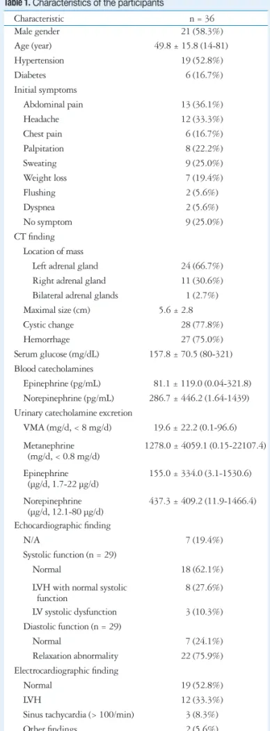
Table 1. Characteristics of the participants
Characteristic n = 36
Male gender 21 (58.3%)
Age (year) 49.8 ± 15.8 (14-81)
Hypertension 19 (52.8%)
Diabetes 6 (16.7%)
Initial symptoms
Abdominal pain 13 (36.1%)
Headache 12 (33.3%)
Chest pain 6 (16.7%)
Palpitation 8 (22.2%)
Sweating 9 (25.0%)
Weight loss 7 (19.4%)
Flushing 2 (5.6%)
Dyspnea 2 (5.6%)
No symptom 9 (25.0%)
CT finding Location of mass
Left adrenal gland 24 (66.7%) Right adrenal gland 11 (30.6%) Bilateral adrenal glands 1 (2.7%) Maximal size (cm) 5.6 ± 2.8
Cystic change 28 (77.8%)
Hemorrhage 27 (75.0%)
Serum glucose (mg/dL) 157.8 ± 70.5 (80-321) Blood catecholamines
Epinephrine (pg/mL) 81.1 ± 119.0 (0.04-321.8) Norepinephrine (pg/mL) 286.7 ± 446.2 (1.64-1439) Urinary catecholamine excretion
VMA (mg/d, < 8 mg/d) 19.6 ± 22.2 (0.1-96.6) Metanephrine
(mg/d, < 0.8 mg/d) 1278.0 ± 4059.1 (0.15-22107.4) Epinephrine
(µg/d, 1.7-22 µg/d) 155.0 ± 334.0 (3.1-1530.6) Norepinephrine
(µg/d, 12.1-80 µg/d) 437.3 ± 409.2 (11.9-1466.4) Echocardiographic finding
N/A 7 (19.4%)
Systolic function (n = 29)
Normal 18 (62.1%)
LVH with normal systolic
function 8 (27.6%)
LV systolic dysfunction 3 (10.3%) Diastolic function (n = 29)
Normal 7 (24.1%)
Relaxation abnormality 22 (75.9%) Electrocardiographic finding
Normal 19 (52.8%)
LVH 12 (33.3%)
Sinus tachycardia (> 100/min) 3 (8.3%)
Other findings 2 (5.6%)
CT: computerized tomography, VMA: vanillylmandelic acid, N/A: not assessed, LVH: left ventricular hypertrophy
Normal finding was the most common (19, 52.8%). LV hyper- trophy was found in 12 (33.3%), sinus tachycardia (> 100/min) in 3 (8.3%) and other findings (2 nonspecific ST segment change and 1 supraventricular tachycardia) in 5.6%. Because there was no clinical symptoms and electrocardiographic ab- normality, echocardiographic exam was not done in 7 patients.
Of total 29 patients, most common finding was normal LV sys- tolic function in 18 patients. Majority of patients (22, 75.9%) underwent echocardiographic examination showed relaxation abnormality.
Three patients showed transient LV systolic dysfunction (2 with inverted Takotsubo type cardiomyopathy and 1 with dif- fuse hypokinesia pattern). Common presenting symptoms were new onset chest discomfort and dyspnea. However, there was no relationship between the presence of LV systolic dys- function with tumor size or presence of necrosis in it. Their electrocardiographic examinations demonstrated sinus tachy- cardia without evidence of ST segment change. All three pa- tients admitted to the emergency department and cared in the intensive care unit. Their clinical profiles were summarized in the Table 2.
Case 1
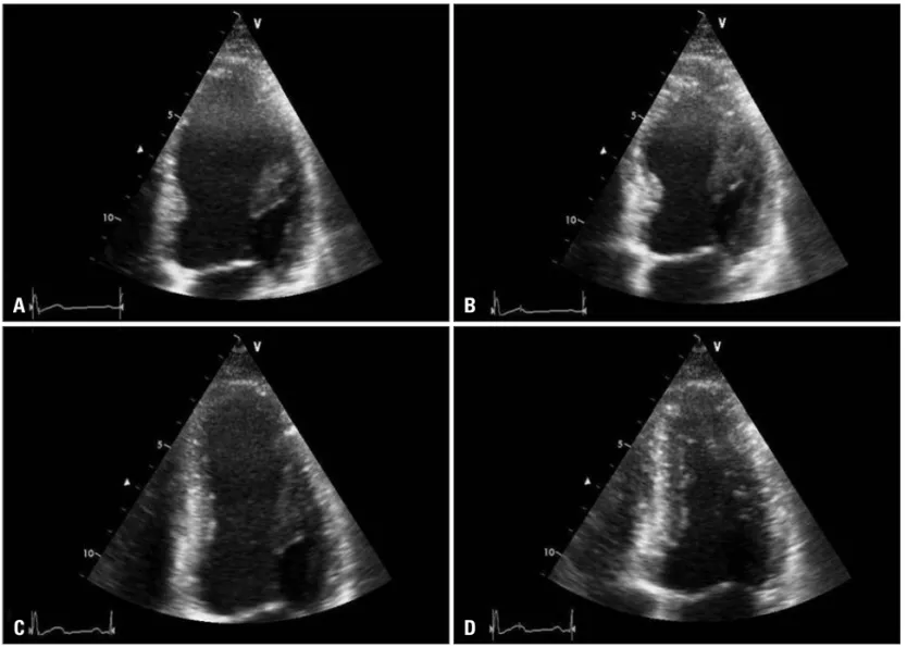
A 32-year-old man visited our emergency department with palpitation and squeezing chest pain. His initial blood pressure was 190/112 mmHg and electrocardiogram showed sinus tachycardia of 120/min. The initial electrocardiogram revealed slight ST-segment elevation in lead II, III, and aVF. Echocar- diography demonstrated a midventricular dilatation of the LV
and decreased LV ejection fraction of 35% (Fig. 1A and B).
However, the motion of the LV apex was preserved. Serum lev- els of cardiac biomarkers were increased. The serum level of N- terminal-pro-B type natriuretic peptide (NT-pro-BNP) was in- creased up to 733.3 pg/mL (normal range: < 200 pg/mL). The coronary arteries were free of any organic stenosis and LV an- giogram showed severe hypokinesia in the mid to basal portion with preserved LV apical wall motion. The serum and 24-hour urine studies were compatible with pheochromocytoma. An abdominal CT showed an approximately 7.8 × 8.3 × 10.0 cm large septated cystic mass with irregular wall enhancement of the right adrenal gland and scintigraphy scan with 123I-MIBG showed huge round uptake in the right adrenal gland. The patient was treated with oral furosemide, an oral alpha blocker and intravenous nitrate infusion. The low dose of beta blocker was initiated after alpha blockade. The follow up echocardiog- raphy after three days of treatment showed marked improve- ment of LV systolic function and decreased LV dimension.
The LV ejection fraction was 59%, with no regional wall mo- tion abnormalities (Fig. 1C and D).
Case 2
A 41-year old man presented to the emergency room due to new-onset dyspnea. His initial blood pressure was 180/100 mmHg and heart rate was 140/min. The initial electrocardio- gram demonstrated sinus tachycardia. Chest radiography showed pulmonary edema which improved after 3 days (Fig. 2).
Serum levels of cardiac biomarkers were increased. NT-pro- BNP was markedly increased up to 34,489 pg/mL. Echocar-
Table 2. Clinical profiles of the patients with transient left ventricular dysfunction
Case1 Case 2 Case 3
Gender / Age (year) Male / 32 Male / 41 Female / 49
Initial symptoms Palpitation, Chest discomfort Dyspnea Dyspnea, Chest discomfort
Initial chest X-ray finding Normal Pul edema Pul edema
Initial EchoCG finding Inverted-Takotsubo cardiomyopathy Inverted-Takotsubo cardiomyopathy Global hypokinesia
Time to normalization of EchoCG finding 3 days 3 days 3 days
Blood catecholamine
Epinephrine (pg/mL) N/A < 15 209.2
Norepinephrine (pg/mL) N/A < 25 299.8
Urinary catecholamine excretion
VMA (mg/d) 31.9 41.7 25.6
Metanephrine (mg/d) 22107.4 11.2 7.2
Epinephrine (µg/d) 1530.6 422.2 604.1
Norepinephrine (µg/d) 1038.8 921.0 699.6
NT-Pro BNP (pg/ mL) 733.3 34489 2458
Cardiac enzymes
CK (U/L) 138 3606 228
CK-MB (ng/mL) 3.84 109.3 20.5
Troponin-I (ng/mL) 1.74 21.56 6.3
EchoCG: echocardiography, VMA: vanillylmandelic acid, NT-Pro BNP: N-terminal Pro B-type natriuretic peptide, CK: creatinine kinase, Pul edema:
pulmonary edema
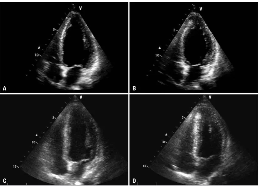
diography showed severe LV dysfunction and a contractile ab- normality, consisting of akinesis of the basal and mid ventricular segments and hyperkinesis of the apical segments (Fig. 3A and B). The patient was admitted at intensive care unit and intravenous administration of dobutamine, furosemide, and
digoxin. His symptoms were improved in one day despite of sporadic surge of blood pressure and pulse rate. The 24-hour urine study was compatible with pheochromocytoma. The ab- dominal CT revealed about 4.9 × 5.7 × 4.5 cm sized right ad- renal cystic mass with hemorrhage. Follow up echocardiogra-
Fig. 1. An apical four-chamber view of the left ventricle in the emergency department is shown at end-diastole (A) and end-systole (B). At end-systole, mid- and basal ventricular ballooning was noted. The follow-up echocardiographic study was performed after three days of treatment. The left ventricular dimension was decreased and ballooning had disappeared (C at end-diastole and D at end-systole).
A
C
B
D
Fig. 2. Chest radiography shows pulmonary edema which improved after 3 days of treatment. A: On admission. B: After treatment.
A B
phy, repeated after two days, demonstrated normalized LV systolic function and regional wall motion abnormalities (Fig.
3C and D). The elective laparoscopic right adrenalectomy was performed after 4 weeks of therapy including alpha and beta blockers.
Case 3
A 49-year-old woman with hypertension and diabetes melli- tus was admitted to emergency room with dyspnea and chest discomfort. She complained palpitation and recurrent headache for several months. Her initial blood pressure was 160/114
Fig. 3. An apical four-chamber view of the left ventricle in the emergency department is shown at end-diastole (A) and end-systole (B). Apical wall motion is normal. However, mid- and basal ventricular ballooning is noted. The follow-up echocardiographic study performed after three days of treatment demonstrates decreased left ventricular size and disappeared mid- and basal ventricular ballooning (C at end-diastole and D at end-systole).
A
C
B
D
Fig. 4. Chest radiography demonstrates marked pulmonary edema which normalized after 3 days of treatment. A: On admission. B: After treatment.
A B
mmHg and heart rate was 100/min. The initial electrocardio- gram demonstrated sinus tachycardia. Chest radiography showed marked pulmonary edema (Fig. 4). Because the patient showed severe hypoxemia, she was intubated and cared with ventilator in the intensive care unit. Her condition improved dramatically after 1 day and ventilator was removed after 2 days. Cardiac enzymes were slightly elevated and NT-pro-BNP increased up to 2,458 pg/mL. Echocardiography showed severe LV systolic dysfunction with an estimated ejection fraction of 20% and severe hypokinesia of all segments. A 24-hour urinary catecholamine excretion was compatible with pheochromocyto- ma. Follow up echocardiography, repeated after two days, dem- onstrated normalized LV systolic function and regional wall motion abnormalities. Cardiac catheterization was performed and angiographically normal epicardial coronary arteries. The patient was hemodynamically stabilized with conventional treatment. The abdominal CT scan revealed about 3.3 × 6.2 sized tumor of the left adrenal gland and 131I-MIBG scintigra- phy showed a hot-uptake lesion of the same location.
Discussion
The prevalence of catecholamine cardiomyopathy associated with pheochromocytoma is about 10% (3 of 29 patients) in our study. The patterns of cardiac involvement are 2 inverted- Takotsubo cardiomyopathies and 1 diffuse severe hypokinesia.
All three patients showed complete recovery with inotropic support and conventional treatment within 3 days.
Pheochromocytomas are rare catecholamine producing tu- mors typically located in the adrenal gland or along the sym- pathetic ganglia. Due to secretion of catecholamines, they usually present with a classic triad of headache, tachycardia and sweating.7)
Shub et al.8) reported echocardiographic patterns in 26 pa- tients with pheochromocytoma. In their data, most common echocardiographic finding was normal or increased systolic function (80% of the patients). The result of our study is con- sistent with this study. However, relaxation abnormality is the most common finding in our study (22, 75.8%). The increased incidence of diastolic dysfunction may be associated with in- jury of myocytes or increased oxidative stress by circulating catecholamines.9)10)
It has also been reported to cause cardiac manifestations in- cluding a transient, reversible cardiomyopathy (catecholamine cardiomyopathy).11) Shaw et al.11) firstly reported echocardio- graphic feature of catecholamine cardiomyopathy associated with pheochromocytoma in the late 1980s. It resembles Ta- kotsubo cardiomyopathy (transient apical ballooning syndrome) presenting with hyperkinesis of the basal segments and apical hypokinesis.3)12-14) The LV systolic dysfunction usually recovers completely within several days. Proposed potential mecha- nisms of transient LV ballooning include micro-vascular dys- function of the coronary arteries, multivessel epicardial spasm, impaired fatty acid metabolism, myocarditis and catechol-
amine-mediated myocardial dysfunction.15)16) Although the mechanism of the association between sympathetic stimula- tion and myocardial stunning is still unknown, several pro- posed mechanisms have been proposed including coronary ar- terial spasm17) and direct injury to myocytes.9) The apex is more vulnerable to a sudden increase in circulating catechol- amine levels may be due to different distribution of sympa- thetic nerves18) and dissimilar density of sympathetic nerves in the heart.19) More recently, a variant of Takotsubo cardiomyop- athy, the ‘inverted-Takotsubo cardiomyopathy’ has been de- scribed.15) It distinguished by dysfunction of the basal and mid-ventricular segments with preserved function of the api- cal segments. Cases of inverted-Takotsubo cardiomyopathy as- sociated with pheochromocytoma were also reported.20-22) Two of our cases showed inverted-Takotsubo cardiomyopathy which recovered completely within 3 days. The variations in segmen- tal involvement regardless of coronary anatomy in patients with excessive catecholamine levels may suggest different sus- ceptibility to sympathetic stimulation from individual to in- dividual.20)
Despite this study firstly described the prevalence of tran- sient LV systolic dysfunction with echocardiographic exami- nation, this study has many limitations. First, this study is a retrospective study with review of their medical records. The incidence and pattern of LV systolic dysfunction could not be assessed exactly. However, all three patients with LV systolic dysfunction complained new onset dyspnea and chest discom- fort led to visiting emergent department. Their serum NT-Pro BNP levels were elevated and echocardiographic examination revealed the presence of LV systolic dysfunction. Moreover, 9 of total patients had no presenting symptoms suggesting pres- ence of LV systolic dysfunction. Second, echocardiographic ex- amination was not performed in all participants. Echocardio- graphic examination was not performed in the patients without clinical symptoms and with normal electrocardiographic find- ings (7, 19.4%). Pheochromocytoma is a very rare tumor and the detection is difficult especially without presenting symp- toms. To evaluate the exact incidence and pattern of catechol- amine cardiomyopathy associated with pheochromocytoma, prospective study will be needed.
In conclusion, of total 36 patients with pheochromocytoma, 3 showed transient LV systolic dysfunction (catecholamine cardiomyopathy associated with pheochromocytoma). Two of their echocardiographic patterns were inverted-Takotsubo car- diomyopathy and 1 was diffuse LV hypokinesia. Their echo- cardiographic abnormalities were normalized with conven- tional treatment within 3 days.
References
1. Sheps SG, Jiang NS, Klee GG, van Heerden JA. Recent developments in the diagnosis and treatment of pheochromocytoma. Mayo Clin Proc 1990;65:88-95.
2. Bravo EL, Gifford RW Jr. Current concepts. Pheochromocytoma: diagno-
sis, localization and management. N Engl J Med 1984;311:1298-303.
3. Wood R, Commerford PJ, Rose AG, Tooke A. Reversible catechol- amine-induced cardiomyopathy. Am Heart J 1991;121:610-3.
4. Bybee KA, Prasad A. Stress-related cardiomyopathy syndromes. Circula- tion 2008;118:397-409.
5. Gerlo EA, Sevens C. Urinary and plasma catecholamines and urinary catecholamine metabolites in pheochromocytoma: diagnostic value in 19 cases.
Clin Chem 1994;40:250-6.
6. Moyer TP, Jiang NS, Tyce GM, Sheps SG. Analysis for urinary cate- cholamines by liquid chromatography with amperometric detection: method- ology and clinical interpretation of results. Clin Chem 1979;25:256-63.
7. Stein PP, Black HR. A simplified diagnostic approach to pheochromocyto- ma. A review of the literature and report of one institution’s experience.
Medicine (Baltimore) 1991;70:46-66.
8. Shub C, Cueto-Garcia L, Sheps SG, Ilstrup DM, Tajik AJ. Echocar- diographic findings in pheochromocytoma. Am J Cardiol 1986;57:971-5.
9. Bolli R, Marbán E. Molecular and cellular mechanisms of myocardial stunning. Physiol Rev 1999;79:609-34.
10. Yamanaka O, Yasumasa F, Nakamura T, Ohno A, Endo Y, Yoshimi K, Miura K, Yamaguchi H. “Myocardial stunning”-like phenomenon during a crisis of pheochromocytoma. Jpn Circ J 1994;58:737-42.
11. Shaw TR, Rafferty P, Tait GW. Transient shock and myocardial impair- ment caused by phaeochromocytoma crisis. Br Heart J 1987;57:194-8.
12. Schuiki ER, Jenni R, Amann FW, Ziegler WH. A reversible form of apical left ventricular hypertrophy associated with pheochromocytoma. J Am Soc Echocardiogr 1993;6:327-31.
13. Takizawa M, Kobayakawa N, Uozumi H, Yonemura S, Kodama T, Fukusima K, Takeuchi H, Kaneko Y, Kaneko T, Fujita K, Honma Y, Aoyagi T. A case of transient left ventricular ballooning with pheochro- mocytoma, supporting pathogenetic role of catecholamines in stress-induced cardiomyopathy or takotsubo cardiomyopathy. Int J Cardiol 2007;114:
e15-7.
14. Lassnig E, Weber T, Auer J, Nömeyer R, Eber B. Pheochromocytoma crisis presenting with shock and tako-tsubo-like cardiomyopathy. Int J Car- diol 2009;134:e138-40.
15. Hurst RT, Askew JW, Reuss CS, Lee RW, Sweeney JP, Fortuin FD, Oh JK, Tajik AJ. Transient midventricular ballooning syndrome: a new variant. J Am Coll Cardiol 2006;48:579-83.
16. Tsuchihashi K, Ueshima K, Uchida T, Oh-mura N, Kimura K, Owa M, Yoshiyama M, Miyazaki S, Haze K, Ogawa H, Honda T, Hase M, Kai R, Morii I; Angina Pectoris-Myocardial Infarction In- vestigations in Japan. Transient left ventricular apical ballooning without coronary artery stenosis: a novel heart syndrome mimicking acute myocardial infarction. Angina Pectoris-Myocardial Infarction Investigations in Japan.
J Am Coll Cardiol 2001;38:11-8.
17. Lacy CR, Contrada RJ, Robbins ML, Tannenbaum AK, Moreyra AE, Chelton S, Kostis JB. Coronary vasoconstriction induced by mental stress (simulated public speaking). Am J Cardiol 1995;75:503-5.
18. Pierpont GL, DeMaster EG, Cohn JN. Regional differences in adrener- gic function within the left ventricle. Am J Physiol 1984;246:H824-9.
19. Kawano H, Okada R, Yano K. Histological study on the distribution of autonomic nerves in the human heart. Heart Vessels 2003;18:32-9.
20. Kim EM, Park JH, Park YS, Lee JH, Choi SW, Jeong JO, Seong IW. Catecholamines may play an important role in the pathogenesis of tran- sient mid- and basal ventricular ballooning syndrome. J Korean Med Sci 2008;23:898-902.
21. Kim S, Yu A, Filippone LA, Kolansky DM, Raina A. Inverted-Ta- kotsubo pattern cardiomyopathy secondary to pheochromocytoma: a clinical case and literature review. Clin Cardiol 2010;33:200-5.
22. Kim TS, Chu EH, Kang HH, Chun SW, Cho EJ, Kim JH. A case of reversal of takotsubo cardiomyopathy in patient with pheochromocytoma. J Cardiovasc Ultrasound 2007;15:50-4.