An Unruptured Anterior Communicating Artery Aneurysm with Bilateral Infraoptic Anterior Cerebral Arteries. Case Report and Review of the Literature
Michelle H. Chua1, Ajith J. Thomas2, Matthew R. Fusco2, Christopher S. Ogilvy2
1Harvard Medical School, 2Department of Neurosurgery, Beth Israel Deaconess Medical Center, Brain Aneurysm Institute, Harvard Medical School, Boston, MA, United States
Variations of the anterior cerebral artery-anterior communicating artery complex are commonly identified in aneurysm surgery. An infraoptic course of the anterior cerebral artery is exceedingly rare. Robison first de- scribed this anomaly from an anatomic dissection in 1959. A unilateral anomalous infraoptic anterior cerebral artery is more common than anomalies of bilateral infraoptic anterior cerebral arteries. We present the case of an unruptured aneurysm at the anterior communicating artery in a patient with bilateral infraoptic anterior cerebral arteries, identified by computed tomography angiography and verified during surgery. Implications for aneurysm formation and surgical treatment are discussed.
J Cerebrovasc Endovasc Neurosurg.
2014 December;16(4):368-373 Received : 4 November 2014 Revised : 11 December 2014 Accepted : 16 December 2014 Correspondence to Christopher S. Ogilvy Department of Neurosurgery, Beth Israel Deaconess Medical Center, 110 Francis Street, Suite 3B Boston, MA 02215, USA
Tel : 1-617-632-7246 Fax : 1-617-632-0949
E-mail : [email protected] ORCID : http://orcid.org/0000-0001-7538-8095
This is an Open Access article distributed under the terms of the Creative Commons Attribution Non- Commercial License (http://creativecommons.org/li- censes/by-nc/3.0) which permits unrestricted non- commercial use, distribution, and reproduction in any medium, provided the original work is properly cited.
Keywords Anterior cerebral artery, Aneurysm, Vascular surgical procedure
INTRODUCTION
The A1 segment of the anterior cerebral artery (ACA) usually arises from the internal carotid artery (ICA) bifurcation and extends anteromedially above the optic nerve or chiasm to join the anterior commu- nicating artery (ACoA). Variations of the ACA-ACoA complex are commonly identified in aneurysm surgery. An infraoptic course of the ACA is exceed- ingly rare. Robinson first described this anomaly from an anatomic dissection in 1959.12) A unilateral anom- alous infraoptic ACA is more common than anomalies of bilateral infraoptic ACAs.4) We report on the case of an unruptured AcoA aneurysm in a patient with bilateral infraoptic ACAs. Implications for aneurysm formation and surgical treatment are discussed.
CASE REPORT
Our patient developed symptoms of episodic left arm twitching. His family history included significant intracranial aneurysms. Imaging demonstrated a 5-mm ACoA aneurysm which projected posteriorly with early bifurcations of the ICA bilaterally (Fig. 1, Fig. 2). The A1 segments were identified as they trav- ersed the pituitary fossa in an unusually low and lat- erally directed course, indicating their infraoptic nature.
Discussion with the patient resulted in the decision to proceed with clip obliteration of the aneurysm due to significant family history and extreme anxiety re- garding the diagnosis. A standard right pterional cra- niotomy was performed and as the optic nerve was exposed for CSF drainage, the infraoptic course of the
A B
C D
Fig. 1. Preoperative computed tomography angiogram demonstrates the ACoA aneurysm (A) and low bifurcation of the internal carotid artery (B, C). Reconstructed images show the aneurysm, which projects posteriorly (D).
ipsilateral A1 became visible. The contralateral A1 was then exposed and showed the same course across the skull base (Fig. 3). Once proximal control was de- fined, the ACoA was exposed along with the bilateral A2 segments. As predicted by the preoperative CTA,
the aneurysm projected posteriorly. Both A2 segments projected anteriorly and swept around the base of the lesion. Each of the recurrent arteries of Huebner arose from its typical location in the proximal A2 segment.
Proximal control with bilateral A1 temporary clipping
A B
Fig. 2. Axial (A) and sagittal (B) imaging demonstrate the aneurysm, which projects posteriorly.
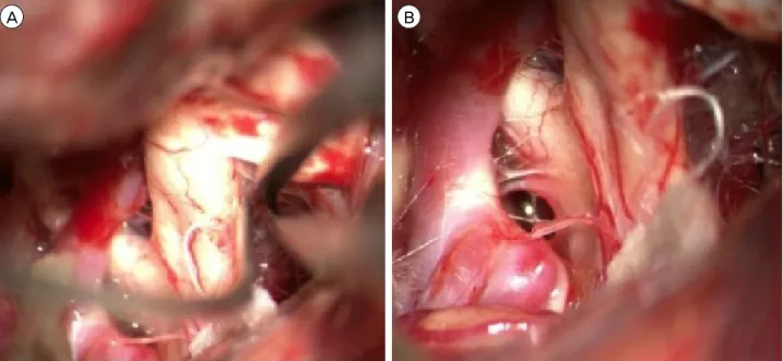
A B
Fig. 3. View from right pterional craniotomy shows an infraoptic A1 segment. The ACA projects medially underneath the optic nerve and arises anterior to the optic chiasm (A) in the pre-chiasmatic space (B).
was performed and final aneurysm dissection with neck preparation was achieved. The aneurysm was clipped uneventfully, the temporary A1 clips were re- moved, and ICG video angiography was used to identify aneurysm obliteration and preservation of the parent vessels. The patient was discharged home on
post-operative day 2. Overall post-operative course was uneventful.
DISCUSSION
An infraoptic ACA is associated with a low bifurca-
Author Year Neurological disease and treatment Patient age and
gender Bilateral or unilateral
infraoptic A1 Direction of aneurysm
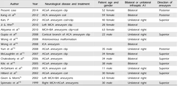
Present case 2014 ACoA aneurysm clip 52 female Bilateral Posterior
Kang et al9) 2012 ACA aneurysm coil 59 female Bilateral Posterior
Kan, P 2012 ACoA aneurysm coil+clip 40 female Unilateral right Superior
Ji & Ahn8) 2010 Left MCA aneurysm clip 28 female Bilateral
Akiyama et al1) 2010 MCA+BA aneurysms clip+coil 63 female Unilateral right
Gupta et al6) 2008 Cortical branch of ACA aneurysm clip 22 male Unilateral right Superior
Wong et al14) 2008 Arteriovenous malformation Unilateral right
Wong et al14) 2008 ICA aneurysm Bilateral
Yurt et al15) 2008 ACoA aneurysm clip 35 male Unilateral right Posterior
McLaughlin et al11) 2007 ACoA aneurysm clip 34 female Bilateral Superior
Chakraborty et al3) 2006 ACoA aneurysm 34 male Bilateral Superior
Kilic et al10) 2005 ACoA aneurysm clip 38 male Bilateral Superior
Al-Qahtani et al2) 2003 ACA aneurysm coil 11 male Unilateral right Superior
Hillard et al7) 2002 ACoA aneurysm coil 30 female Unilateral right Superior
Given & Morris5) 2002 Left MCA+BA aneurysms 63 female Unilateral right
Spinnato et al13) 1999 Right MCA+ACoA aneurysms 30 male Unilateral right Superior ACoA = anterior communicating artery; ACA = anterior cerebral artery; MCA = middle cerebral artery; ICA = internal carotid artery; BA = basilar artery
Table 1. Infraoptic anterior cerebral arteries reported in the literature
tion of the ICA and suggests abnormal development of the circle of Willis.13) This has been associated with other coexisting vascular anomalies including normal ACoA complex with an additional infraoptic vessel that forms an anastomosis between the ICA and ACA;
absent ipsilateral or contralateral A1; early bifurcation of the ICA at the level of the ophthalmic artery; and termination of an accessory infraoptic ACA in normal frontal brain parenchyma in the presence of a normal ACoA complex.
Bilateral infraoptic ACAs are less common than uni- lateral anomalies.8) One report describes an associated gyral abnormality.11) The anomalous ACA usually arises from the intra-dural ICA near the level of the ophthalmic artery, as it did in the presented case, or rarely from the extra-dural ICA.9)
The most important clinical association of such an anomaly is the high incidence of intracranial aneur- ysms of the ACA-ACoA complex.5)8)14) Recognition of this anomaly is important in planning surgery for ACA-ACoA complex aneurysms in order to identify
proximal arterial control of the aneurysm. Failure to account for this anomaly might result in unnecessary dissection along and possible damage to the optic ap- paratus or inferior frontal lobe during aneurysm repair.
In the case of this anomaly, surgical treatment of the aneurysm at the ACoA may be difficult because the proximal ACA may be obscured by the optic nerve.
In addition, the altered course of the A1 may change the orientation of the ACoA complex (and thus its im- portant perforators which normally project posteri- orly) or the Artery of Heubner as it arises from the proximal A2 or distal A1. Careful radiographic evalu- ation and understanding of variations of the ACA are essential for surgery. In this case, pre-operative knowledge of these anatomic variants enabled early identification of the ICA bifurcation under the optic nerve and unnecessary dissection along the inferior frontal lobe or optic apparatus was avoided. In this case, given the early medial infraoptic course of the A1 arteries, establishment of proximal control was comparatively easy.
Pre-operative evaluation of the ACoA complex anat- omy is critical in the clipping of an ACoA aneurysm (as it is for any aneurysm location). Exposure of aneurysms located more than 1 cm above the skull base will be more difficult due to increased need for frontal lobe retraction as well as possibly interhemi- spheric dissection and gyrus rectus resection just to visualize the aneurysm. In addition, clipping of poste- riorly projecting ACoA aneurysms is typically more difficult as the aneurysm projects in the same direc- tion as the important perforator arteries. A posterior projecting aneurysm is often found in cases of an in- fraoptic A1 given the course of the ACA from inferior to superior as it turns under the optic nerve towards the ACoA complex (Table 1). However, in the re- ported case, the unusually anterior location of the ACoA complex actually made aneurysm exposure easier. In addition, the perforating vessels from the ACoA were pushed above the lesion, making safe surgical clipping without compromise of the asso- ciated perforating arteries more likely.
CONCLUSION
Safety in surgical clipping of intracranial aneurysms is achieved through many steps. Thorough pre-oper- ative evaluation of the aneurysm as well as its parent vasculature is critical to achieving good surgical results.
An infraoptic course of A1 from an early ICA bifurca- tion is rare; bilateral infraoptic A1 courses are even rarer. A pre-disposition to ACoA aneurysms appears pos- sible with an infraoptic A1 course given the previous reports of association as well as this case. Identification of this anomaly pre-operatively can enhance surgical safety in aneurysm clipping and unnecessary and po- tentially dangerous dissection can be avoided.
STATEMENT OF CONTRIBUTORSHIP
MHC and MRF drafted the manuscript. AJT and CSO revised the draft manuscript.
Disclosure
This authors have no personal financial or institu- tional interest in any of the materials or devices de- scribed in this article.
REFERENCES
1. Akiyama Y, Okada T, Hayashi N, Yokoi T. Infraoptic course of the anterior cerebral artery originating from the extradural internal carotid artery associated with contralateral internal carotid artery agenesis and multi- ple intracerebral aneurysms. Neurol Med Chir (Tokyo).
2010;50(11):984-7.
2. Al-Qahtani S, Tampieri D, Brassard R, Sirhan D, Mellanson D. Coil embolization of an aneurysm associated with an infraoptic anterior cerebral artery in a child. AJNR Am J Neuroradiol. 2003 May;24(5):990-1.
3. Chakraborty S, Fanning NF, Lee SK, Terbrugge KG.
Bilateral infraoptic origin of anterior cerebral arteries: a rare anomaly and its embryological and clinical significance.
Interv Neuroradiol. 2006 Jun;12(2):155-9.
4. Fujimoto S, Murakami M. Anomalous branch of the internal carotid artery supplying circulation of the an- terior cerebral artery. Case report. J Neurosurg. 1983 Jun;58(6):941-6.
5. Given CA 2nd, Morris PP. Recognition and importance of an infraoptic anterior cerebral artery: case report.
AJNR Am J Neuroradiol. 2002 Mar;23(3):452-4.
6. Gupta V, Chugh M, Vaishya S. Infraoptic azygous ante- rior cerebral artery. Neurol India. 2008 Oct-Dec;56(4):487-8.
7. Hillard VH, Musunuru K, Nwagwu C, Das K, Murali R, Zablow B, et al. Treatment of an anterior communi- cating artery aneurysm through an anomalous anasto- mosis from the cavernous internal carotid artery. J Neurosurg. 2002 Dec;97(6):1432-5.
8. Ji C, Ahn JG. Infraoptic course of both anterior cerebral arteries. J Korean Neurosurg Soc. 2010 Jan;47(1):71-3.
9. Kang HJ, Lee YS, Suh SJ, Lee JH, Ryu KY, Kang DG.
A ruptured aneurysm at the infraoptic azygous anterior cerebral artery with the contralateral internal carotid ar- tery agenesis treated by Y-stent assisted coil embolization.
J Cerebrovasc Endovasc Neurosurg. 2012 Sep;14(3):237-42.
10. Kilic K, Orakdogen M, Bakirci A, Berkman Z. Bilateral internal carotid to anterior cerebral artery anastomosis with anterior communicating artery aneurysm: technical case report. Neurosurgery. 2005 Oct;57(4 Suppl):E400;
discussion E400.
11. McLaughlin N, Bojanowski MW. Infraoptic course of an- terior cerebral arteries associated with abnormal gyral segmentation. Case report. J Neurosurg. 2007 Aug;107(2) :430-4.
12. Robinson LR. An unusual human anterior cerebral artery.
J Anat. 1959 Jan;93(1):131-3.
13. Spinnato S, Pasqualin A, Chioffi F, Da Pian R. Infraoptic course of the anterior cerebral artery associated with an anterior communicating artery aneurysm: anatomic case
report and embryological considerations. Neurosurgery.
1999 Jun;44(6):1315-9.
14. Wong ST, Yuen SC, Fok KF, Yam KY, Fong D. Infraoptic anterior cerebral artery: review, report of two cases and an anatomical classification. Acta Neurochir (Wien). 2008 Oct;150(10):1087-96.
15. Yurt A, Uçar K, Ozer F, Oran I, Arda N. A rare em- bryologic variation: anterior communicating artery aneur- ysm associated with carotid-anterior cerebral artery anas- tomosis or infraoptic course of the anterior cerebral artery.
Clin Med Case Rep. 2008 Sep;1:123-5.