Annals of Surgical Treatment and Research 209
pISSN 2288-6575 • eISSN 2288-6796 http://dx.doi.org/10.4174/astr.2014.87.4.209 Annals of Surgical Treatment and Research
CASE REPORT
Small bowel diverticulosis in patient with early gastric cancer
Pyeong Su Kim, Eun-Joo Jung, Ho Yoon Bang
Department of Surgery, Konkuk University Medical Center, Konkuk University School of Medicine, Seoul, Korea
INTRODUCTION
Diverticula of the jejunum and ileum are rare in adults [1]. In the small intestine, duodenal diverticula are nearly five times more common than jejunoileal diverticula [2]. The majority of patients with small bowel diverticula remain symptom free, yet, acute complications may happen which include obstruction, gastrointestinal bleeding, diverticulitis and perforation. Chro- nic symptoms are also reported in these patients, which in- clude intermittent abdominal pain, flatulence, diarrhea and constipation [3].
In South Korea, the second most common organ cancer develops in the stomach [4]. Resectable treatment options are subtotal or total gastrectomy, which are based on the site of the tumor. Reconstruction options after subtotal gastrectomy are gastroduodenostomy or gastrojejunostomy.
In the literature, there are case reports of gastric cancer accompanied Meckel’s diverticulum or duodenal diverticulum.
But, combination of gastric cancer and multiple diverticula of duodenum and jejunum is not reported. Here, we report a case of early gastric cancer which had duodenal and jejunal diverticula.
CASE REPORT
A 67-year-old woman was admitted to Konkuk University Medical Center with the chief complaint of chronic diarrhea and a significant weight loss of 19 kg over 2 months. The patient also complained of general weakness and dizziness along without abdominal pain. Past medical history was otherwise unremarkable but her surgical history showed that the patient had undergone appendectomy 19 years prior to this presentation and balloon dilatation of a duodenal stricture 4 years before her current presentation which was not found to be of a known origin.
Her laboratory tests at the time of admission showed elevated
Received December 13, 2013, Revised January 7, 2014, Accepted January 14, 2014
Corresponding Author: Ho Yoon Bang
Department of Surgery, Konkuk University Medical Center, Konkuk University School of Medicine, 120 Neungdong-ro, Gwangjin-gu, Seoul 143-729, Korea
Tel: +82-2-2030-7598, Fax: +82-2-2030-7346 E-mail: [email protected]
Copyright ⓒ 2014, the Korean Surgical Society
cc Annals of Surgical Treatment and Research is an Open Access Journal. All articles are distributed under the terms of the Creative Commons Attribution Non- Commercial License (http://creativecommons.org/licenses/by-nc/3.0/) which permits unrestricted non-commercial use, distribution, and reproduction in any medium, provided the original work is properly cited.
Jejunal and ileal diverticula are rare in adults. Duodenal diverticula are five times more prevalent than jejunoileal diverticula. Most patients are asymptomatic. However, chronic symptoms including intermittent abdominal pain, flatulence, diarrhea and constipation are seen in 10%–30% of patients. Gastric cancer is the second most common cancer in South Korea and here we report a case of early gastric cancer with multiple duodenal and jejunal diverticula. A 67-year-old woman was admitted to Konkuk University Medical Center with chronic diarrhea and weight loss of 19 kg over 2 months.
Following gastroduodenoscopy, we identified adenocarcinoma of the lower body of the stomach. On abdominopelvic computed tomography, diverticula of duodenum and jejunum were found. Patient underwent distal gastrectomy and gastroduodenostomy with lymphadenectomy. She was discharged on the tenth postoperative day without complications.
[Ann Surg Treat Res 2014;87(4):209-212]
Key Words: Small Intestinal diverticulosis, Stomach neoplasms, Malabsorption syndromes
210
Annals of Surgical Treatment and Research 2014;87(4):209-212
WBC count of 11,190/mm3 (normal range, 4,000–10,000/mm3) and low hemoglobin level of 9.6 g/dL (normal range, 12.0–16.0 g/dL). We also found low albumin level of 1.6 g/dL (normal range, 3.3–5.3 g/dL) and 2.9-mmol/L low potassium level (normal
range, 3.5–5.5 mmol/L). The patient had prolonged prothrombin time of 23.7 seconds (normal range, 11.7–14.7 seconds) and prolonged activated partial thromboplastin time of 75.2 seconds (normal range, 29.0–45.0 seconds). Urinalysis found 3 WBC in high-power field (negative is normal) and bacteria. Based on these findings, we had initial impressions of protein losing gastroenteropathy and urinary tract infection.
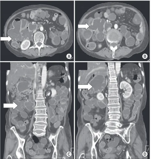
In gastroduodenoscopy, we identified a 5-mm ulcerative lesion on the greater curvature of the lower body of the stomach and duodenal diverticula on second and third parts of the duodenum (Fig. 1). The lesion of the stomach was endo- scopically clipped. Pathology study of the endoscopic biopsy confirmed a moderately differentiated adenocarcinoma. Abdo- minopelvic computed tomography (APCT) revealed multiple diverticula in duodenum and jejunum. Small bowel loops were slightly dilated with fluid and there was no evidence of inflammation around diverticula (Fig. 2). APCT did not find lymphadenopathy or distant metastasis, therefore, the clinical stage of this gastric cancer was cT1N0M0.
During the surgery, no gastric tumor could be palpated but endoscopic clip was palpated. There was no peritoneal seed-
Fig. 2. Abdominopelvic com- puted tomography findings.
Arrows point to the diverticula in duodenum and jejunum. (A, B) Axial view, (C, D) coronal view.
Fig. 1. Diverticula in the second and third portions of the duodenum on gastroduodenoscopy.
Annals of Surgical Treatment and Research 211 ing or metastatic lesion. The observable site of pylorus was
displaced proximally due to the previous ulcer scar (Fig. 3A).
We found diverticula with 2- to 3-cm size on second and third parts of the duodenum (Fig. 3B). In a distance of about 150 cm from the ligament of Treitz to the jejunum, we identified 20–30 diverticula on the mesenteric border of the small bowel which had a median size of 3–4 cm (Fig. 3C, D). Distal gastrectomy and gastroduodenostomy with lymphadenectomy concluded the operation.
Pathology report confirmed moderately differentiated adeno- carcinoma with mucosal invasion. There was no metastasis in the 21 retrieved lymph nodes. Final TNM staging was T1N0M0, stage Ia.
The patient recovered without complications and was dis- charged on the 10th postoperative day.
DISCUSSION
Diverticula of the jejunum and ileum are not commonly seen
in adults; occurring in approximately 1%–2% of the population [1]. Duodenal diverticula are five times more prevalent than jejunoileal diverticula [2]. Duodenal diverticula are divided into primary congenital diverticula and secondary acquired pseudodiverticula. Congenital diverticula of the duodenum are very rare in which all layers of the intestinal wall are herniated [5]. The etiology of small bowel pseudodiverticula is unclear but it is believed that motor dysfunction or intestinal dyskinesia leads to the formation of a diverticulum [6]. Following the increase in intraluminal pressures, the mucosa and submucosa protrude through the weakest site of the muscularis layer [6]. In general, the weakest site of the bowel wall is on the mesenteric border [6].
Patients with small bowel diverticula are typically asymp- tomatic. However, acute complications such as obstruction, gastrointestinal bleeding, diverticulitis, and perforation can occur which may need emergent surgical intervention. The treatment of choice in these settings is segmental resection of small intestine harboring the diverticulum [7,8]. Chronic Fig. 3. Intraoperative findings showing diverticula in duodenum and jejunum. (A) Previous ulcer scar (arrow). (B) Diverticula in the second portion of the duodenum (arrows). (C) Jejunal diverticula on the mesenteric border of the small bowel. (D) Jejunal diverticula become smaller as we move distally until the diverticula disappear (arrow: terminal end of diverticula).
Pyeong Su Kim, et al: Small bowel diverticulosis
212
Annals of Surgical Treatment and Research 2014;87(4):209-212
1. Longo WE, Vernava AM 3rd. Clinical implications of jejunoileal diverticular disease. Dis Colon Rectum 1992;35:381-8.
2. Akhrass R, Yaffe MB, Fischer C, Ponsky J, Shuck JM. Small-bowel diverticulosis:
perceptions and reality. J Am Coll Surg 1997;184:383-8.
3. Choi JJ, Ogunjemilusi O, Divino CM.
Diagnosis and management of diverticula in the jejunum and ileum. Am Surg 2013;
79:108-10.
4. Jung KW, Won YJ, Kong HJ, Oh CM, Seo HG, Lee JS. Cancer statistics in Korea:
incidence, mortality, survival and preva- lence in 2010. Cancer Res Treat 2013;45:1- 14.
5. Thorson CM, Paz Ruiz PS, Roeder RA, Sleeman D, Casillas VJ. The perforated duodenal diverticulum. Arch Surg 2012;
147:81-8.
6. Woods K, Williams E, Melvin W, Sharp K.
Acquired jejunoileal diverticulosis and its complications: a review of the literature.
Am Surg 2008;74:849-54.
7. Koger KE, Shatney CH, Dirbas FM, Mc-
Clena than JH. Perforated jejunal diver ti- cula. Am Surg 1996;62:26-9.
8. Lee BG, Park SB, Byun CG, Koh YT, Suh DY, Park DS, et al. Localized peritonitis due to perforation of multiple jejunal diver ticula. J Korean Surg Soc 2004;67:75- 8.
9. Kassahun WT, Fangmann J, Harms J, Bartels M, Hauss J. Complicated small- bo wel diverticulosis: a case report and review of the literature. World J Gastroen- terol 2007;13:2240-2.
REFERENCES
symptoms such as intermittent abdominal pain, flatulence, diarrhea, and constipation are reported in 10%–30% of patients with jejunoileal diverticula [3]. Conservative management is recommended when acute complication are not seen [8].
The reason for malabsorption is not clear but stasis in diverticular lumen is hypothesized to be the cause. Stasis results in an overgrowth of coliform organisms similar to the blind loop syndrome. It leads to vitamin B12 deficiency and consequently megaloblastic anemia. Steatorrhea could also develop. These complex pathophysiologic changes lead to malabsorption [1,6]. Medical treatment is the first step of management in patients with malabsorption including our presented case. These treatment measures include admini- stration of oral antibiotics (e.g., metronidazole), high-protein low-residue diet and vitamin supplementation [1,2,9]. Surgical treatment is reserved for patients who are intractable to medical treatment or who have serious malabsorption or steatorrhea.
It is widely accepted that the surgical treatment of early gastric cancer on the lower body of the stomach is distal gas-
trectomy with lymphadenectomy. After radical resection, surgeon decides the method of anastomosis which could be either gastroduodenostomy or gastrojejunostomy. This decision is difficult to make in patients with small bowel diverticula. Duodenal or jejunal diverticula around the site of the anastomosis may lead to complications such as anastomosis leakage, bleeding, ulcer and perforation which develop after direct contact with incompletely digested food. In our presented case, gastroduodenostomy was performed. There were two reasons behind this decision. First, gastrojejunostomy could become more complicated because stasis in the afferent loop can increase the intraluminal pressure in the diverticula and cause more diverticular complications. Second, this anastomosis method is closer to physiologic states.
CONFLICTS OF INTEREST
No potential conflict of interest relevant to this article was reported.