2020 by Korean Society for Surgery of the Hand, Korean Society for Microsurgery, and Korean Society for Surgery of the Peripheral Nerve. All Rights reserved.
This is an open-access article distributed under the terms of the Creative Commons Attribution license (http://creativecommons.org/licenses/by/4.0/), which permits unrestricted use, distribution, and reproduction in any medium, provided the original work is properly cited.
무릎주위 피부연조직결손의 재건: 증례 보고
장남, 신현우, 윤근철
성균관대학교 의과대학 강북삼성병원 성형외과
Reconstruction of Soft-Tissue Defect Around the Knee Using a Pedicled Reverse-Flow
Anterolateral Thigh Flap: A Case Report
Nam Jang, Hyun Woo Shin, Kun Chul Yoon
Department of Plastic and Reconstructive Surgery, Kangbuk Samsung Hospital, Sungkyunkwan University School of Medicine, Seoul, Korea
Coverage of traumatic soft-tissue defects around the knee is a challenging problem for reconstructive surgeons though many reconstructive options are available. We planned to use a pedicled reverse-flow anterolateral thigh (ALT) flap using the distal branch of the descending branch of the lateral circumflex femoral artery (LCFA) for pedicle length extension in a patient with the ALT perforator branch originating from the proximal portion of the descending branch of LCFA. We present the successful use of a pedicled reverse-flow ALT flap to cover a soft tissue defect around the knee.
Key Words: Lower extremity, Surgical flaps, Reconstructive surgical procedures
Introduction
Soft tissue coverage around the knee after trauma remains a challenge for plas- tic and reconstructive surgeons. The anterolateral thigh (ALT) flap was first de- scribed in 1984 by Song et al. [1] and has become the most commonly used per- forator flap due to its versatility, ease of use, and minimal donor site morbidity [2,3]. Perfusion of the ALT flap is based on the musculocutaneous or septocuta- neous perforators of the descending branch of the lateral circumflex femoral ar- tery (LCFA) [4], which connects distally with the lateral superior genicular artery or the profunda femoral artery [4]. The pedicled reverse-flow ALT flap was first described by Zhang [5], who used a retrograde vascular pedicle to prevent active bleeding from the distal end of the descending branch of the LCFA. In the de- scribed case, we planned a pedicled reverse-flow ALT flap for pedicle length ex- tension with the distal descending branch of the LCFA to address possible insuffi- cient pedicle length because the perforator emerged from a proximal portion of the descending branch.
Case Report
A 69-year-old female had a left tibia plateau open fracture with large degloving pISSN 2586-3290 · eISSN 2586-3533
Arch Hand Microsurg 2020;25(1):55-59 https://doi.org/10.12790/ahm.19.0038
Case Report
Received: July 29, 2019 Revised: November 4, 2019 Accepted: November 26, 2019 Corresponding author:
Hyun Woo Shin
Department of Plastic and Reconstructive Surgery, Kangbuk Samsung Hospital, Sungkyunkwan University School of Medicine, 29, Saemunan-ro, Jongno-gu, Seoul 03181, Korea
Tel: +82-2-2001-2178 Fax: +82-2-2001-2177 E-mail: [email protected] ORCID:
https://orcid.org/0000-0003-4396-3395
injury resulting from a traffic accident. After several surgical debridements, the resultant defect measured 20.0 cm×15.0 cm with significant patella tendon exposure (Fig. 1). Prior to sur- gery, a perforator pedicled propeller flap using a musculocuta-
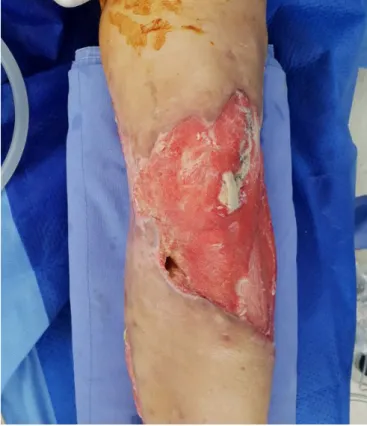
neous or septocutaneous perforator branched from the distal portion of the thigh was planned. Doppler mapping of perfora- tors was performed before flap elevation. The method used was similar to the conventional method used to harvest an ALT flap, that is, with the identified perforator in the center, a 12.0 cm× 4.0 cm skin paddle was designed to cover the exposed patella tendon of the anterolateral knee. A search for cutaneous perforators was performed in the suprafascial and subfascial plane, but we failed to identify a perforator branched from dis- tal portion of thigh intraoperatively. The only musculocutane- ous perforator found branched at the proximal descending branch of the LCFA 12.0 cm above the lateral edge of the patel- la. So, the preoperative plan was changed to a pedicled re- verse-flow ALT flap. The identified perforator was dissected in a retrograde fashion, and the proximal pedicle was clamped us- ing a vascular clamp and the flap pedicle was isolated distally along the descending branch until a pivot point was reached 7 cm above the knee joint that allowed sufficient pedicle length without tension. Intramuscular dissection of the perforator with skeletonization of the descending branch helped increase the degree of flap advancement, and the pedicled reverse-flow ALT flap was elevated and transposed to cover the exposed pa- tella tendon. The pedicle length was 10 cm (Fig. 2A, 2B). The donor site was closed primarily and the other skin defect area around the knee without tendon exposure was covered with a split thickness skin graft from the right thigh. The wound healed completely without any complication and remarkable limitation of range of movement after 3 months (Fig. 3).
Written informed consents were obtained.
Fig. 1. A soft tissue (20.0 cm×15.0 cm) defects above the knee following several debridement, with significant patella tendon exposure on anterolateral side of the knee.
Fig. 2. (A) A pedicled reverse-flow anterolateral thigh flap (12.0 cm×4.0 cm sized) was harvested using musculocutaneous perforator of descending branch of the lateral circumflex femoral artery (LCFA). Descending branch of the LCFA and the perforator to the flap was dissected intramuscularly. (B) The pivot point was determined at about 8 cm above the knee joint and the pedicled reverse-flow anterolateral thigh flap was advanced to cover the patella tendon exposure area.
A B
long enough to reach suprapatellar defects and are capable of resurfacing the whole knee. However, it is technically difficult to dissect ALT perforator flaps, especially when they are small, because perforating arteries exhibit wide anatomic variations.
In 1998, Kimata et al. [8] classified the perforators of ALT flaps into 8 types according to the branching pattern of the perfora- tor vessel.
The pedicled reverse-flow ALT flap is based on perforators of the descending branch of LCFA. Zhang [5] introduced the con- cept of a pedicled reverse-flow ALT flap in 1990. However, de- spite its several advantages and low donor site morbidity, the pedicled reverse-flow ALT flap has not been widely adopted, possibly because of wide anatomic variations of the vascular pedicle and a high risk of venous congestion [9]. In addition, reverse-flow based flaps are prone to congestion in peripheral portions of flaps, and this is followed by partial necrosis, be- cause of insufficient venous drainage caused by the resistance of venous valves. In a recent study, a modified method was de- scribed that reduces venous congestion, wherein the venae comitantes is anastomosed to the proximal stump in an antero- grade manner [7,10]. Wong et al. [9] reported sixteen cases of pedicled reverse-flow ALT flaps. Of 16 cases, venous conges- tion was noted in 8 cases, 3 of which were salvaged with venous supercharging with the long saphenous vein. Two that were not venous supercharged underwent partial flap necrosis and all other flaps healed without complications. In our case, there was no sign of severe venous congestion, and thus, no additional venous augmentation was performed.
Originally, we planned a perforator pedicled propeller flap to cover the soft tissue defect around the left knee based on pre- operative doppler mapping findings, but intraoperatively, we were unable to find the perforator near the midpoint of the thigh. We only found a musculocutaneous perforator from a proximal region of the descending branch of the LCFA, which prevented use of a propeller flap. Accordingly, the reconstruc- tion plan was changed to a pedicled reverse-flow ALT flap with pedicle length extension to enable the elevated flap to reach the knee tendon extension area. To achieve this, the proximal branch of the LCFA was clamped and its distal branch was dis- sected to increase flap mobility sufficiently to reach the ex- posed knee tendon area without excessive tension.
Due to anatomic variations of ALT flap perforators, surgeons might encounter unplanned situations during surgery. If a planned flap cannot be applied, a decision must be made whether to adopt an alternative reconstructive method or some other method in same operative field. Notably, ALT flap pedicle lengthening might be required in same operative field and may Fig. 3. Photograph of the flap at 3 months of follow-up. Satis-
factory healing and contour achieved.
Discussion
Coverage of traumatic soft-tissue defects around the knee re- mains a challenging problem for reconstructive surgeons be- cause of a lack of an adequate recipient vessel in anterior, lateral knee area and the need for a thin, pliable knee joint coverage.
Various options have been utilized to provide soft tissue recon- struction for these defects. The use of a free flap is not always preferable in this region, due to high donor site morbidity, long surgical time, and difficulties associated with selecting an ap- propriate recipient vessel positioned deeply around the knee.
Pedicled muscle flaps have been the workhorse for coverage of defects around the knee for decades. The gastrocnemius mus- cle flap has proved to be the most reliable, safest, and easiest surgical option [6]. However, perforator flaps have recently rev- olutionized soft tissue reconstruction around the knee, due to low donor morbidity and available pedicle length. Gravvanis et al. [7] demonstrated the use of a pedicled reverse-flow ALT flap for knee reconstruction and considered it super to the gastroc- nemius muscle flap, because of its greater size, shape, and flexi- bility, better color and texture match, and less bulkiness. Fur- thermore, pedicled reverse-flow ALT flap have arcs of rotation
aid reconstruction, and recent studies have described ALT flap pedicle length extension in operative fields [10]. The pedicled reverse-flow ALT flap is a good option that enables relatively straightforward pedicle extension within the same operative field when securing an adequate pedicle length is difficult be- cause of a proximal location of the perforator in the descending branch.
As described in the present case, successful soft tissue recon- struction around the knee is possible using a pedicled re- verse-flow ALT flap, especially when the perforator in the de- scending branch of the LCFA emerges from proximal thigh.
Conflicts of Interest
The authors have nothing to disclose.
References
1. Song YG, Chen GZ, Song YL. The free thigh flap: a new free flap concept based on the septocutaneous artery. Br J Plast Surg. 1984;37:149-59.
2. Koshima I, Nanba Y, Tsutsui T, Takahashi Y, Itoh S. Perforator flaps in lower extremity reconstruction. Handchir Mikrochir Plast Chir. 2002;34:251-6.
3. Koshima I, Nanba Y, Tsutsui T, Takahashi Y. New anterolater- al thigh perforator flap with a short pedicle for reconstruction
of defects in the upper extremities. Ann Plast Surg. 2003;
51:30-6.
4. Zhou G, Zhang QX, Chen GY. The earlier clinic experience of the reverse-flow anterolateral thigh island flap. Br J Plast Surg.
2005;58:160-4.
5. Zhang G. Reversed anterolateral thigh island flap and myocu- taneous flap transplantation. Zhonghua Yi Xue Za Zhi. 1990;
70:676-8.
6. Chung YJ, Kim G, Sohn BK. Reconstruction of a lower ex- tremity soft-tissue defect using the gastrocnemius muscuload- ipofascial flap. Ann Plast Surg. 2002;49:91-5.
7. Gravvanis AI, Iconomou TG, Panayotou PN, Tsoutsos DA.
Medial gastrocnemius muscle flap versus distally based an- terolateral thigh flap: conservative or modern approach to the exposed knee joint? Plast Reconstr Surg. 2005;116:932-4.
8. Kimata Y, Uchiyama K, Ebihara S, Nakatsuka T, Harii K. Ana- tomic variations and technical problems of the anterolateral thigh flap: a report of 74 cases. Plast Reconstr Surg. 1998;102:
1517-23.
9. Wong JK, Deek N, Hsu CC, Chen HY, Lin CH, Lin CH. Ver- satility and “flap efficiency” of pedicled perforator flaps in lower extremity reconstruction. J Plast Reconstr Aesthet Surg.
2017;70:67-77.
10. Huang YH, Hsieh TY, Lai CS, Lin SD, Chang KP. In situ pedi- cle lengthening of the anterolateral thigh flap. Plast Reconstr Surg. 2014;133:85e-7e.
전가쪽 대퇴부 역류 유경피판을 이용한 무릎주위 피부연조직결손의 재건: 증례 보고
장남, 신현우, 윤근철
성균관대학교 의과대학 강북삼성병원 성형외과
무릎주위의 외상성 피부연조직결손의 재건은 많은 재건방법들의 선택이 가능함에도 재건의들에게 있어 여전히 어려운 문제이다. 우리는 전가쪽 대퇴부 천공지 피판을 계획함에 있어, 가쪽 대퇴회선동맥의 하행가지의 근위부에서 천공지가 기시하는 환자들의 경우 가쪽 대퇴회선동맥의 먼쪽 가지 혈관을 이용한 전가쪽 대퇴부 역류 유경피판을 통해 혈관경의 길이 연장을 계획하였다. 우리는 본 증례보고를 통하여 전가쪽 대퇴부 역류 유경피판을 통한 무릎주위 피부연조직결손의 성공적 재건을 보고하고자 한다.
색인단어: 하지, 수술용 플랩, 재건 수술
접수일 2019년 7월 29일 수정일 2019년 11월 4일 게재확정일 2019년 11월 26일 교신저자 신현우
03181, 서울시 종로구 삼성로 29, 성균관대학교 의과대학 강북삼성병원 성형외과 TEL 02-2001-2178 FAX 02-2001-2177 E-mail [email protected] ORCID https://orcid.org/0000-0003-4396-3395