484 Ann Dermatol Letter to the Editor
Received July 27, 2011, Revised January 26, 2012, Accepted for publication February 1, 2012
Corresponding author: Enver Turan, M.D., Department of Dermatology, Ministry of Health Hospital, Gültepe mah. Eflatun cad. No: 1 Batman/Turkey. Tel: +9005053323929 Fax: +900488 221 30 68, E-mail: enverturan@gmail.com
This is an Open Access article distributed under the terms of the Creative Commons Attribution Non-Commercial License (http://
creativecommons.org/licenses/by-nc/3.0) which permits unrestricted non-commercial use, distribution, and reproduction in any medium, provided the original work is properly cited.
2. Mashiah J, Brenner S. Possible mechanisms in the induction of vitiligo-like hypopigmentation by topical imiquimod. Clin Exp Dermatol 2008;33:74-76.
3. Dahl MV. Imiquimod: a cytokine inducer. J Am Acad Dermatol 2002;47(4 Suppl):S205-208.
4. Moretti S, Spallanzani A, Amato L, Hautmann G, Gallerani I,
Fabiani M, et al. New insights into the pathogenesis of vitiligo: imbalance of epidermal cytokines at sites of lesions.
Pigment Cell Res 2002;15:87-92.
5. Al-Dujaili Z, Hsu S. Imiquimod-induced vitiligo. Dermatol Online J 2007;13:10.
http://dx.doi.org/10.5021/ad.2012.24.4.484
Delayed Diagnosis of Squamous Cell Carcinoma of the Scrotum in a Patient with Behçet’s Disease
Enver Turan, M.D., Berker Buyukgural, M.D.
1, Ozgur Ilhan Celik, M.D.
2, Alaaddin Akay, M.D.
3, Gul Turkcu, M.D.
2Departments of Dermatology, 1Plastic Surgery, 2Pathology, 3Urology, Ministry of Health Batman Regional Government, Batman, Turkey
Dear Editor:
Behçet’s disease (BD), a chronic inflammatory disease, is characterized by oral apthae and genital ulcer, arthritis, cutaneous lesions, such as non-bacterial folliculitis, and erythema nodosum, as well as ocular, gastrointestinal and neurological manifestations. Although genital ulcers are histologically similar to oral aphthae, they are deeper than oral aphthous ulcers and are a major criterion for diag- nosis. We present a case of scrotal squamous cell carcino- ma (SCC) in a patient diagnosed with BD.
A 39-year-old male patient was admitted to our clinic with recurrent oral aphthae and a deep ulcer, covering the scrotum and perineal regions. The patient had been suffer- ing from recurrent oral aphthous lesions for almost 11 years, and he had been medicated with 1.5 mg/day of colchicine, with a diagnosis of BD. Over the preceding 2 years, the genital ulcer had gradually enlarged and deepe- ned. During this period, the patient received topical treat- ment and systemic antibiotics therapy; however, he was lost to follow-up.
Dermatological examination revealed a large and painless ulcer, extending from the scrotum to perineal region, which was approximately 8 cm in diameter (Fig. 1). This
ulcer had an indurated floor and also sharp and irregular edges. No growth was observed in the bacterial and fungal cultures of the genital ulcer. No inguinal lymphadenopathy was noted and syphilis tests were negative.
The ulcer had a foul odored heavy discharge; it was considered as a secondary bacterial infection and systemic antibiotics therapy was initiated. After the treatment of wound infection, the lesion was excised with a margin of at least 1 cm of normal skin. The surgical specimen showed neoplastic proliferation of squamous differentia- ted tumour cells in the epidermis (Fig. 2). The test of the tumor cells for anti-human papilloma viruses (HPVs) antibodies was negative; furthermore, HPVs DNA was not detected by direct polymerase chain reaction (PCR) method in excisional tissue specimen. In addition, abdominal, chest and pelvic computed tomography scan showed no evident distant metastasis, including the regional lymph nodes. Six months after surgical treatment, local recurrence or metastasis was not detected yet.
Genital ulcers in BD are sometimes large and deeply located; however, they heal with scarring in 4 weeks1. Our patient had been suffering from genital ulcer for two
Vol. 24, No. 4, 2012 485
Letter to the Editor
Fig. 1. Well-demarcated large destructive ulcers on the scrotum and perineal area.
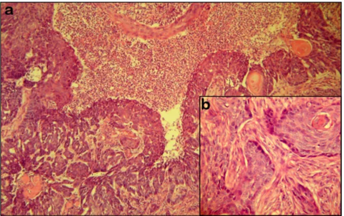
Fig. 2. (a) Histological picture of squamous cell carcinoma:
Neoplastic proliferation of squamous differentiated tumour cells with dyskeratotic and atypical keratinocytes in the epidermis (H&E, ×200). (b) Moderately differentiated tumour cells fill and expand papillary and reticular dermis (H&E, ×400).
years. The ulcer had a nodulo-ulcerative appearance and its base was highly indurated. The clinical features of the ulcer and its unresponsiveness to treatment led us to investigate other possible causes of genital ulcer. We doubted whether the SCC developed on the chronic genital ulcer of BD or emerged directly as SCC, unrelated with genital ulcer. In fact, SCC was diagnosed at first by us during his first appointment at our clinic. However, we suggest that the lesion occurred directly as SCC, due to infiltration of the whole ulcer region and the rapid progression of the lesion.
Skin tumours in BD are rarely observed2. In situ SCC development on a chronic vulvar ulcer of a female patient with BD was previously reported. The authors had poin- ted out that long-term systemic corticosteroid treatment could contribute to the development of SCC in this genital ulcer, which was resistant to treatment3. Satolli et al.4 reported a Merkel cell carcinoma in a patient with BD, and mentioned that the immunosuppresives may lead to the development of cancer. In our case, there was no predisposition for the development of SCC, such as
immunosuprressive drugs, arsenic exposure, irradiation, burn scarring, and HPV infection. We suppose that the presence of SCC together with BD is coincidental. Like BD, the other dermatoses, composed of various com- ponents, should be followed carefully, and differential diagnoses of signs and symptoms should be made.
Especially, all clinical features of a genital ulcer should be noted and unusual causes should not be forgotten.
REFERENCES
1. Mat MC, Goksugur N, Engin B, Yurdakul S, Yazici H. The frequency of scarring after genital ulcers in Behçet's synd- rome: a prospective study. Int J Dermatol 2006;45:554-556.
2. Mansur AT, Kocaayan N, Serdar ZA, Alptekin F. Giant oral ulcers of Behçet's disease mimicking squamous cell carcino- ma. Acta Derm Venereol 2005;85:532-534.
3. Hata H, Aoyagi S, Iitani MM, Homma E, Shimizu H. Squamo- us cell carcinoma in a chronic genital ulcer in Behçet's disease. Acta Derm Venereol 2010;90:539-540.
4. Satolli F, Venturi C, Vescovi V, Morrone P, De Panfilis G.
Merkel-cell carcinoma in Behçet's disease. Acta Derm Vene- reol 2005;85:79.