ISSN 2234-3806 • eISSN 2234-3814
http://dx.doi.org/10.3343/alm.2015.35.5.494
Phenotypic and Genotypic Analysis of Anti-
Tuberculosis Drug Resistance in Mycobacterium tuberculosis Isolates in Myanmar
Wah Wah Aung, Ph.D.1, Phyu Win Ei, M.Med.Sc.1, Wint Wint Nyunt, M.Med.Sc.2, Thyn Lei Swe, M.Med.Sc.2, Thandar Lwin, M.Phil.2, Mi Mi Htwe, M.S.1, Kyung Jun Kim, B.S.3 Jong Seok Lee, Ph.D.4, Chang Ki Kim, M.D.5, Sang Nae Cho, Ph.D.6, Sun Dae Song, M.D.4, and Chulhun L. Chang, M.D.3,7
Advanced Molecular Research Centre1, Department of Medical Research, Yangon, Myanmar; National Tuberculosis Program2, Nay Pyi Taw, Myanmar;
Department of Laboratory Medicine3, Pusan National University Yangsan Hospital, Yangsan; International Tuberculosis Research Center4, Changwon; Korean Institute of Tuberculosis5, Osong; Yonsei University College of Medicine6, Seoul; Convergence of Biomedical Science and Technology7, Pusan National University Yangsan Hospital, Yangsan, Korea
Background: Tuberculosis (TB) is one of the most serious health problems in Myanmar.
Because TB drug resistance is associated with genetic mutation(s) relevant to responses to each drug, genotypic methods for detecting these mutations have been proposed to overcome the limitations of classic phenotypic drug susceptibility testing (DST). We ex- plored the current estimates of drug-resistant TB and evaluated the usefulness of geno- typic DST in Myanmar.
Methods: We determined the drug susceptibility of Mycobacterium tuberculosis isolated from sputum smear-positive patients with newly diagnosed pulmonary TB at two main TB centers in Myanmar during 2013 by using conventional phenotypic DST and the Geno- Type MTBDRplus assay (Hain Lifescience, Germany). Discrepant results were confirmed by sequencing the genes relevant to each type of resistance (rpoB for rifampicin; katG and inhA for isoniazid).
Results: Of 191 isolates, phenotypic DST showed that 27.7% (n=53) were resistant to at least one first-line drug and 20.9% (n=40) were resistant to two or more, including 18.3%
(n =35) multidrug-resistant TB (MDR-TB) strains. Monoresistant strains accounted for 6.8% (n =13) of the samples. Genotypic assay of 189 isolates showed 17.5% (n =33) MDR-TB and 5.3% (n=10) isoniazid-monoresistant strains. Genotypic susceptibility re- sults were 99.5% (n=188) concordant and agreed almost perfectly with phenotypic DST (kappa=0.99; 95% confidence interval 0.96-1.01).
Conclusions: The results highlight the burden of TB drug resistance and prove the useful- ness of the genotypic DST in Myanmar.
Key Words: Mycobacterium tuberculosis, Drug, Resistance, Genotype, Myanmar
Received: January 27, 2015 Revision received: April 3, 2015 Accepted: June 22, 2015
Corresponding author: Chulhun L. Chang Department of Laboratory Medicine, Pusan National University School of Medicine, 20 Geumo-ro, Mulgum-eup, Yangsan 626-770, Korea
Tel: +82-55-360-1878 Fax: +82-55-360-1880 E-mail: [email protected]
Co-corresponding author: Wah Wah Aung Advanced Molecular Research Centre, Department of Medical Research, No. 5, Ziwaka Road, Dagon P.O., Yangon 11191, Myanmar
Tel: +95-1-375447 Fax: +95-1-251514
E-mail: [email protected]
© The Korean Society for Laboratory Medicine This is an Open Access article distributed under the terms of the Creative Commons Attribution Non-Commercial License (http://creativecom- mons.org/licenses/by-nc/3.0) which permits unrestricted non-commercial use, distribution, and reproduction in any medium, provided the original work is properly cited.
INTRODUCTION
Tuberculosis (TB) is a major global health problem and the fore- most cause of death from a single infectious agent, Mycobacte- rium tuberculosis. Nearly nine million new cases of TB and 1.4
million deaths occurred worldwide in 2011. With the emergence of multidrug-resistant (MDR) and extensively drug-resistant (XDR) strains, TB has become an even greater threat [1]. Myan- mar is one of the 22 countries with high-burden TB and among 27 countries with high-burden MDR-TB. An estimated 180,000
new TB cases occurred in Myanmar in 2011, and the propor- tion of MDR-TB was 4.2% among these cases and 10.0%
among previously treated cases according to the second nation- wide drug-resistant TB survey (2007-2008) [1].
The identification of resistant strains through drug susceptibil- ity testing (DST) and subsequent appropriate treatment might be the most effective strategy for controlling the spread of drug- resistant TB. The phenotypic DST widely used in developing countries is a proportion method that uses fixed concentrations of first-line TB drugs: isoniazid (INH), rifampicin (RIF), etham- butol (EMB), and streptomycin (SM). This method is simple and cost-effective; however, the results require a long time to obtain and can be unreliable in some diagnostic examination settings.
Because TB drug resistance is associated with mutation(s) in the genes relevant to responses to each drug, genotypic meth- ods for detecting mutations in these genes have been proposed to overcome the limitations of phenotypic DST [2]. A promising genotypic DST, the line-probe assay (LiPA), was endorsed by WHO in its 2008 policy statement for use with smear-positive pulmonary specimens [3]. Commercially available LiPAs for the rapid detection of INH and RIF resistance are based on se- lected numbers of commonly reported mutations. One LiPA, GenoType MTBDRplus (Hain Lifescience, Nehren, Germany), was designed for simultaneous detection of the most important rpoB mutations, which confer RIF resistance, and katG and inhA mutations, which confer high-level INH resistance [4].
We used both phenotype-based conventional DST and a commercially available genotypic DST (GenoType MTBDRplus) to investigate TB drug resistance in patients with newly diag- nosed pulmonary TB in Myanmar to obtain more accurate infor- mation on drug resistance, evaluate the usefulness of GenoType MTBDRplus, and characterize genetic alterations specific to TB in Myanmar.
METHODS
1. Patient enrollment and sample collection
A cross-sectional descriptive study was carried out at the Latha TB Diagnostic Centre (Yangon) and Mandalay Regional TB Cen- tre (Mandalay) in Myanmar from January to August 2013. The WHO case definition was applied, in which a new TB case was defined as a sputum- smear positive patient who had never re- ceived treatment for TB or who had taken TB drugs for less than one month [1]. Two sputum samples were collected from each patient after obtaining written informed consent. The fol- lowing definition was applied to determine smear-positive TB:
one or more initial sputum smear examinations (direct smear microscopy) positive for acid-fast bacilli (AFB) or one AFB-posi- tive sputum examination plus radiographic abnormalities con- sistent with active pulmonary TB as determined by a clinician [1]. Demographic characteristics were recorded in the pro forma when patients were enrolled.
2. Study patients
During the study period, 212 sputum samples from newly diag- nosed pulmonary TB patients were subjected to culture. The mean age of the patients was 39 ±16.4 yr, and 128 (60.4%) were males. After excluding 21 contaminated or unculturable samples, 191 M. tuberculosis isolates comprising 142 (74.3%) from the Yangon region and 49 (25.7%) from the Mandalay re- gion were subjected to phenotypic and genotypic DST.
3. Isolation of M. tuberculosis and phenotypic DST
Isolation of M. tuberculosis from sputum samples and DST with conventional culture was performed at the National TB Refer- ence Laboratory (NTRL; Yangon, Myanmar) and Upper Myan- mar TB Laboratory (Mandalay, Myanmar). M. tuberculosis was isolated from sputum samples according to the WHO method [5]. Sputum samples were decontaminated with N-acetyl-L-cys- teine sodium hydroxide. After centrifugation at a speed of 3,000- 3,500g for 15 min, the pellet was suspended in 1 mL 1×phos- phate-buffered saline, inoculated on two Lowenstein-Jensen (L- J) media slants, and incubated at 37°C for 6-8 weeks depending on the time required for the organisms to become evident. My- cobacterial growth was monitored every week. The M. tuberculo- sis isolates were identified according to growth rate and colony morphology. The Capilia TB test (Tauns, Numazu, Japan), an immunochromatographic assay that uses a monoclonal antibody to detect MPB64 antigen, was used to differentiate M. tubercu- losis complex from non-tuberculous mycobacteria.
Phenotypic DST was carried out on confirmed M. tuberculo- sis isolates. The test was performed on L-J media containing INH (0.2 μg/mL), RIF (40 μg/mL), SM (4 μg/mL), and EMB (2 μg/mL) according to the WHO-recommended proportional method for all primary isolates [6]. Inocula were cultured in a 37°C incubator for 6 weeks, and the results were interpreted as susceptible or resistant. The standard criterion of the proportion method for classifying a strain as resistant was the ratio of the number of colonies obtained on drug-containing medium to the number of colonies obtained on drug-free medium (growth of ≥ 1% of colonies). Any-drug resistance was defined as resistance to one or more first-line drugs. Monoresistance was defined as
resistance to only one of the four drugs.
4. Genotypic detection of INH and RIF resistance
Genotypic detection of INH and RIF resistance was carried out at Pusan National University Yangsan Hospital (Yangsan, Ko- rea). The GenoType MTBDRplus, a reverse hybridization line probe assay, was performed according to the manufacturer’s recommendations. Multiplex PCR was used to amplify the genes responsible for drug resistance, including katG, inhA, and rpoB, and the resulting biotin-labeled amplicons were hybrid- ized to DNA probes that affixed to the membrane. DNA probes for each gene consisted of wild-type (WT) and mutant (MUT) samples. Resistance to INH and RIF was defined as either (1) missing WT probe signal with MUT probe signal, or (2) missing WT probe signal.
5. Confirmation of discrepant results between phenotypic and genotypic DSTs
The genotypic and phenotypic DST results were compared.
Further examinations were performed with additional pheno- typic DST or DNA sequencing (rpoB for RIF and katG and inhA for INH) when the results disagreed. Each discordant gene re- gion was amplified with PCR, and direct sequencing of PCR products was carried out by Genotech (Daejeon, Korea). The sequencing results were analyzed with the CLC Main Work- bench (CLC bio, Aarhus, Denmark) at the International Tuber- culosis Research Center (Changwon, Korea).
6. Statistical analysis
The results were analyzed with the SPSS statistical software package, version 16 (SPSS Inc., Chicago, IL, USA). Drug-sus- ceptible and drug-resistant cases were documented as the per- centages of the total study population, and the drug-resistance pattern in newly diagnosed patients was determined. The resis-
tance results of phenotypic and genotypic DST were tested for kappa agreement [7].
7. Ethics approval
This study was approved by the Ethics Review Committee, De- partment of Medical Research, Yangon, Myanmar.
RESULTS
1. Drug-resistance patterns of M. tuberculosis isolates from newly diagnosed patients
Of the 191 M. tuberculosis isolates, 27.7% (n=53) were resis- tant to at least one first-line drug, and 40 (20.9%) were resistant to two or more, including 35 (18.3%) classified as MDR-TB strains. Monoresistant strains accounted for 6.8% (n=13), in- cluding 2.1% (n =4) INH-monoresistant strains and 4.7%
(n=9) SM-monoresistant strains. The total resistance rates were as follows: INH (44; 23.0%), RIF (35; 18.3%), EMB (15; 7.9%), and SM (41; 21.5%).
2. Gene mutation patterns in drug-resistant strains
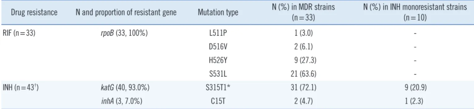
Among the 191 tested strains, two had a PCR inhibitor and were excluded from genotypic analysis. Genotypic assay of the remaining 189 samples showed 17.5% (n =33) MDR-TB strains and 5.3% (n =10) INH-monoresistant strains. Among the 33 RIF-resistant isolates, the S531L mutation, which is the most prevalent, was present in 63.6% (n=21). Of the 43 INH- resistant strains, 93.0% (n=40) had a mutation at the S315T1 (AGC→ACC) codon of katG (Table 1).
3. Comparison of phenotypic and genotypic TB drug susceptibility patterns
After isolates with discordant phenotypic and genotypic DST re- sults were confirmed with additional phenotypic DST or the se-
Table 1. Distribution of mutation patterns in rpoB, katG, and inhA among drug-resistant Mycobacterium tuberculosis isolates Drug resistance N and proportion of resistant gene Mutation type N (%) in MDR strains
(n=33) N (%) in INH monoresistant strains (n=10)
RIF (n=33) rpoB (33, 100%) L511P 1 (3.0) -
D516V 2 (6.1) -
H526Y 9 (27.3) -
S531L 21 (63.6) -
INH (n=43†) katG (40, 93.0%) S315T1* 31 (72.1) 9 (20.9)
inhA (3, 7.0%) C15T 2 (4.7) 1 (2.3)
*S315T1 means AGC→ACC/Ser→Thr exchange; †includes 33 INH-resistant isolates that were also resistant to RIF, composing 33 MDR isolates.
Abbreviations: INH, isoniazid; MDR, multidrug resistant; RIF, rifampicin.
quencing of discordant genes, genotypic DST results were 99.5% (188/189) concordant with the phenotypic DST results.
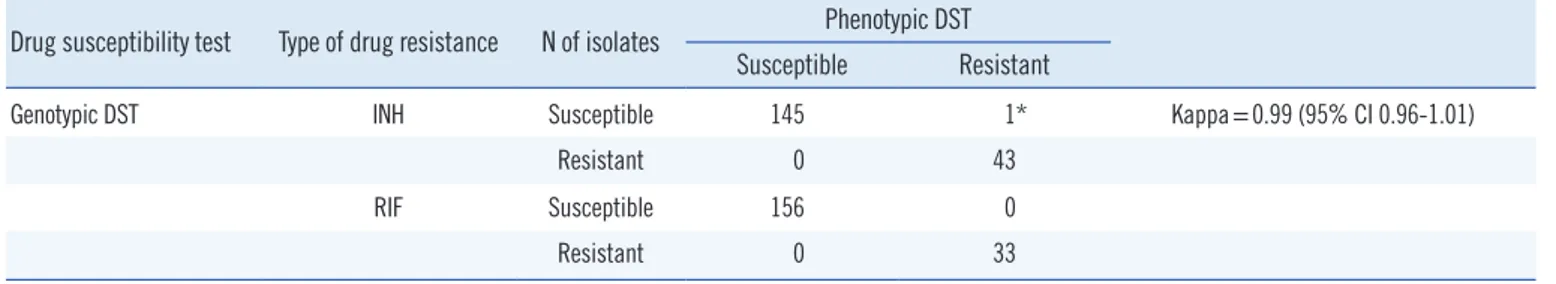
One isolate, which was resistant to INH on phenotypic DST and susceptible to INH in the GenoType MTBDRplus assay, was confirmed as resistant without any mutation in the target genes (katG or inhA). The genotypic DST had almost perfect agree- ment with the phenotypic DST (kappa=0.99; 95% confidence interval [CI] 0.96-1.01); Table 2). The 17 discordant phenotype and genotype results are listed in Table 3.
DISCUSSION
Drug resistance is a key cause of treatment failure in TB, partic- ularly when strains are resistant to the primary drugs, INH and RIF, which results in the development of MDR-TB. Managing the increasing number of MDR-TB cases is a crucial part of the STOP TB strategy and a component of all TB control programs [8]. MDR-TB strains can be transmitted in the community, re- placing susceptible strains and consequently making first-line regimens inadequate for achieving high cure rates. Drug resis- Table 2. Comparison of phenotypic and genotypic drug susceptibility tests
Drug susceptibility test Type of drug resistance N of isolates Phenotypic DST Susceptible Resistant
Genotypic DST INH Susceptible 145 1* Kappa=0.99 (95% CI 0.96-1.01)
Resistant 0 43
RIF Susceptible 156 0
Resistant 0 33
*isolate no. 1998 in Table 3.
Abbreviations: DST, drug susceptibility testing; INH, isoniazid; RIF, rifampicin; CI, confidence interval.
Table 3. Confirmation of 17 discordant Mycobacterium tuberculosis isolates via additional phenotypic drug susceptibility test and DNA se- quencing
Isolate No. First phenotypic DST Genotypic DST (GenoType
MTBDRplus assay) Second phenotypic DST DNA sequencing Final interpretation of
results
RIF INH RIF INH RIF INH RIF (rpoB) INH (katG/inhA)
381 R R S S - - NM NM Concordant*
606 S S S R S R - - Concordant*
674 R R S S - - NM NM Concordant*
677 R R S S - - NM NM Concordant*
779 R R S S - - NM NM Concordant*
1417 R R S R - - NM katG: 315AGC to ACC Concordant*
1419 R S S S S S NM NM Concordant*
1542 S S S R S R NM inhA: -15 C to T Concordant*
1864 S S R R R R 531TCG to TTG katG:315AGC to ACC Concordant*
1865 S S R R S R 531TCG to TTG katG:315AGC to ACC Concordant†
1868 S R S S NM NM Concordant*
1998 S R S S S R NM NM Discordant‡
2215 S S R S R R 531TCG to TTG inhA: -15 C to T Concordant§
U3 S S R R R R - - Concordant*
U5 S S R R R R 531TCG to TTG KatG-315AGC to ACC Concordant*
U9 S R S S S S - - Concordant*
U10 S S S R S R - - Concordant*
*first DST error; †both first and second DST error; ‡mutation other than those in target genes might be involved; §GenoType MTBDRplus assay test error.
Abbreviations: DST, drug susceptibility testing; INH, isoniazid; NM, no mutation; R, resistant; RIF, rifampicin; S, susceptible.
tance in new cases has been used to evaluate recent transmis- sion and can be used as a proxy for acquired resistance in a previously treated group.
In Myanmar, the second nationwide drug resistance survey (2007-2008) showed an MDR-TB rate of 4.2% among new pa- tients and 10.0% among previously treated patients [1]. Previ- ous studies carried out at the Yangon Divisional TB Center, a major referral center in Myanmar, during 1994-2004 found that the MDR-TB and any-drug resistance rates were 1.25-4.2%
and 15.8-35.3%, respectively [9-12].
The present study showed a high rate of MDR-TB (18.3%) and any-drug resistance rate of 27.7% among new TB cases as determined with a phenotypic method. These rates were much higher than those reported in previous nationwide and TB cen- ter-based studies. According to the 2012 Annual Report of the Myanmar National TB Program, Yangon had the highest TB case detection rate (112%), and many TB patients living in Yan- gon had one of the statistically significant risk factors for MDR- TB (odds ratio [OR]=3.0; 95% CI=1.5 5.8; P =0.014). In the present study, 74.3% of tested MTB isolates were from Yangon, which may account for the high drug-resistance rate [13]. INH resistance was most common. The individual anti-drug resis- tance pattern was similar to that reported in previous studies abroad and in Myanmar [9-12, 14].
The MDR-TB rate in Myanmar is among the highest in South- east Asia [1], and the present study showed a high MDR-TB rate among new smear-positive pulmonary TB patients. The drug resistance results of newly diagnosed patients may reflect the primary resistance to first-line TB drugs in Myanmar, al- though primary resistance is difficult to determine because pa- tients may not know or may deny that they have had previous TB treatment. The high prevalence of drug resistance in Myan- mar highlights the need for a control program to improve case management and strengthen TB control strategies.
Drug resistance in M. tuberculosis develops as a result of random mutations in the genes responsible for resistance to each drug [15, 16]. In the case of RIF, 95-98% of resistance develops because of mutations in the 81-bp core region (RIF resistance-determining region or hot spot) of the β subunit of the RNA polymerase gene (rpoB), which results in peptide al- terations at codon 531 or codon 526. The most common muta- tions are S531L and H526Y [14, 15]. The molecular basis of INH resistance is more complex and is associated with genes such as katG, inhA, ahpC, and oxyR [17]. Approximately 50- 95% of INH-resistant strains contain mutations in codon 315 of katG, whereas 20-35% contain mutations in the inhA regulatory
region [4].
In the present study, among the 33 RIF-resistant isolates found with genotypic DST, the S531L mutation was the most common at 63.6% (n=21). The H526Y mutation was the sec- ond most common, accounting for 27.3% (n=9) of MDR strains.
Of the 43 INH-resistant strains, 93.02% (n =40; 31 MDR strains and nine INH-monoresistant strains) had a mutation in the S315T1 region of katG, and only 6.97% (n =3; two MDR strains and one monoresistant strain) had a mutation in the C15T region of inhA. This finding was consistent with the previ- ous results [15, 16]. The high prevalence of katG mutations accounts for a high proportion of INH resistance in countries with high TB prevalence, presumably as a result of the trans- mission of these strains in a high-burden setting [18]. No other mutation in ahpC or oxyR was observed in this study, which suggests that those mutations are infrequently involved in INH resistance in Myanmar.
The overall concordance between phenotypic and genotypic DST was 99.5% (188/189) after the confirmation of these sam- ples with additional phenotypic DST and DNA sequencing. Sim- ilar results were reported in previous studies, with values rang- ing from 88.9% to 100% [19-22]. When we analyzed the phe- notypic and genotypic results with kappa statistics, the geno- typic assay showed almost perfect agreement with phenotypic DST (kappa=0.99; 95% CI 0.96-1.01). The one discordant iso- late was susceptible to INH in the GenoType MTBDRplus assay but resistant to INH without any mutation in the target genes.
Mutation other than that in the target gene might be involved in the resistance in this isolate. The high MDR-TB rate, high accu- racy of genotypic DST, and common drug resistance-mutation pattern found in the present study suggested that genotypic DST should be more widely used as a routine test to determine drug susceptibility in newly diagnosed TB patients. Genotypic DST may also lead to the rapid detection of MDR-TB cases and be invaluable in preventing the transmission of drug-resistant strains in Myanmar. In conclusion, this study highlighted the high prevalence of drug resistance among new pulmonary TB cases in Myanmar and the usefulness of genotypic DST for de- termining drug susceptibility in TB patients and for the rapid and accurate diagnosis of TB in Myanmar.
Authors’ Disclosures of Potential Conflicts of Interest
No potential conflicts of interest relevant to this article were re- ported.
Acknowledgments
This study was supported financially by the Korea International Co-operation Agency (KOICA); the Basic Science Research Pro- gram through the National Research Foundation of Korea (NRF) funded by the Ministry of Education, Science and Technology (2012R1A1A2004593); and an NRF Grant funded by the Inter- national Funds for Science technology (2013K1A3A9A010 43836). Research at the International Tuberculosis Research Center (ITRC) was made possible by continued support from the Ministry for Health, Welfare and Family Affairs of the Repub- lic of Korea.
REFERENCES
1. World Health Organization. Global Tuberculosis Report 2012. WHO/
HTM/TB/2012.6. Geneva: WHO, 2012.
2. Agonafir M, Lemma E, Wolde-Meskel D, Goshu S, Santhanam A, Girm- achew F, et al. Phenotypic and genotypic analysis of multidrug-resistant tuberculosis in Ethiopia. Int J Tuberc Lung Dis 2010;14:1259-65.
3. World Health Organization. Policy Statement. Molecular line probe as- says for rapid screening of patients at risk of multidrug-resistant tuber- culosis (MDR-TB). Geneva: WHO, 2008. http://www.who.int/tb/fea- tures_archive/policy_statement.pdf (Updated on 27 June 2008).
4. Hillemann D, Weizenegger M, Kubica T, Richter E, Niemann S. Use of the genotype MTBDR assay for rapid detection of rifampin and isonia- zid resistance in Mycobacterium tuberculosis complex isolates. J Clin Microbiol 2005;43:3699-703.
5. World Health Organization. Laboratory services in tuberculosis control.
Culture Part III 1998; WHO/TB/98.258. Geneva: WHO, 1998.
6. World Health Organization. Guidelines for surveillance of drug resis- tance in tuberculosis. WHO/CDS/CSR/RMD/2003. Geneva: WHO, 2003. Available from http://whqlibdoc.who.int/publications/2003/
9241546336.pdf.
7. Viera AJ and Garrett JM. Understanding interobserver agreement: the kappa statistic. Fam Med 2005;37:360-3.
8. World Health Organization. Management of MDR-TB: a field guide.
WHO/HTM/TB/2008.402a. Geneva: WHO, 2009.
9. Ti T, Lwin T, Phyu S, Aung WW, Khaing TMM, Min A, et al. Magnitude of the MDR-TB problem among patients attending Yangon Divisional TB Centre, Myanmar. Myanmar Med J 2007;50:2-7.
10. Phyu S, Ti T, Jureen R, Hmun T, Myint H, Htun A, et al. Drug-resistant
Mycobacterium tuberculosis among new tuberculosis patients,Yangon, Myanmar. Emerg Infect Dis 2003; 9:274-6.
11. Ti T, Lwin T, Mar TT, Maung W, Noe P, Htun A, et al. National anti-tu- berculosis drug resistance survey, 2002 in Myanmar. Int J Tuberc Lung Dis 2006;10:1111-6.
12. Aung WW, Ti T, Than KK, ThidaM, Nyein MM, Htun YY, et al. Study of drug resistant cases among new pulmonary tuberculosis patients at- tending a tuberculosis center, Yangon, Myanmar. Southeast Asian J Trop Med Public Health 2007;38:104-10.
13. National Tuberculosis Programme, Myanmar. Annual Report 2012.
Ministry of Health, Myanmar: NTP, October 2014.
14. Bai GH, Park YK, Choi YW, Bai JI , Kim HJ, Chang CL, et al. Trends of anti-tuberculosis drug resistance in Korea, 1994-2004. Int J Tuberc Lung Dis 2007;11:571-6.
15. Telenti A, Imboden F, Marchesi F, Lowrie D, Cole S, Colston MJ, et al.
Detection of rifampicin-resistance mutations in Mycobacterium tuber- culosis. Lancet 1993;341:647-50.
16. Ozturk CE, Sanic K, Kaya D, Ceyhan I. Molecular analysis of isoniazid, rifampin and streptomycin resistance in Mycobacterium tuberculosis isolates from patients with tuberculosis in Düzce, Turkey. Jpn J Infect Dis 2005;58:309-12.
17. Ahmad S and Mustafa AS. Molecular diagnosis of drug resistant tuber- culosis. Kuwait Med J 2001;33:120-6. Available from http://www.re- searchgate.net/profile/Abu_Mustafa/publication/230667668_Molecu- lar_diagnosis_of_drug-resistant_tuberculosis.
18. Baker LV, Brown JT, Maxwell O, Gibson AL, Fang Z, Yates MD, et al.
Molecular analysis of isoniazid-resistant Mycobacterium tuberculosis isolates from England and Wales reveals the phylogenetic significance of the ahpC-46A polymorphism. Antimicrob Agents Chemother 2005;
49:1455-64.
19. Hillemann D, Rüsch-Gerdes S, Richter E. Evaluation of the GenoType MTBDRplus assay for rifampin and isoniazid susceptibility testing of Mycobacterium tuberculosis strains and clinical specimens. J Clin Mi- crobiol 2007;45:2635-40.
20. Causse M, Ruiz P, Gutierrez JB, Zerolo J, Casal M. Evaluation of new GenoType MTBDRplus for detection of resistance in cultures and direct specimens of Mycobacterium tuberculosis. Int J Tuberc Lung Dis 2008;
12:1456-60.
21. Huyen MN, Tiemersma EW, Lan NT, Cobelens FG, Dung NH, Sy DN, et al. Validation of the GenoTypenMTBDRplus assay for the diagnosis of multidrug resistant tuberculosis in South Vietnam. BMC Infect Dis 2010;10:149.
22. Tessema B, Beer J, Emmrich F, Sack U, Rodloff AC. Analysis of gene mutations associated with isoniazid, rifampicin and ethambutol resis- tance among Mycobacterium tuberculosis isolates from Ethiopia. BMC Infect Dis 2012;12-37.