https://doi.org/10.4174/astr.2018.94.3.142 Annals of Surgical Treatment and Research
In vivo porcine training model of laparoscopic common bile duct repair with T-tube insertion under the situation of iatrogenic common bile duct injury
Eun Young Kim1, Tae Ho Hong2
1Department of Trauma and Surgical Critical Care, Seoul St. Mary’s Hospital, College of Medicine, The Catholic University of Korea, Seoul, Korea
2Department of Surgery, Seoul St. Mary’s Hospital, College of Medicine, The Catholic University of Korea, Seoul, Korea
INTRODUCTION
Since the 1990s, laparoscopic cholecystectomy (LC) has replaced the open cholecystectomy and become the gold stan- dard procedure for symptomatic cholelithiasis or benign gall- bladder (GB) disease such as GB polyp. However, common bile duct (CBD) injury during the operation still occurs in 0.5%–0.7%
of LC procedures [1], even with experts who are specialized for hepatobiliary surgery. CBD injury can result in the fatal out- comes such as bile peritonitis, sepsis and death. Progression to the biliary stricture can result in recurrent cholangitis, obstruc- tive jaundice, or biliary sepsis.
In many cases of CBD injury found intraoperatively, lapar- oscopic CBD repair accompanying with T-tube insertion is preferred as the definite management for this complication. The single-stage procedure does not require further intervention.
However, proficiency of the operator for laparoscopic CBD mani- pulation is necessary.
For those who are novices at the technique, surgical train ing model for laparoscopic CBD manipulation would be advan- tageous. Practicing the specific procedure or operation should be required prior to the actual clinical situation [2]. A hands-on training model could be a very feasible and effective educational tool to allow surgeons to acquire the specific surgical technique Purpose: We introduce a training porcine model for laparoscopic common bile duct (CBD) repair with T-tube insertion. The model could be the feasible training tool for a surgeon learning hepatobiliary surgery.
Methods: Totally laparoscopic CBD repair with T-tube insertion was performed on 9 pigs by 9 trainees naïve in hepatobiliary surgery. Similar to the situation of iatrogenic injury, CBD was transected by laparoscopic scissors at the middle part about 1 cm in length, and the transected CBD was repaired through end-to-end anastomosis with T-tube insertion. A secureness of anastomosis was confirmed by saline leakage test and all animals were sacrificed after the surgery.
Results: All novice surgeons completed operations successfully without complications. Total mean operative time was 85 ± 1.7 minutes and the mean time spent performing the CBD repair with T-tube insertion was 71 ± 3 minutes. There was no bile leakage after primary anastomosis in all animals.
Conclusion: This porcine training model for laparoscopic CBD repair with T-tube insertion could be a feasible and effective training tool for surgeons with little experience in laparoscopic hepatobiliary surgery.
[Ann Surg Treat Res 2018;94(3):142-146]
Key Words: Training model, CBD injury, T-tube, Laparoscopy
Reviewed January February March April May June July August September October November December
Received June 1, 2017, Revised July 24, 2017, Accepted August 17, 2017 Corresponding Author: Tae Ho Hong
Department of Surgery, Seoul St. Mary’s Hospital, College of Medicine, The Catholic University of Korea, 222 Banpo-daero, Seocho-gu, Seoul 06591, Korea
Tel: +82-2-2258-2876, Fax: +82-2-595-2822 E-mail: [email protected]
ORCID code: https://orcid.org/0000-0003-3864-8104
Copyright ⓒ 2018, the Korean Surgical Society
cc Annals of Surgical Treatment and Research is an Open Access Journal. All articles are distributed under the terms of the Creative Commons Attribution Non- Commercial License (http://creativecommons.org/licenses/by-nc/4.0/) which permits unrestricted non-commercial use, distribution, and reproduction in any medium, provided the original work is properly cited.
or skills [3,4].
Here, we report the development and use of a porcine training model of laparoscopic CBD repair with T-tube inser- tion. The feasibility of the model as an effective training tool for trainees who are not expert in hepatobiliary surgery is demon- strated. We also added the several tricks and tact to conduct this technique more efficiently and simply.
METHODS
Study design
All experimental operations were performed in the lapar- oscopic training center and the current study was approved by the institutional animal care and use committee of the hospital.
Three-month-old female pigs 31 to 35 kg in weight under- went bowel enema at midnight the day of surgery and were fasted for 24 hours before the surgery. The operative procedures were performed by general surgery residents.
They had performed fewer than 15 cases of LC and had no experience with laparoscopic CBD manipulation. Prior to the operation, residents were educated about the whole process of the opera tion through a video review. Surgery was supervised by a sur geon specialized in hepato-biliary pancreas surgery with ex perience of over 100 cases of laparoscopic CBD manipulation. The operation time, intraoperative blood loss, and CBD diameter were recorded. To confirm the security of anastomosis for bile leakage at the anastomosis site, a saline solution of indocyanine green was flushed through T-tube. The whole operation time and the average time to perform the CBD repair with T-tube insertion using the laparoscopic suture were accurately calculated through the video decipher and operative records.
Operative technique
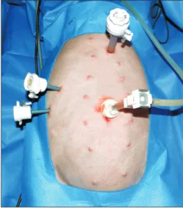
Under general anesthesia, 4 trocars were placed as the same manner in the standard LC [5]; 10-mm trocar at the umbilicus for the laparoscope and three 5-mm trocars were used for the retrac tion of GB, with the the surgeon’s right and left hand for working channels, respectively (Fig. 1). After establishment of
pneu moperitoneum with 12-mmHg carbon dioxide, the pig was placed in the reverse Trendelenburg position tilted to the left.
The operation was initiated by liver retraction through the direct suture to lift up the hepatic lobe adjoined to the GB and the hepatoduodenal ligament was dissected carefully to expose the CBD. The anatomical structure of porta hepatis in porcine has several characteristic features in common with those in humans [6,7]. The porcine cystic duct is buried in the adjacent hepatic parenchyme deeply that makes it hard to distinguish from the surroundings. The diameter of the porcine CBD is 2.3–
3.0 mm, which is a bit smaller than that of humans. However, it runs to the right side of hepatic artery and lies in front of portal vein, as in humans [8]. This enables dissection of porcine CBD as is done in humans.
The Hartman’s pouch of the GB was retracted superiorly and laterally by the first assistant using the laparoscopic gras per to facilitate the dissection. After the identification of anato mical structure around the CBD, the CBD was transected by lapar- oscopic scissors at the midpart and a segment of CBD about 1 cm in length was resected, similar to common cases of iatro-
Fig. 1. Trocar locations are the same as with standard lapar
os copic cholecystectomy.
1 cm
Proximal end of transected CBD
Distal end of transected CBD
A B
1 cm
Proximal end of transected CBD
Distal end of transected CBD
Fig. 2. Intracorporeal view of lapar oscopic common bile duct (CBD) transection. (A) CBD was transected by laparoscopic sci
ssors at the middle part about 1 cm in length. (B) The view of tran sectd CBD which is similar to the common cases of iatrogenic CBD transection.
genic CBD transection (Fig. 2).
The first step of CBD repair used 2 stay sutures without making the knot on both corners of the CBD resection line to sustain the tension of suture lines during the anastomosis.
The stay sutures were taken out separately; the stay suture on the right side was taken out from the body through the lateral port, whereas the left one was laid in the peritoneal cavity with a laparoscopic clip on tips to help sustain the tensile force appropriately during the CBD repair by lifting it. After making the stay sutures, the posterior side of CBD incision was closed with 2 interrupted stitches using absorbable 5-0 multifilament sutures from to right to left side. A latex rubber 8F T-tube was inserted after cutting the T limb to an adequate size. The 2 short limbs were about 1.0 and 1.5 cm long, respectively; the longer limb was inserted toward the distal side of CBD and the shorter limb was introduced upwards for the prevention of the dislocation of tube. When the tube was positioned in
place, the anterior side of CBD incision was closed from right to left using about 2 or 3 interrupted sutures snugly around the T-tube. After closure of CBD incision, stay sutures at both edge of anastomosis line were tied. The opposite end of T-tube was brought out through the minimal incision in the upper abdominal wall and the bile leakage test was performed (Fig.
3). If there was any evidence of leakage, such as the flow of greenish fluid from the anastomosis site, an additional suture was applied. After the establishment of anastomosis security, the cystic duct was divided and standard LC was performed.
After operation, all animals were sacrificed.
RESULTS
Nine domestic pigs underwent the operation during the study period. Surgery was performed by nine junior residents who had not performed laparoscopic CBD exploration. The pre operative characteristics of the animals are provided in Table 1. Total mean operative time was 85 ± 1.7 minutes (range, 84–86 minutes). To estimate the consumed time in more detail, the mean time spent performing the CBD repair with T-tube insertion was calculated as 71 ± 3 minutes (range, 68–74 minutes). After deducting the time spent for T-tube manipulation, total anastomosis time for CBD repair was 67.5 ± 1.5 minutes (range, 66–69 minutes). The average number of knots needed to complete the anastomosis was 7.4 ± 0.2 (range, 6–8).
Bile leakage after primary anastomosis occurred in 1 case (11.1%) which needed 1 additional suture on the anterior aspects after anastomosis. There was no significant intraoperative bleeding requiring fluid resuscitation or which caused inst ability of vital signs during the operation. All operators com pleted the task successfully. The surgical outcomes are also described in Table 1.
Fig. 3. The bile leakage test was done to confirm the secu
reness of anastomosis after closure of common bile duct in
ci sion. A salin solution of the dye (arrowhead) was flushed through the Ttube to rule out leakage.
Table 1. Preoperative characteristics and operative outcomes of porcine common bile duct (CBD) repair with Ttube insertion
Case
No. Age (mo)/
sex Weight
(kg)
Operative outcomes Total operative
time (min)
CBD repair with Ttube manipulation
time (min)
Total anastomosis time for CBD repair
time (min)
Total count of knot to complete the
anastomosis
Postoperative complication
1 3/F 22.0 578 30 100 7
2 3/F 21.3 395 21 75 7
3 3/F 22.4 415 25 75 7
4 3/F 31.9 347 16 125 8
5 3/F 23.5 335 15 50 8
6 3/F 31.9 347 16 125 8
7 3/F 23.5 335 15 50 7
8 3/F 23.5 335 15 50 8
9 3/F 23.5 335 15 50 7
DISCUSSION
Iatrogenic CBD injury during LC still occurs despite of the remarkable progress in surgical techniques and instruments, and can involve expert and novice surgeon alike. Laparoscopic CBD repair with T-tube insertion is a useful solution to this complication, and can permit the intraoperative, single-stage man age ment without additional intervention in the post opera- tive period. The procedure can be performed in cases with partial CBD resection or cases having segmental CBD resection that can be repaired through primary end-to-end repair without ten sion [9,10]. However, the surgical outcome is influenced by opera tor proficiency and skill. The approach is challenging for a novice surgeon. As a solution, a well-designed training animal model could be an effective tool for practice and mastery of the method. Gaining experience in the surgery and handling the necessary equipment could equip surgeons to better and more safely perform the operation in actual clinical situations.
Pigs have very similar characteristics with humans in terms of the internal organ or vascular structures [7,8,11]. Porcine organs are relatively comparable in size or shape with humans than those of other mammals including the rat, dogs, and rabbit. These properties are beneficial for application as the surgical training model, and pigs are been widely used for this pur pose, especially for laparoscopic procedures [12]. The living porcine model has the several advantages that an artificial material or nonliving model cannot meet. This living model maximally simulates the human situation because of ongoing respiration and pulsatile movements of vascular structures.
The tactile sense of the manipulated tissue is almost the same as human tissue, which heightens the value of the simulation [13,14]. These characteristics make the living animal model opti- mal for the evaluation of the safety or feasibility of a surgical tech nique [15].
How ever, despite of these advantages of living porcine model, there are a number of problems. The model is relatively costly, since specialized animal care by veterinarian or additional equip ment can be needed. Therefore, limited medical resources can preclude repeated use of the porcine model.
With the accumulated experience through the training, authors have devised several tracks and tact which help to im- prove efficiency of training. The present data demonstrate the feasibility of the model and the satisfactory training results, especially regarding the clinical callowness of the opera tor.
Several trivial, but ingenious, tips are helpful. Firstly, the optimal length of thread used in laparoscopic suture is bet- ween 10 and 15 cm. This length permits the delicate motion for intracorporeal suture by laparoscopic instruments in the limited space. It also helps to keep the appropriate tension during the suturing without loosening of thread or knots that causes the thread to get tangled. Secondly, the type of thread is
important. We used 5-0 absorbable multifilament sutures in our model; this suture is characterized by its structure that several suture filaments have been twisted into one thread. This feature would be favorable for the stability of knot during the suture motioning. Additionally, it has the soft and flexible char- ac teristics to better enable knot-making by a novice surgeon.
Regarding the repair method, the stay sutures were on the both edge of CBD anastomosis line at first; then we started to make the repair at the posterior wall. This method could prevent the CBD lumen to become narrow as the anastomosis pro gressed. The width of CBD lumen was maintained con- sistently during the anastomosis in our experiments, and opera- tor could feel more comfortable during the operation with out the visual interference that helps the suturing more accurately and firmly. After making stay sutures, only 2 additional sutures were needed to make the posterior anastomosis; this procedure resisted the intraluminal pressure and maintained the proper level of flow patency when the bile flow was reopened. The handling of the relatively smaller size of the porcine CBD might pose some technical difficulty. However, we feel that this difficulty in training could act as the advantage rather in the actual clinical situation that could be influenced by a lot of variables. In addition, we inserted the proximal limb of T-tube before placing the distal limb because it is much easier than the insertion of a distal limb before the proximal limb. Authors also suppose that the rubber material is better for the T-tube because it is more flexible than silastic one, which would be helpful to handle or place the tube into CBD.
The surgical outcomes in this article were attained from the limited number of cases. Because one operator could not practice the same experiments repeatedly, we failed to analyze the training effect according to the chronological order.
However, even allowing for these limitations, our results about surgical technique seemed to be very favorable and feasible in terms of anastomosis time, the anastomotic security and the completeness of performance especially considering the fact that all operators had no prior experience with this technique.
Based on these results, we consider that our training model could act as the effective and reliable practice tool preceded before the implementation of laparoscopic CBD repair with T-tube insertion to the patient in operating room.
In conclusion, the training model for laparoscopic CBD repair using a living porcine model with the application of several tricks as described above could be a feasible training model for a surgeon with no experience in the laparoscopic CBD sur gery before. To confirm the training effect of this model in more detail, a further study involving more cases and repeated per- for mance of operation by each trainee should be done.
CONFLICTS OF INTEREST
No potential conflict of interest relevant to this article was reported.
REFERENCES
1. Dagash H, Chowdhury M, Pierro A. When can I be proficient in laparoscopic surgery?
A systematic review of the evidence. J Pediatr Surg 2003;38:720-4.
2. Schoffl H, Hager D, Hinterdorfer C, Dunst KM, Froschauer S, Steiner W, et al. Pulsatile perfused porcine coronary arteries for microvascular training. Ann Plast Surg 2006;57:213-6.
3. Pettigrew RA, Burns HJ, Carter DC. Evalu- ating surgical risk: the importance of tech nical factors in determining outcome.
Br J Surg 1987;74:791-4.
4. Wu JS, Strasberg SM, Luttmann DR, Meininger TA, Talcott MR, Soper NJ.
Lapar oscopic hepatic lobectomy in the por cine model. Surg Endosc 1998;12:232- 5.
5. Hua J, Lin S, Qian D, He Z, Zhang T, Song Z. Primary closure and rate of bile leak fol lowing laparoscopic common bile duct ex ploration via choledochotomy. Dig Surg
2015;32:1-8.
6. Cameron BH, O’Regan PJ, Anderson DL.
A pig model for advanced laparoscopic biliary procedures. Surg Endosc 1994;8:
1423-4.
7. Watson DI, Treacy PJ, Williams JA.
Developing a training model for lapar- oscopic common bile duct surgery. Surg Endosc 1995;9:1116-8.
8. Ferrer J, Scott WE 3rd, Weegman BP, Suszynski TM, Sutherland DE, Hering BJ, et al. Pig pancreas anatomy: implications for pancreas procurement, preservation, and islet isolation. Transplantation 2008;
86:1503-10.
9. Strasberg SM, Callery MP, Soper NJ.
Lapar oscopic surgery of the bile ducts.
Gastrointest Endosc Clin N Am 1996;6:81- 105.
10. Strasberg SM, Hertl M, Soper NJ. An analysis of the problem of biliary injury during laparoscopic cholecystectomy. J
Am Coll Surg 1995;180:101-25.
11. Lee JS, Hong TH. In vivo porcine training model for laparoscopic Roux-en-Y chole do- cho jejunostomy. Ann Surg Treat Res 2015;
88:306-10.
12. Sanchez A, Otano N, Rodriguez O, Sanchez R, Benitez G, Schweitzer M.
Laparoscopic common bile duct ex plo ra- tion four-task training model: construct vali dity. JSLS 2012;16:10-5.
13. Leida Z, Ping B, Shuguang W, Yu H. A ran- domized comparison of primary closure and T-tube drainage of the common bile duct after laparoscopic choledochotomy.
Surg Endosc 2008;22:1595-600.
14. Lee JS, Hong TH. The rat chole do cho jeju- nostomy model for microsurgical training.
Ann Surg Treat Res 2016;90:246-9.
15. Palter VN, Orzech N, Aggar wal R, Okrainec A, Grantcharov TP. Resident per- cep tions of advanced laparoscopic skills train ing. Surg Endosc 2010;24:2830-4.