Laparoscopic Removal of a Gastric Trichobezoar in an 8-Year-Old Girl
- a Case Report -
최규석․최병호1․박진영
경북대학교 의학전문대학원 외과학교실, 소아과학교실1
이 논문은 2010년 6월 10일 대구에서 개최된 제 26 회 대한소아외과학회 춘계학술대회에서 구연되었음.
접수일 : 10 / 7 / 28 게재승인일 : 10 / 8 / 31
교신저자:박진영, 700-721 대구광역시 중구 삼덕 2 가 50번지 경북대학병원 외과
Tel : 053)420-5612, Fax : 053)421-0510 E-mail: [email protected]
INTRODUCTION
Trichobezoars are a rare type of bezoar formed from ingested human hair as well as hair from sources such as dolls, stuffed toys, blankets, and/or carpets. They are usually encountered in the stomach, but may also be found in the small intestine, colon, and Meckel’s diverticulum1. Tricho- bezoars are usually encountered in young women with mental retardation, pica, or psychiatric problems who have trichotillo- mania and trichophagia2-6. Trichobezoars can cause abdominal pain, bowel obstruc- tion, gastric ulceration, perforation, and gastrointestinal bleeding2,5. With the advent of minimally invasive surgery, laparoscopic removal of large gastric trichobezoars has become feasible with
smaller incisions and better cosmesis. We describe here an 8-year-old girl with trichotillomania and trichophagia and a huge gastric trichobezoar, which was removed by laparoscopic surgery.
CASE REPORT
An 8-year-old girl was admitted to our institution due to a 4 day history of vomiting, epigastric pain and palpable mass. She had a history of trichotillomania and trichophagia over the previous 2 months. Her parents noticed that she pulled out her hair when she was left alone at home, but she did not receive medical attention. On physical examination, a huge, firm nontender mobile mass was palpated in her epigastrium. Her hair looked normal but there was a bald spot on her scalp. Initial laboratory parameters were within normal limits. An upper gastrointestinal series showed a large intraluminal filling defect
Fig. 1. Upper gastrointestinal series showing a huge intraluminal mass.
Fig. 2. Contrast enhanced abdominal CT scan showing a large intraluminal mass with entrapped air in the stomach.
Fig. 3. Sites of laparoscopic port placement.
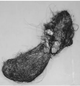
Fig. 4. Trichobezoar after removal.
in the stomach (Fig. 1), and an abdominal CT scan revealed a large intraluminal mass with entrapped air in the stomach (Fig. 2). The mass was diagnosed as a trichobezoar by endoscopy. Two endo- scopic attempts to fragment and remove the bezoar under sedation failed, and she was referred to the surgical service for
laparoscopy. A 10 mm periumbilical port was made and pneumoperitoneum was established and set to an insufflation pressure of 10 mm Hg. Three additional 5 mm ports were placed in the right upper, right lower and left lower quadrants (Fig.
3). After placement of stay sutures, a longitudinal anterior gastrotomy over the
gastric antrum was made using a harmonic scalpel (Ethicon, Johnson &
Johnson Corp., Somerville, NJ, USA). An auto Suture Endocatch II Specimen Pouch (Tyco Healthcare, Auckland, New Zealand) was inserted and a 15 × 6 cm trichobezoar was placed in a plastic bag and retrieved through an extended periumbilical port site for a camera (Fig. 4). After extraction, the gastric incision site was closed with continuous 2-0 Vicryl sutures (Ethicon, Johnson & Johnson Corp., Somerville, NJ, USA) in a single layer. Inspection of the entire intestinal tract revealed no other bezoars. Her postoperative course was uneventful. She was referred to a pediatric psychiatrist.
DISCUSSION
Trichobezoars are impactions of ingested hairs in the stomach and intestine, resulting from the chronic ingestion of human hair as well as hair from dolls, stuffed toys, and blankets. Trichobezoars gradually enlarge as ingested hairs accumulate within the stomach. A gastric trichobezoar with a long tail extending into the small intestine can lead to symptoms of intestinal obstruction, known as the Rapunzel syndrome2,7.
Trichobezoars are more common in children than in adults, with up to 90 % occurring in girls younger than 20 years2,3.
Males are rarely affected. They have been associated with mental retardation, pica, and trichotillomania. Although rare, an underlying functional or mechanical ob- struction of the gastrointestinal tract, such as gastric surgery or gastric dysmotility, may predispose to bezoar formation3,5.
Symptoms of trichobezoar develop insidiously and are often vague and nonspecific; they may include abdominal pain, nausea, vomiting, early satiety, and weight loss. These symptoms may be complicated by gastric ulceration, perforation, gastrointestinal bleeding, acute pancreatitis, obstructive jaundice, or hypochromic anemia2,5,8.
On physical examination, a huge, firm, nontender, freely movable mass can be palpated in the epigastrium or left upper quadrant of the abdomen. Alopecia and halitosis may be present. Trichobezoars are diagnosed through careful history taking and various radiographic methods.
Plain abdominal radiography may show a mottled radiotransparent heterogeneous mass that appears similar to a food-filled stomach9. An upper gastrointestinal series with barium may reveal the presence of a freely mobile intraluminal mass. Abdo- minal CT scan may show a low-density intraluminal mass containing air bubbles and exhibiting a characteristic mottled appearance9. However, endoscopy is the diagnostic method of choice because it
allows the bezoar to be visualized and distinguished, directing further manage- ment. Trichobezoars often require surgical removal because in many cases it is not possible to chemically soften or endoscopically fragment, and eventually remove, the tightly interlaced hairs5.
Gastric trichobezoars can be removed by laparoscopic surgery10. Nirasawa et al11 first used this approach on a 7-year-old girl who underwent laparoscopic gastric incision and removal of the trichobezoar by lower abdominal incision. In this patient, however, there was a possibility of spillage in the abdominal cavity and incisional wound contamination during extraction. To avoid these problems, we placed the trichobezoar in a laparoscopic endo-bag and retrieved it through an extended periumbilical port site for a camera.
Following trichobezoar removal, proper psychiatric consultation and management are necessary to prevent recurrence.
Trichotillomania is considered a disorder of impulse control4. This disorder may be present in otherwise normal children as a benign transient habit disorder, but it is frequently accompanied by comorbid obsessive-compulsive, mood and anxiety disorder.
The findings reported here suggest that laparoscopic removal of gastric tricho- bezoar is an effective treatment option
that avoids laparotomy and results in rapid recovery and a reduced likelihood of wound-related morbidity.
REFERENCES
1. Canavese F, Maiullari E, Castantino S, Rosina F, Gesmundo R, Cortese MG, et al.: A gastric trichobezoar: a report of clinical case with anomalous presentation.
Pediatr Med Chir 16:289-91, 1994
2. Palanivelu C, Rangarajan M, Senthilkumar R, Madankumar MV: Trichobezoars in the stomach and ileum and their laparoscopy-assisted removal: a bizarre case. Singapore Med J 48:e37-9, 2007 3. Shami SB, Jararaa AAM, Hamade A,
Ammori BJ: Laparoscopic removal of a huge gastric trichobezoar in a patient with trichotillomania. Surg Laparosc Endosc Percutan Tech 17:197-200, 2007 4. Meyer-Rochow GY, Grunewald B:
Laparoscopic removal of a gastric trichobezoar in a pregnant woman. Surg Laparosc Endosc Percutan Tech 17:129-32, 2007
5. Antoine DB, Vera VN, Yvant V: Huge gastric trichobezoar in a 10-year-old girl:
Case report with emphasis on endoscopy in diagnosis and therapy. J Pediatr Gastroenterol Nutr 28:513-5, 1999
6. Lynch KA, Feola PG, Guenther E:
Gastric trichobezoar: An important cause of abdominal pain presenting to the pediatric emergency department. Pediatric Emergency Care 19:343-7, 2003
7. Silva AP, Carvalho J, Pinho R, Fernandes S, Sousa P, Fraga J: Giant gastric and duodenal trichobezoar.
Endoscopy 39;Suppl:E12-3, 2007
8. Ripolles T, Garcia-Aguayo J, Martinez
MJ, Gil P: Gastrointestinal bezoars:
Sonographic and CT characteristics. Am J Roentgenol 177:65-9, 2001
9. Ciampa A, Moore BE, Listerud RG, Kydd D, Kim RD: Giant tricho- phytobezoar in a pediatric patient with trichotillomania. Pediatr Radiol 33:219-20, 2003
10. Song KY, Choi BJ, Kim SN, Park CH:
Laparoscopic removal of gastric bezoar.
Surg Laparosc Endosc Percutan Tech 17:42-4, 2007
11. Nirasawa Y, Mori T, Ito Y, Tanaka H, Seki N, Atomi Y: Laparoscopic removal of a large gastric trichobezoar. J Pediatr Surg 33:663-5, 1998
Laparoscopic Removal of a Gastric Trichobezoar in an 8-Year-Old Girl
- a Case Report-
Gyuseog Choi, M.D., Byungho Choe, M.D.1, Jinyoung Park, M.D.
Department of Pediatric1, Department of Pediatric Surgery, School of Medicine, Kyungpook National University, Taegu, Korea
Gastric trichobezoars are commonly observed in young women with trichotillomania and trichophagia. We encountered an 8-year-old girl who had trichotillomania and trichophagia with abdominal pain and a mass, which was diagnosed as a large gastric trichobezoar. On physical examination, a huge, firm nontender mobile mass was palpated in her epigastrium. An upper gastrointestinal series and abdominal computed tomography (CT) scan showed a large mass in the stomach. Endoscopic removal was tried but failed. Laparoscopic removal was therefore performed. The trichobezoar was successfully retrieved through a gastrotomy and removed through an extended umbilical trocar incision. This case demonstrates that laparoscopic removal of large gastric trichobezoars is feasible and safe without a large abdominal incision.
(J Kor Assoc Pediatr Surg 16(1):43~48), 2010.
Index Words:Trichobezoar, Girl, Laparoscopic removal
Correspondence:Jinyoung Park, M.D, Department of Surgery, Kyungpook National University Hospital, 50 Samduk-2Ga, Chung-gu, Taegu 700-721, Korea
Tel : 053)420-5612, Fax : 053)421-0510 E-mail: [email protected]