Uterine Artery Embolization in Patients with Postpartum Hemorrhage: Clinical Efficacy and Safety of Treatment with N-Butyl-2-Cyanoacrylate
산후출혈 환자에서의 자궁동맥 색전술:
N-Butyl-2-Cyanoacrylate를 이용한 치료의 임상적 유용성 및 안전성
Yohan Kwon, MD1 , Young Ho So, MD2* , Byoung Jae Kim, MD3, Sun Min Kim, MD3, Young Ho Choi, MD2, Min Hoan Moon, MD2
1Department of Radiology, Seoul National University Hospital, Seoul, Korea Departments of 2Radiology, 3Obstetrics and Gynecology, Seoul National University Seoul Metropolitan Government Boramae Medical Center, Seoul, Korea
Purpose To evaluate the clinical efficacy and safety of uterine artery embolization (UAE) using N-butyl-2-cyanoacrylate (NBCA) in patients with postpartum hemorrhage (PPH).
Materials and Methods From February 2010 to May 2018, 14 patients (age: 28–39 years;
mean: 33 years) underwent UAE using NBCA among 82 patients with PPH. Medical records were retrospectively reviewed to evaluate the patients characteristics, cause of PPH, emboli- zation procedure, and outcomes.
Results Angiograms revealed extravasation (n = 10) or pseudoaneurysm (n = 4) in all patients.
The causes of PPH were hysterotomy or hysterectomy related arterial injury (n = 11), cervical laceration (n = 2), and abnormal placentation (n = 1). UAE was performed with NBCA in all pa- tients. Additional UAE with gelatin sponge particles was performed in two patients. Additional non-uterine artery embolization was performed in three patients. Coagulopathy was found in five (35.7%) patients. The technical and clinical success rates were 92.9% and 85.7%, respec- tively. One patient died from multi-organ failure eight days after UAE. One patient with abnor- mal placentation had pelvic organ ischemia due to multiple pelvic artery embolization.
Conclusion UAE using NBCA is safe and effective for the patients with PPH showing extravasa- tion or pseudoaneurysm.
Index terms Embolization, Therapeutic; Postpartum Hemorrhage; Cyanoacrylates; Uterine Artery; Aneurysm, False
Received August 3, 2018 Revised November 1, 2018 Accepted November 12, 2018
*Corresponding author Young Ho So, MD Department of Radiology, Seoul National University Seoul Metropolitan Government Boramae Medical Center, 20 Boramae-ro 5-gil,
Dongjak-gu, Seoul 07061, Korea.
Tel 82-2-870-2535 Fax 82-2-870-3863 E-mail [email protected] This is an Open Access article distributed under the terms of the Creative Commons Attribu- tion Non-Commercial License (https://creativecommons.org/
licenses/by-nc/4.0) which permits unrestricted non-commercial use, distri-bution, and reproduc- tion in any medium, provided the original work is properly cited.
ORCID iDs Young Ho So https://
orcid.org/0000-0002-1508-6869 Yohan Kwon
https://
orcid.org/0000-0001-9502-386X
INTRODUCTION
Obstetric hemorrhage is one of the major causes of maternal mortality and is responsible for about 25% of maternal deaths (1). Initial conservative treatment methods for postpartum hemorrhage (PPH) include administration of uterotonic agents, uterine massage, vaginal packing, and transfusion (2). Recently, intrauterine balloon tamponade has been attempted, and the success rate was reported to be approximately 83% (3). However, when the conserva- tive treatments fail, surgery or transcatheter arterial embolization (TAE) should be consid- ered as the next treatment option.
Since uterine artery embolization (UAE) was introduced, it has been accepted as one of the most effective treatments for intractable PPH, with high success rates (4-6). The most commonly used material in UAE for PPH is gelatin sponge particles, and these help in spon- taneous resolution within three to six weeks. Gelatin sponge particles were chosen due to the concern about the negative impact on fertility owing to permanent devascularization of the uterus (7). However, when treating patients with coagulopathy or hemodynamic instabil- ity, gelatin sponge particles are often not effective and conversion to a permanent embolic material should be considered (8, 9).
N-butyl-2-cyanoacrylate (NBCA) has emerged as a potent permanent embolic material in the treatment of acute hemorrhage of various etiologies because of its rapid intravascular polymerization (10-12). It has gained increasing attention, especially in embolization for co- agulopathic patients (13). However, the use of NBCA is generally avoided due to its rapid po- lymerization as well as permanent embolic property.
In case of PPH, previous reports suggest that NBCA was effective in patients with coagu- lopathy and pseudoaneurysm or extravasation (14-17). However, there are only a few reports of the use of NBCA in patients with PPH.
Therefore, the purpose of our study was to evaluate the efficacy and safety of UAE using NBCA in patients with PPH.
MATERIALS AND METHODS
PATIENT SELECTION AND BASIC CHARACTERISTICS
Our Institutional Review Board approved this retrospective study, and the requirement for informed consent was waived. We retrospectively reviewed the medical records of 82 pa- tients who underwent TAE for PPH from February 2010 to May 2018. Of these, 14 (17.1%) pa- tients (age range: 28–39 years; mean age: 33 years) underwent UAE using NBCA. The inclu- sion criteria for the use of NBCA were the visualization of extravasation or pseudoaneurysm on angiography and the achievement of selective catheterization of the microcatheter at the bleeding focus. The initial laboratory findings and basic characteristics of these patients are listed in Table 1.
EMBOLIZATION PROCEDURE
Before the patients were transferred to the angiography room, dual-phase computed to- mographic (CT) angiography was performed in possible cases with a slice thickness of 1.3
mm. One of two interventional radiologists (each with 10 and 13 years of experience) per- formed the TAE. A 5F vascular sheath was introduced via the right or left common femoral artery using the Seldinger technique. In patients with a weak femoral arterial pulse, an arte- rial puncture was performed using a Micropuncture© Access Set (Cook Medical, Blooming- ton, IN, USA), under ultrasound guidance. When the focus of bleeding was identified on CT angiography, pelvic aortography was not performed. In the cases where CT scan was not performed, pelvic aortography was performed with a 5F pigtail catheter (Cook Medical) to evaluate pelvic arterial anatomy and identify the bleeding focus. After the bleeding focus was confirmed, selective arteriography and UAE were performed using a 1.98–2.3F microcathe- ter (Asahi Masters Parkway Soft; Asahi, Nagoya, Japan; Progreat, Terumo, Tokyo, Japan; Mi- croferret, Cook Medical). When the extravasation or pseudoaneurysm was noticed and selec- tive catheterization was achieved, NBCA was used. A mixture of NBCA and iodized oil (Lipiodol, Guerbet, Aulnay-Sous-Bois, France), mixed in a ratio of 1:1 to 1:3, was used. After UAE, the obstetrician performed a vaginal examination to ensure adequate control of bleed- ing. In the event of persistent vaginal bleeding, arteriography including aortography above the level of renal arteries was performed to detect another bleeding source. If another bleed- ing focus was identified, additional embolization was performed.
DATA ANALYSIS AND DEFINITION
We evaluated clinical and angiographic findings, procedure-related complications, and Table 1. Basic Characteristics and Laboratory Findings of the Patients Who Underwent Uterine Artery Em- bolization Using N-Butyl-2-Cyanoacrylate
Variables Values
Age (mean [range], years) 33 [28–39]
Hb level (mean ± SD) 7 ± 2.4
Delivery type (%)
Vaginal delivery 4 (28.6)
Cesarean delivery 10 (71.4)
Major causes of PPH (%)
Hysterotomy or hysterectomy related arterial injury 11 (78.6)
Cervical laceration 2 (14.3)
Abnormal placentation 1 (7.1)
Onset of PPH (%)
Primary 11 (78.6)
Secondary 3 (21.4)
Conservative treatment (%)
Uterotonic agents 11 (78.6)
Intrauterine balloon tamponade (Bakri) 2 (14.3)
Coagulopathy 5 (35.7)
Hemodynamic instability (%)
Stable 3 (21.4)
Unstable 11 (78.6)
Hb = hemoglobin, PPH = postpartum hemorrhage, SD = standard deviation
technical and clinical success rates. Primary PPH was defined as blood loss of more than 500 mL in the first 24 hours following birth (2). Secondary PPH was defined as blood loss of more than 500 mL after the first day and up to six weeks after birth (2). Hemodynamic insta- bility was defined as systolic blood pressure < 90 mm Hg and fluid resuscitation requirement
> 2000 mL or requirement of a blood transfusion of > 4 units within 24 hours (18, 19). We de- fined technical success as complete embolization of the target arteries, and clinical success as the cessation of bleeding on vaginal examination without need for additional surgical management during the patient’s admission period. We defined patients with a prothrombin ratio greater than 1.5 or a platelet count of less than 80000/µL as coagulopathic (15, 20). Com- plications were categorized into major or minor types. Major complications included those requiring prolonged hospitalization, causing permanent adverse sequelae, or death. Minor complications were those requiring no treatment or treatment by an interventionist in an an- giography room without prolonged hospitalization or adverse consequences (21).
RESULTS
Out of 82 patients, 14 (17.1%) underwent UAE using NBCA. Before transfer to the angiogra- phy room, uterotonic agents were administrated in 11 patients. Two patients were trans- ferred to the emergency room after hysterectomy performed at other hospitals. We found primary and secondary PPH in 11 (78.6%) and 3 (21.4%) patients, respectively, out of the 14.
In two patients, intrauterine balloon (Bakri, Cook Medical) tamponade was attempted at the discretion of the obstetrician. The initial hemoglobin levels ranged from 3.7 g/dL to 9.8 g/dL (mean: 7 g/dL), and coagulopathy was present in 5 (35.7%) patients. The major causes of PPH were hysterotomy- or hysterectomy-related arterial injury (n = 11) (Fig. 1), cervical laceration (n = 2) (Fig. 2), and abnormal placentation (n = 1). In all the patients, UAE was performed with NBCA. Additional UAE with gelatin sponge particles was performed due to combined uterine atony in two patients. Additional embolization for inferior epigastric artery (IEA) was
Fig. 1. A 34-year-old woman pre- sented with postpartum hemor- rhage after cesarean delivery.
A. Pelvic aortography shows ex- travasation of contrast material (arrowhead) at the proximal por- tion of the uterine artery (arrow).
B. A radiograph obtained imme- diately after embolization shows a microcatheter advanced into the uterine artery (arrow) and N- butyl-2-cyanoacrylate filled in the pseudoaneurysm and uter- ine artery (arrow heads).
A B
performed due to abdominal wall bleeding in two patients. One patient with abnormal pla- centation underwent multiple pelvic artery embolizations. Patient characteristics, laboratory and angiographic findings, treatment details, and outcomes are summarized in Tables 1-3.
Eight of the patients underwent CT angiography. In these patients, extravasation from the cervical artery (n = 1), extravasation from the uterine artery (n = 4), and uterine artery pseu- doaneurysm (n = 3) were observed. Additionally, abdominal wall bleeding was observed in two patients.
The technical and clinical success rates were 92.9% (13/14) and 85.7% (12/14), respectively.
Among five patients with coagulopathy, we observed one technical and two clinical failures.
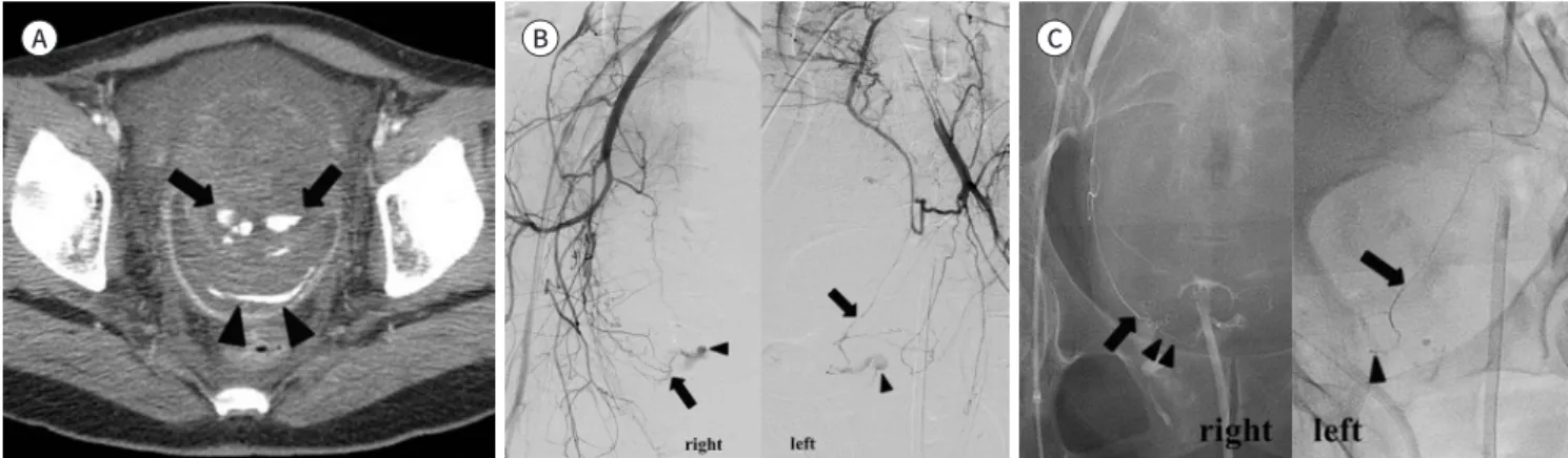
Immediate hysterectomy was performed in two patients at the obstetrician’s discretion. One patient who survived cardiopulmonary resuscitation died from multi-organ failure eight days after UAE. One patient with abnormal placentation that was a combination of placenta Fig. 2. A 38-year-old woman presented with postpartum hemorrhage after vaginal delivery.
A. Axial CT scan shows contrast material extravasation at the posterior aspect of the lacerated uterine cervix (arrows). Extravasated contrast material pooling in the posterior fornix of the vagina (arrowheads).
B. Bilateral internal iliac arteriography showing extravasation (arrowheads) from bilateral cervical arteries (arrows).
C. Post-embolic spot image showing the microcatheter advanced into the cervical artery (arrows) and the casted N-butyl-2-cyanoacrylate at the bleeding focus (arrowheads).
A B C
Table 2. Angiographic Findings and the Results of Embolization
Parameters Values (%)
Angiographic finding
Extravasation 10 (71.4)
Pseudoaneurysm 4 (28.6 )
Embolized arteries
Uterine artery (including two cervical artery) 14
Inferior epigastric artery 2
Vaginal artery 1
Vesical artery 1
Internal pudendal artery 1
Technical success 13/14 (92.9)
Clinical success 12/14 (85.7)
Clinical success in coagulopathy 3/5 (60)
Major complication 1 (7.1)
previa and placenta percreta underwent multiple pelvic artery embolizations as well as UAE.
This patient underwent repeated embolization for multiple pelvic arteries due to deteriorat- ed vital signs three hours after the initial embolization. Despite repeated embolization, the patient underwent a hysterectomy due to persistent vaginal bleeding. This patient under- went a colostomy and skin flap surgery in the bilateral buttock area due to pelvic ischemia.
DISCUSSION
Permanent embolic materials have not been commonly used in patients with PPH be- Table 3. Supplementary Information of the Patients, Treatment Details, and Outcomes
Pt.
No.
Age (Y)
Type of Delivery
Onset of
Bleeding Cause of PPH
Initial Hb Level (g/dL)
Coagulopathy
Embolized Arteries Using NBCA
Non-Uterine Artery Embolization
Hysterec- tomy
Technical Success
Clinical Success
1 31 Vaginal Primary Cervical laceration 8.7 No CA None No Success Success
2 38 Vaginal Primary Cervical laceration 6.8 Yes CA None No Success Success
3 34 Cesarean Secondary Hysterotomy related arterial injury 8.5 Yes UA None No Success Success 4 39 Cesarean Primary Hysterotomy related
arterial injury 9.8 No UA None No Success Success
5 31 Cesarean Secondary Hysterotomy related arterial injury 8.2 No UA None No Success Success 6 30 Cesarean Primary Hysterectomy
related arterial
injury 8.4 No UA None Yes* Success Success
7 28 Cesarean Primary Hysterotomy related
arterial injury 9.7 No UA None No Success Success
8 31 Cesarean Secondary Hysterotomy related arterial injury,
uterine atony 9.1 No UA None No Success Success
9 30 Cesarean Primary Hysterotomy related
arterial injury 3.9 Yes UA None Yes Success Fail
10 34 Vaginal Primary
Hysterectomy related arterial injury, abdominal wall bleeding
6.9 No UA, IEA Yes Yes* Success Success
11 35 Cesarean Primary Hysterotomy related
arterial injury 5.2 No UA None No Success Success
12 33 Cesarean Primary Placenta previa with
percreta 3.7 Yes UA Yes Yes Fail Fail
13 32 Cesarean Primary
Hysterotomy related arterial injury, abdominal wall bleeding, uterine atony
8.4 Yes UA Yes No Success Success
14 36 Vaginal Primary Hysterotomy related
arterial injury 8.8 No UA None No Success Success
*Hysterectomy was performed in another hospital.
CA = cervical artery, Hb = hemoglobin, IEA = inferior epigastric artery, NBCA = N-butyl-2-cyanoacrylate, PPH = postpartum hemorrhage, Pt = patient, UA = uterine artery
cause of concerns regarding fertility. Nevertheless, these materials have been recently ap- plied in PPH. In example, use of NBCA was first reported by Pelage et al. (22). They used NBCA in patients with pseudoaneurysm. Since then, a few articles have reported the use of NBCA in patients with PPH who showed extravasation or pseudoaneurysm, and the success rates were 67.9–92.3% (14, 15). In our study, we performed UAE using NBCA and achieved high technical (92.9%) and clinical success (85.7%) rates with low complication rates.
In our cases, we observed uterine artery rupture or pseudoaneurysm in 11 patients. In most cases, the lesions usually occurred at the proximal portion of the ascending segment of the uterine artery, which made it possible to use NBCA because there was no need to ad- vance the microcatheter to the distal uterine artery through the tortuous ascending segment of the uterine artery. However, in one case of uterine fundal bleeding, we advanced the mi- crocatheter through the tortuous segment and performed successful UAE (Fig. 3). When the microcatheter is not wedged into the vessel, NBCA runs through the vessel with arterial flow until it is polymerized. In previous reports, when NBCA was diluted to 50% with Lipiodol, the polymerization time of the 50% of the NBCA and Lipiodol mixture was six to ten seconds in the in-vitro experiments that could be replicated in the in-vivo environments under vessel wall interaction (23, 24). In our experience, NBCA did not advance rapidly in the uterine ar- tery because of its tortuosity. Therefore, in TAE of the uterine artery, it should be considered that the distance traversed by NBCA might be shorter than expected.
In our study, there were two cases of cervical artery bleeding. In these cases, considering the complex pelvic arterial anatomy and collateral arteries that developed during pregnancy, embolization of the bleeding focus as well as its proximal and distal segments was needed for successful embolization (25). Therefore, we chose NBCA as an embolic material in these cases. The extent of embolization with NBCA could be controlled by wedging the catheter into the target vessel, as NBCA is a liquid embolic material. By doing this, we could embolize the bleeding vessel without repeated embolization.
Fig. 3.A 31-year-old woman presented with postpartum hemorrhage after cesarean delivery.
A. Right internal iliac arteriography showing extravasation of contrast material at the uterine fundus (arrow).
B. A radiograph showing the microcatheter advanced into the far distal uterine arterial branch through the tortuous segment (arrows).
C. Post-embolic arteriography showing the casted N-butyl-2-cyanoacrylate at the bleeding segment without reflux to the proximal or distal uterine artery (arrows).
A B C
In our study, additional embolization for IEA was performed in two patients due to surgical wound bleeding. These bleeding foci were detected on CT angiography before the emboliza- tion procedures and these might have been missed if not for the CT scan. Therefore, detec- tion of the bleeding focus with a CT scan is important for successful hemostasis in patients with PPH (26).
One patient experienced pelvic ischemia after repeated embolization. However, consider- ing that the NBCA was used only for UAE, this could be deemed unrelated to the use of NBCA. In this patient, to our knowledge, pelvic ischemia resulted from multiple pelvic arte- rial embolizations with gelatin sponge particles. We performed embolization with NBCA only when the selection of the target artery was successfully achieved. By doing this, we could avoid unexpected embolization of the vessels that we did not want to embolize perma- nently. Therefore, we conclude that the occurrence of complication was determined, not by the kind of embolic material, but by the extent and number of arteries that were embolized.
In our study, we observed clinical failure in two (14.3%) patients. These patients were in coagulopathy at the time of UAE. Of these, one patient underwent UAE with NBCA, but the persistent vaginal bleeding was due to severe disseminated intravascular coagulation (DIC).
Another patient showed multiple and massive bleeding foci which could not be controlled by embolization due to placenta previa combined with placenta percreta. Therefore, our study reveals that the possibility of clinical failure increases in patients with coagulopathy combined with multiple bleeding sources or severe DIC.
Our study has some limitations that warrant consideration. It was limited first by its retro- spective nature and second by the small number of patients. In particular, we observed coag- ulopathy in five patients, and they showed a relatively lower success rate (3/5, 60%) than that reported in previous studies (67.9–90.5%) (14, 15). With more cases, we could have clearly identified the factors contributing to failure in coagulopathic patients. In addition, future prospective studies are needed for the appropriate selection of patients with PPH for whom the use of NBCA will be suitable.
In conclusion, UAE using NBCA is safe and effective in patients with PPH showing extrava- sation or pseudoaneurysm.
Conflicts of Interest
The authors have no potential conflicts of interest to disclose.
REFERENCES
1. Khan KS, Wojdyla D, Say L, Gülmezoglu AM, Van Look PF. WHO analysis of causes of maternal death: a sys- tematic review. Lancet 2006;367:1066-1074
2. Devine PC. Obstetric hemorrhage. Semin Perinatol 2009;33:76-81
3. Revert M, Cottenet J, Raynal P, Cibot E, Quantin C, Rozenberg P. Intrauterine balloon tamponade for man- agement of severe postpartum haemorrhage in a perinatal network: a prospective cohort study. BJOG 2017;124:1255-1262
4. Pelage JP, Le Dref O, Mateo J, Soyer P, Jacob D, Kardache M, et al. Life-threatening primary postpartum hemorrhage: treatment with emergency selective arterial embolization. Radiology 1998;208:359-362 5. Ganguli S, Stecker MS, Pyne D, Baum RA, Fan CM. Uterine artery embolization in the treatment of post-
partum uterine hemorrhage. J Vasc Interv Radiol 2011;22:169-176
6. Park JK, Shin TB, Baek JC, Shin JK, Choi WJ, Lee SA, et al. Failure of uterine artery embolization for control-
ling postpartum hemorrhage. J Obstet Gynaecol Res 2011;37:971-978
7. Gonsalves M, Belli A. The role of interventional radiology in obstetric hemorrhage. Cardiovasc Intervent Radiol 2010;33:887-895
8. Tanahashi Y, Goshima S, Kondo H, Ando T, Noda Y, Kawada H, et al. Transcatheter arterial embolization for primary postpartum hemorrhage: predictive factors of need for embolic material conversion of gelatin sponge particles to N-butyl cyanoacrylate. Cardiovasc Intervent Radiol 2017;40:236-244
9. Matsubara S, Sato T, Nakata M. Vaginal artery embolization with a permanent embolic agent for intracta- ble postpartum hemorrhage. J Obstet Gynaecol Res 2011;37:377-378
10. Koo HJ, Shin JH, Kim HJ, Kim J, Yoon HK, Ko GY, et al. Clinical outcome of transcatheter arterial emboliza- tion with N-butyl-2-cyanoacrylate for control of acute gastrointestinal tract bleeding. AJR Am J Roentgen- ol 2015;204:662-668
11. Song HH, Won YD, Kim YJ. Transcatheter N-butyl cyanoacrylate embolization of pseudoaneurysms. J Vasc Interv Radiol 2010;21:1508-1511
12. Yoo DH, Jae HJ, Kim HC, Chung JW, Park JH. Transcatheter arterial embolization of intramuscular active hemorrhage with N-butyl cyanoacrylate. Cardiovasc Intervent Radiol 2012;35:292-298
13. Yonemitsu T, Kawai N, Sato M, Tanihata H, Takasaka I, Nakai M, et al. Evaluation of transcatheter arterial embolization with gelatin sponge particles, microcoils, and n-butyl cyanoacrylate for acute arterial bleed- ing in a coagulopathic condition. J Vasc Interv Radiol 2009;20:1176-1187
14. Kim GM, Yoon CJ, Seong NJ, Kang SG, Kim YJ. Postpartum haemorrhage from ruptured pseudoaneurysm:
efficacy of transcatheter arterial embolisation using N-butyl-2-cyanoacrylate. Eur Radiol 2013;23:2344- 2349
15. Park KJ, Shin JH, Yoon HK, Gwon DI, Ko GY, Sung KB. Postpartum hemorrhage from extravasation or pseu- doaneurysm: efficacy of transcatheter arterial embolization using N-butyl cyanoacrylate and comparison with gelatin sponge particle. J Vasc Interv Radiol 2015;26:154-161
16. Kanematsu M, Watanabe H, Kondo H, Goshima S, Kato H, Furui T, et al. Postpartum hemorrhage in coagu- lopathic patients: preliminary experience with uterine arterial embolization with N-butyl cyanoacrylate. J Vasc Interv Radiol 2011;22:1773-1776
17. Lee WH, Yang SB, Goo DE, Kim YJ, Lee JM, Kang CH, et al. Uterine artery embolization: the interventional treatment of female genital diseases. J Korean Soc Radiol 2017;76:1-9
18. Huang YS, Chang CC, Liou JM, Jaw FS, Liu KL. Transcatheter arterial embolization with N-butyl cyanoacry- late for nonvariceal upper gastrointestinal bleeding in hemodynamically unstable patients: results and predictors of clinical outcomes. J Vasc Interv Radiol 2014;25:1850-1857
19. Dohan A, Eveno C, Dautry R, Guerrache Y, Camus M, Boudiaf M, et al. Role and effectiveness of percutane- ous arterial embolization in hemodynamically unstable patients with ruptured splanchnic artery pseudoa- neurysms. Cardiovasc Intervent Radiol 2015;38:862-870
20. Aina R, Oliva VL, Therasse E, Perreault P, Bui BT, Dufresne MP, et al. Arterial embolotherapy for upper gas- trointestinal hemorrhage: outcome assessment. J Vasc Interv Radiol 2001;12:195-200
21. Sacks D, McClenny TE, Cardella JF, Lewis CA. Society of Interventional Radiology clinical practice guide- lines. J Vasc Interv Radiol 2003;14:S199-S202
22. Pelage JP, Soyer P, Repiquet D, Herbreteau D, Le Dref O, Houdart E, et al. Secondary postpartum hemor- rhage: treatment with selective arterial embolization. Radiology 1999;212:385-389
23. Wang BH, Boulton M, Lee DH, Pelz DM, Lownie SP. A systematic characterization of the factors influencing polymerization and dynamic behavior of n-butyl cyanoacrylate. J Neurointerv Surg 2018;10:150-155 24. Takasawa C, Seiji K, Matsunaga K, Matsuhashi T, Ohta M, Shida S, et al. Properties of N-butyl cyanoacry-
late-iodized oil mixtures for arterial embolization: in vitro and in vivo experiments. J Vasc Interv Radiol 2012;23:1215-1221
25. Pelage JP, Le Dref O, Soyer P, Jacob D, Kardache M, Dahan H, et al. Arterial anatomy of the female genital tract: variations and relevance to transcatheter embolization of the uterus. AJR Am J Roentgenol 1999;172:989-994
26. Kim JE, So YH, Kim BJ, Kim SM, Choi YH, Sung CK. Postpartum hemorrhage from non-uterine arteries:
clinical importance of their detection and the results of selective embolization. Acta Radiol 2018;59:932- 938
산후출혈 환자에서의 자궁동맥 색전술:
N-Butyl-2-Cyanoacrylate를 이용한 치료의 임상적 유용성 및 안전성
권요한1 · 소영호2* · 김병재3 · 김선민3 · 최영호2 · 문민환2
목적 산후출혈 환자에서 N-butyl-2-cyanoacrylate (이하 NBCA)를 이용한 자궁동맥 색전술 의 임상적 유용성 및 안전성을 평가하고자 하였다.
대상과 방법 2010년 2월부터 2018년 5월까지 산후출혈로 자궁동맥 색전술을 시행받은 82 명의 환자 중, NBCA를 이용하여 자궁동맥 색전술을 시행받은 14명(나이: 28~39세; 평균:
33세)을 대상으로 하였다. 후향적 의무기록 분석을 통하여 환자의 특성, 산후출혈의 원인, 색전술, 결과를 분석하였다.
결과 혈관조영술에서 혈액 누출은 10명, 가성동맥류는 4명에서 관찰되었다. 산후출혈의 원 인은 자궁절개 또는 자궁절제술 후 동맥손상(n = 11), 자궁경관열상(n = 2), 전치태반(n = 1)이었다. 모든 환자의 자궁동맥 색전술에서 NBCA가 이용되었다. 2명의 환자에서 젤라틴 스폰지 입자를 이용한 추가적인 자궁동맥 색전술을 시행하였다. 3명의 환자에서 자궁동맥 외 동맥 색전술을 시행하였다. 5명(35.7%)의 환자에서 혈액응고병증이 관찰되었다. 색전 술의 기술적 성공률은 92.9%, 임상적 성공률은 85.7%였다. 색전술을 시행받은 환자 중 1명 은 다발성 장기부전으로 인해 색전술 시행 8일 후에 사망하였다. 전치태반을 가진 1명의 환 자는 여러 골반동맥 색전술로 인해 골반장기의 허혈성 손상이 유발되었다.
결론 혈관 외 혈액 누출 및 가성동맥류를 보이는 산후출혈 환자에서 NBCA를 이용한 자궁동 맥 색전술은 임상적으로 유용하고 안전하였다.
1서울대학교병원 영상의학과, 서울특별시 보라매병원 2영상의학과, 3산부인과