INTRODUCTION
Chest CT has become a common imaging modality for vari- ous clinical conditions of the lung, chest wall, mediastinum, pleura, and diaphragm (1). Although not routinely used to as- sess breast lesions, the entirety of the breast usually appears on chest CT. With the increasing use of chest CT, incidental breast lesions are being frequently encountered (2-5). Thus, it is impor- tant for clinicians to be familiar with the CT characteristics of breast lesions, which can be referred to mammography or breast ultrasonography (US) for further evaluation if necessary (6).
Several previous studies have reported that the detection rate of incidental breast lesions on chest CT ranges from 0.06% to 7.63% (7-9). The referral rates of incidental breast lesions on chest CT are uncertain and malignancy rates of the referred cases vary with a wide range (9.2% to 70%) (9-12). This sug- gests that effective criteria for referral to diagnostic breast eval- uation are needed for incidental breast lesions found on chest CT, which may provide further characterization of the inciden- tal breast lesions due to its high resolution, large field of view (FOV), and cross-sectional capabilities (13).
There are a few reports that have investigated suspicious CT
Incidental Breast Lesions on Chest CT: Clinical Significance and Differential Features Requiring Referral
흉부 전산화단층촬영에서 우연히 발견된 유방 병변: 임상적 중요성 및 진료 의뢰가 필요한 특징적 영상 소견
Yun Jung Choi, MD
1, Tae Hoon Kim, MD
1, Yoon Jin Cha, MD
2, Eun Ju Son, MD
1, Hye Mi Gweon, MD
1, Chul Hwan Park, MD
1*
Departments of 1Radiology, 2Pathology, Gangnam Severance Hospital, Yonsei University College of Medicine, Seoul, Korea
Purpose: To evaluate the CT features of incidental breast lesions on chest CT and to suggest useful criteria for referral to a specialized breast unit.
Materials and Methods: Between May 2009 and April 2014, enhanced chest CT examination reports containing the key word ‘breast’ were reviewed retrospectively.
Patients who had incidental breast lesion and were referred to a specialized breast unit and then underwent pathological confirmation or follow-up over a 1-year pe- riod were included. Finally, 86 patients (all female, mean age, 48.9 ± 12.6 years) were enrolled. Two radiologists evaluated lesion characteristics, including size, shape, margins, and enhancement. The correlations between the CT features and pathologies were evaluated, and the diagnostic accuracy of CT features in various combinations was assessed.
Results: Among the CT features, irregular shape, non-circumscribed margin, and high contrast enhancement were different between malignant and benign lesions (p <
0.05). The combination of non-circumscribed margin and high contrast enhance- ment had the highest accuracy (97.7%).
Conclusion: Reliable CT features for incidental malignant breast masses are irregular shape, non-circumscribed margin, and high contrast enhancement. The combination of non-circumscribed margin and high contrast enhancement could help distinguish incidental malignant breast lesions and indicate referral to a specialized breast unit.
Index terms Breast Neoplasm
Tomography, X-Ray Computed Incidental Findings
Received May 24, 2018 Revised June 25, 2018 Accepted July 14, 2018
*Corresponding author: Chul Hwan Park, MD Department of Radiology, Gangnam Severance Hospital, Yonsei University College of Medicine, 211 Eonju-ro, Gangnam-gu, Seoul 06273, Korea.
Tel. 82-2-2019-3510 Fax. 82-2-3462-5472 E-mail: [email protected]
This is an Open Access article distributed under the terms of the Creative Commons Attribution Non-Commercial License (https://creativecommons.org/licenses/by-nc/4.0) which permits unrestricted non-commercial use, distri- bution, and reproduction in any medium, provided the original work is properly cited.
J Korean Soc Radiol 2018;79(6):303-310 https://doi.org/10.3348/jksr.2018.79.6.303
characteristics of incidental breast malignancy (3, 12, 14, 15).
Reliable features for breast malignancy on CT include spicula- tion, irregular shape, and rim enhancement (16). However, these CT features are not robust and some remain controversial (10, 11, 17). The aims of this study were to evaluate the CT features of incidentally found breast lesions on chest CT, and to suggest use- ful criteria for referral to a specialized breast unit for assessment.
MATERIALS AND METHODS
Patients
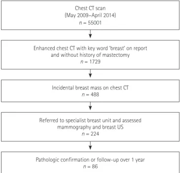
This retrospective study was performed with approval of the Institutional Review Board (3-2017-0114). Given the retrospec- tive nature of the study and the use of anonymized data, require- ments for informed consent were waived. All reports of chest CT examinations from May 2009 to April 2014 containing the key word ‘breast’ were reviewed. Patients with known breast dis- ease, previous history of breast cancer or breast surgery, or who had undergone only non-enhanced CT examination were ex- cluded from the study. Inclusion criteria were patients who ex- hibited incidental breast lesion(s) on enhanced chest CT and were referred to a specialist breast unit for assessment and then underwent pathological confirmation or follow-up over a 1-year period. A total of 55001 chest CT examinations were performed during the study period, and 1729 radiological reports contain- ing the key word ‘breast’ were found. A total of 488 patients with no history of breast disease or breast operation were reported to have an incidental breast lesion on chest CT. Of these 488 pa- tients, 224 (45.9%) were referred to a specialized breast unit; of these 224, 86 underwent pathological confirmation or follow-up for at least 1 year (Fig. 1).
Chest CT Scan Protocol
CT scans were obtained using one of three multi-detector CT scanners: a 16-slice (Somatom Sensation 16; Siemens Medical Solutions, Erlangen, Germany); a 64-slice (Somatom Sensation 64; Siemens Medical Solutions), or a 128-slice (Somatom Defi- nition AS+; Siemens Medical Solutions) device. The chest CT scans were obtained from the lung apices to the level of the ad- renal glands, during a breath hold at the end of inspiration, in the supine position. After acquiring a scout image to determine the FOV, CT scans were obtained after contrast material injec-
tion. Scanning was done by using a helical technique, with a 3 mm or 5 mm reconstruction interval. The exposure parame- ters for the CT scans were 120 kVp and 50-130 mA. Image re- construction for conventional CT scans was obtained by using the scanner’s workstation. All CT scans were transferred to a picture archiving and communication system (Centricity 2.0;
GE Medical Systems, Mount Prospect, IL, USA).
CT Image Analysis
Two radiologists (C.H.P and T.H.K), each with > 10 years of experience with chest CT interpretation, reviewed the chest CT scans and were blinded to the final results of the breast assess- ment. Decisions regarding CT features were determined by consensus. The radiologists evaluated lesion morphology and contrast-enhancement patterns. In the absence of a formal CT lexicon for breast lesions, the modified Breast Imaging and Re- porting Data System (Bi-RADS) terminology for MRI lexicon 2013 was used for the analysis of morphology (18).
The following CT findings were recorded for each lesion:
size, location; shape (round or oval, irregular), margin (well- circumscribed, non-circumscribed), and enhancement ratio (poor enhancement, high contrast enhancement).
To overcome the various contrast media injection protocols, the attenuation ratio between breast masses and the ipsilateral trapezius muscles were calculated and used as the indicator of
Chest CT scan (May 2009–April 2014)
n = 55001
Enhanced chest CT with key word ‘breast’ on report and without history of mastectomy
n = 1729
Referred to specialist breast unit and assessed mammography and breast US
n = 224
Incidental breast mass on chest CT n = 488
Pathologic confirmation or follow-up over 1 year n = 86
Fig. 1. Flow chart of patient selection.
enhancement strength. Regions of interest were drawn in the breast lesion (as large as possible) and the ipsilateral trapezius muscle (larger than 1.5 cm2) and the attenuation (Hounsfield units) was measured. The enhancement ratio of the breast mass was calculated as (attenuation of breast lesion)/(attenuation of trapezius muscle). All lesions were categorized into 1 of 2 groups according to the attenuation ratio as follows: poor en- hancement (attenuation ratio ≤ 1), or high contrast enhance- ment (attenuation ratio > 1).
Final Diagnosis
Eighty-six patients subsequently underwent breast US or mammography for the incidental breast lesion(s). Findings were reported according to the Bi-RADS US or mammography lexicon (19, 20). All mammography and breast US were ana- lyzed by two experienced radiologists (H.M.G and E.J.S). Sono- graphically guided core-needle biopsy or surgery was done for definite diagnosis in 38 (44.2%) patients. The diagnoses in the remaining 48 (55.8%) patients were determined on the basis of follow-up chest CT or breast US for a period of at least 12 months.
Statistical Analysis
Continuous variables were stated as mean ± standard devia- tion, and categorical variables were expressed as frequencies and/or percentages. The Shapiro-Wilk test was performed to evaluate the distribution of continuous data. The independent t-test was used to compare the age and size of benign and malig- nant lesions. The Fisher’s exact test was used to assess potential correlations of various CT features with breast lesion malignan- cy. The sensitivity, specificity, positive predictive value (PPV), negative predictive value (NPV), and overall diagnostic accuracy were calculated for the various criteria using significant CT characteristics to diagnose breast lesions on chest CT scans. A p value of < 0.05 was considered to be statistically significant. All statistical analyses were done using commercially available soft- ware (SPSS version 20, IBM Corp., Armonk, NY, USA).
RESULTS
Assessment Findings
Of a total of 86 breast lesions found on chest CT, the final as-
sessment categories from mammography and breast US were distributed according to BI-RADS as follows: category 1 in 4 le- sions (4.6%), category 2 in 12 lesions (14.0%), category 3 in 42 lesions (48.8%), category 4 in 23 lesions (26.7%), and category 5 in 5 lesions (5.8%). All 28 lesions that were BI-RADS category 4 and 5 underwent sonographically guided biopsy, which re- vealed 13 cancers and 15 benign lesions. One category 0 lesion was designated a category 3 lesion on follow-up breast US. Of the 4 breast lesions detected on chest CT that were BI-RADS category 1, breast mammography and US, one cancer was diag- nosed. This lesion appeared as an 0.8 cm enhancing lesion in chest CT, and was not detected at mammography or US. Two- year follow-up breast US revealed a 1.0 cm irregular hypoecho- ic lesion in the right breast, and the final assessment was BI- RADS category 4. Subsequent tissue sampling and surgery were performed, and diagnosis of a 0.9 cm infiltrative ductal carci- noma was made (Fig. 2). The other three lesions were not de- tected on follow-up mammography or breast US, which sug- gested false-positive lesions on chest CT.
Overall, the malignancy rate of the incidental breast mass at chest CT was 16.3% (14 of 86), 78.6% (11 of 14) was invasive carcinoma [median tumor size, 1.5 cm (range, 0.5–1.9 cm)] and
A B
C D
Fig. 2. A 60-year-old woman underwent chest CT for a medical ex- amination.
A. Axial CT reveals a round, non-circumscribed mass with high con- trast enhancement in the right upper medial quadrant of the breast.
B. Two years later, tumor size increased from 8 mm to 10 mm.
C. Sonography of the right breast at the corresponding location re- veals hypoechoic mass with irregular margin.
D. Pathological examination revealed infiltrative ductal carcinoma with desmoplastic stroma (hematoxylin-eosin stain, × 200).
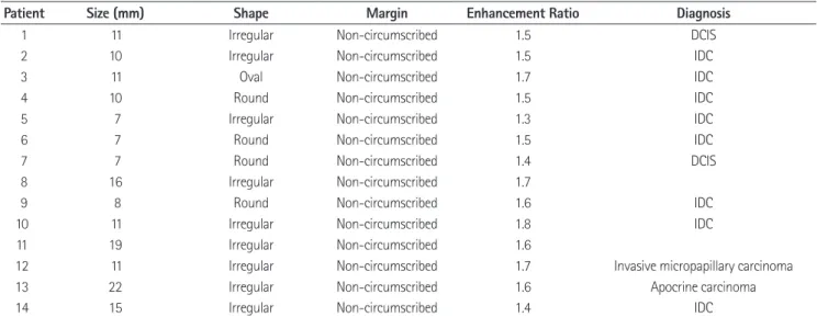
21.4% (3 of 14) were ductal carcinoma in situ (Table 1).
There were 24 biopsy-proven benign lesions among the 86 patients. These included six fibrocystic diseases, 11 fibroadeno- mas, two atypical lobular hyperplasia, two sclerosing adenosis, one intraductal papilloma, one fibrosis and one abscess. The other 48 lesions were followed-up with CT or US and con- firmed to be benign.
Clinical and CT Features of Breast Masses
The clinical and CT features were compared between malig- nant and benign lesions (Table 2). The mean age of patients with malignant breast tumors (56.7 ± 14.6 years) was higher than that of patients with benign breast lesions (47.4 ± 11.7 years) (p = 0.011). The mean size of the incidental malignant breast tumor was 1.2 ± 4.6 cm (range, 0.7-2.2 cm), whereas the Table 1. Pathological Outcomes and Features of Incidental Malignant Breast Lesions on Chest CT
Patient Size (mm) Shape Margin Enhancement Ratio Diagnosis
1 11 Irregular Non-circumscribed 1.5 DCIS
2 10 Irregular Non-circumscribed 1.5 IDC
3 11 Oval Non-circumscribed 1.7 IDC
4 10 Round Non-circumscribed 1.5 IDC
5 7 Irregular Non-circumscribed 1.3 IDC
6 7 Round Non-circumscribed 1.5 IDC
7 7 Round Non-circumscribed 1.4 DCIS
8 16 Irregular Non-circumscribed 1.7
9 8 Round Non-circumscribed 1.6 IDC
10 11 Irregular Non-circumscribed 1.8 IDC
11 19 Irregular Non-circumscribed 1.6
12 11 Irregular Non-circumscribed 1.7 Invasive micropapillary carcinoma
13 22 Irregular Non-circumscribed 1.6 Apocrine carcinoma
14 15 Irregular Non-circumscribed 1.4 IDC
DCIS = ductal carcinoma in situ, IDC = invasive ductal carcinoma
Table 2. Demographic Information and Features of Breast Masses Detected on Chest CT
Feature Lesion
p-Value Malignant (n = 14) Benign (n = 72)
Age (years), mean ± SD 56.7 ± 14.6 47.4 ± 11.7 0.011
Menopausal status, n
Pre 4 (28.6) 44 (61.1) 0.038
Post 10 (71.4) 28 (38.9)
Lesion size (cm), mean ± SD 1.2 ± 4.6 1.1 ± 6.8 0.793
Multiplicity 0.593
Single 14 (100) 65 (90.3)
Multiple 0 (0) 7 (9.7)
Shape < 0.001
Oval 1 (7.1) 56 (77.8)
Round 4 (28.6) 11 (15.3)
Irregular 9 (64.3) 5 (6.9)
Margin < 0.001
Well-circumscribed 0 (0) 68 (94.4)
Non-circumscribed 14 (100) 4 (5.6)
Attenuation ratio < 0.001
Poor enhancement 0 (0) 30 (41.7)
High contrast enhancement 14 (100) 42 (58.3)
Data presented as n (%) unless otherwise indicated.
SD = standard deviation
mean size of incidental benign breast lesions was 1.1 ± 6.8 cm (range, 0.4-4.5 cm). There was no difference between the mean size of malignant and benign lesions (p = 0.793).
Among CT features, the shape, margin, and attenuation ratio were significantly different between malignant and benign le- sions. Of the 14 patients with malignant breast masses, irregular shape was defined in 9 (64.3%) patients, and the other 5 (35.7%) patients exhibited oval or round masses. Of 72 patients with benign breast tumors, 5 (6.9%) exhibited an irregular shape and 67 (93.1%) exhibited oval or round masses. Inciden- tal malignant breast masses exhibited a higher rate of irregular shape (p < 0.001).
All malignant breast masses exhibited a non-circumscribed margin. Well-circumscribed margins were identified in 68 of 72 (94.4%) patients with benign breast masses and only 4 (6.9%) exhibited non-circumscribed margins. Notably, the incidence of non-circumscribed margin was higher in malignant masses than in benign masses (100% vs. 6.9%; p < 0.001). All malig- nant breast masses exhibited high contrast enhancement.
Among benign breast masses, the incidence of high contrast enhancement was similar to that of poor enhancement (58.3%
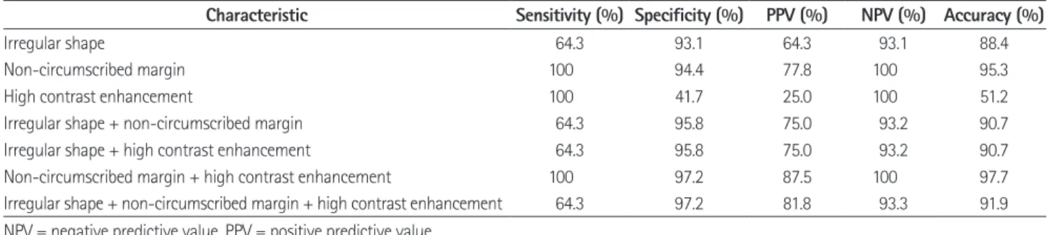
vs. 41.7%). Table 3 summarizes the sensitivity, specificity, PPV, NPV, and accuracy for malignancy in relation to the shape, margin, enhancement ratio, and combinations thereof. A fea- ture of malignancy with high PPV was a non-circumscribed margin (94.4%). The combination of non-circumscribed mar- gin and high contrast enhancement had the highest accuracy (97.7%), followed by non-circumscribed margin, with an accu- racy of only 95.4% (Fig. 3).
DISCUSSION
In our study, we evaluated the CT features of incidental breast lesions on chest CT, and assessed useful CT features for referral to a specialized breast unit. Among the CT features, shape, margin, and attenuation ratio were significantly different be- tween malignant and benign lesions. The combination of non- circumscribed margin and high contrast enhancement had the highest accuracy for malignancy.
In previous studies, most incidental breast lesions have been reported to be benign (2, 21). Therefore, it is important for ra- diologists to be able to discern the differences in typical CT fea- Table 3. Diagnostic Performance in the Differentiation of Malignant from Benign Incidental Breast Lesions
Characteristic Sensitivity (%) Specificity (%) PPV (%) NPV (%) Accuracy (%)
Irregular shape 64.3 93.1 64.3 93.1 88.4
Non-circumscribed margin 100 94.4 77.8 100 95.3
High contrast enhancement 100 41.7 25.0 100 51.2
Irregular shape + non-circumscribed margin 64.3 95.8 75.0 93.2 90.7
Irregular shape + high contrast enhancement 64.3 95.8 75.0 93.2 90.7
Non-circumscribed margin + high contrast enhancement 100 97.2 87.5 100 97.7
Irregular shape + non-circumscribed margin + high contrast enhancement 64.3 97.2 81.8 93.3 91.9 NPV = negative predictive value, PPV = positive predictive value
Fig. 3. An 86-year-old woman underwent CT for a thyroid mass.
A. Axial CT scan reveals a non-circumscribed, irregular mass with high contrast enhancement in the left upper medial quadrant of the breast.
B. Sonography reveals a hypoechoic mass with irregular margin.
C. Pathological examination revealed infiltrative ductal carcinoma (hematoxylin-eosin stain, × 200).
A B C
tures of malignant and benign breast lesions to avoid unneces- sary additional imaging workup. A few studies have evaluated the correlation between CT features and pathological outcomes of incidental breast lesions (9-12, 17). The predictive CT fea- tures suggestive of malignant breast tumors on chest CT in- cluded larger size, irregular margins, spiculation, and rim en- hancement (11, 12, 16). In our study, malignant breast lesions exhibited significantly higher probabilities of irregular shape and non-circumscribed margins. Similarly, well-circumscribed lesions were suggestive of benignity. Careful assessment of the margins and shapes of incidental breast lesions may differenti- ate benign and malignant lesions.
Uematsu et al. (22) reported that attenuation of breast lesions is not useful in the differential diagnosis, and the presence of enhancement alone does not always suggest a malignant tumor.
Another study reported that additional delayed enhancement on CT may be important to differentiate benign from malig- nant lesions (23). However, chest CT was usually obtained for indications other than breast pathologies, and protocols are not optimized for breast imaging; therefore, variation in contrast medium administration and scan time is expected. In this study, we used the attenuation ratio between the breast lesions and ipsilateral trapezius muscles to correct the diversity of en- hancement protocols. High contrast-enhanced masses exhibit- ed high sensitivity (100%), although a low PPV (25.0%) and ac- curacy (51.2%). Our findings suggest that enhancement ratio alone cannot be used as a reliable CT feature of malignancy or benignity. However, a combination of non-circumscribed mar- gin and high contrast enhancement had high sensitivity, speci- ficity, PPV, NPV and accuracy for incidental malignant breast masses (100%, 97.2%, 88.2%, 100%, and 97.7%, respectively).
In the present study, four incidental breast lesions were con- sidered BI-RADS category 1 on initial mammography and breast US. One of these lesions increased in size on follow-up chest CT and was confirmed to be a malignant tumor; the other three lesions were not delineated on follow-up mammography and breast US. Mammography and breast US are currently pre- ferred imaging modalities for breast abnormalities. Compared with mammography and US, CT has several advantages. A breast lesion may be better visualized using contrast-enhanced CT than mammography if the breast lesion is in a dense breast or if the lesion is located adjacent to the chest wall (24). Howev-
er, normal breast glandular tissue could be appeared as a ‘pseu- do-mass’ on CT, and this finding was more common in dense breasts (10). Therefore, chest CT cannot be a standard modality for breast evaluation, and it is important to refer to a specialized breast unit when incidental breast lesion(s) are detected.
There were a few limitations to this study, including its single- center, retrospective design. Second was the potential percep- tion bias of missed or unreported breast lesions. Finally, despite a large number of chest CT examinations were carefully re- viewed with strict inclusion/exclusion criteria, histological eval- uation was absent in one-half of the incidental breast masses.
Thus the malignancy rate of incidental breast lesions would have increased in this study.
In conclusion, the malignancy rate of incidentally found breast lesions on chest CT is not negligible. Reliable features for incidental malignant breast masses on chest CT were irregular shape, non-circumscribed margin, and high contrast enhance- ment. The combination of non-circumscribed margin and high contrast enhancement could help to distinguish incidental ma- lignant breast lesions on routine chest CT and inform referral to a specialized breast unit.
REFERENCES
1. Skinner S. Guide to thoracic imaging. Aust Fam Physician 2015;44:558-562
2. Yi JG, Kim SJ, Marom EM, Park JH, Jung SI, Lee MW. Chest CT of incidental breast lesions. J Thorac Imaging 2008;
23:148-155
3. Monzawa S, Washio T, Yasuoka R, Mitsuo M, Kadotani Y, Hanioka K. Incidental detection of clinically unexpected breast lesions by computed tomography. Acta Radiol 2013;54:374-379
4. Cho EM, Kang H, Shin YG, Yun JH, Oh KS, Park S. Detec- tion of breast abnormalities on enhanced chest CT: corre- lation with breast composition on mammography. J Kore- an Soc Radiol 2017;76:96-103
5. Kim JH, Chang YW, Hwang JH, Kim HH, Lee EH, Yang SB.
Evaluation of the significance of incidental breast lesions detected by chest CT. J Korean Soc Radiol 2013;68:229-235 6. Goldberg PA, White CS, McAvoy MA, Templeton PA. CT ap- pearance of the normal and abnormal breast with mam-
mographic correlation. Clin Imaging 1994;18:262-272 7. Jacobs PC, Mali WP, Grobbee DE, van der Graaf Y. Preva-
lence of incidental findings in computed tomographic screening of the chest: a systematic review. J Comput As- sist Tomogr 2008;32:214-221
8. Shojaku H, Seto H, Iwai H, Kitazawa S, Fukushima W, Saito K. Detection of incidental breast tumors by noncontrast spiral computed tomography of the chest. Radiat Med 2008;26:362-367
9. Hussain A, Gordon-Dixon A, Almusawy H, Sinha P, Desai A.
The incidence and outcome of incidental breast lesions detected by computed tomography. Ann R Coll Surg Engl 2010;92:124-126
10. Porter G, Steel J, Paisley K, Watkins R, Holgate C. Inciden- tal breast masses detected by computed tomography: are any imaging features predictive of malignancy? Clin Radi- ol 2009;64:529-533
11. Bach AG, Abbas J, Jasaabuu C, Schramm D, Wienke A, Sur- ov A. Comparison between incidental malignant and be- nign breast lesions detected by computed tomography: a systematic review. J Med Imaging Radiat Oncol 2013;57:
529-533
12. Lin WC, Hsu HH, Li CS, Yu JC, Hsu GC, Yu CP, et al. Inciden- tally detected enhancing breast lesions on chest computed tomography. Korean J Radiol 2011;12:44-51
13. Harish MG, Konda SD, MacMahon H, Newstead GM. Breast lesions incidentally detected with CT: what the general ra- diologist needs to know. Radiographics 2007;27 Suppl 1:S37-S51
14. Jakanani G, Al-Attar M. RE: incidental breast masses de- tected by computed tomography: are any imaging features predictive of malignancy? Clin Radiol 2009;64:1041-1042 15. Prionas ND, Lindfors KK, Ray S, Huang SY, Beckett LA,
Monsky WL, et al. Contrast-enhanced dedicated breast CT:
initial clinical experience. Radiology 2010;256:714-723 16. Inoue M, Sano T, Watai R, Ashikaga R, Ueda K, Watatani M,
et al. Dynamic multidetector CT of breast tumors: diag- nostic features and comparison with conventional tech- niques. AJR Am J Roentgenol 2003;181:679-686
17. Litmanovich D, Gourevich K, Israel O, Gallimidi Z. Unex- pected foci of 18F-FDG uptake in the breast detected by PET/CT: incidence and clinical significance. Eur J Nucl Med Mol Imaging 2009;36:1558-1564
18. D’Orsi CJ. ACR BI-RADS Atlas: breast imaging reporting and data system. 5th ed. Reston, VA: American College of Radiology 2013.
19. Mendelson E, Böhm-Vélez M, Berg W, Whitman G, Feld- man M, Madjar H. ACR BI-RADS® ultrasound. In D’Orsi CJ, ed. ACR BI-RADS Atlas: breast imaging reporting and data system. 5th ed. Reston, VA: American College of Radiology 2013:35-100
20. Sickles E, D’Orsi C, Bassett L, Appleton C, Berg W, Burnside E. ACR BI-RADS® mammography. In D’Orsi CJ, ed. ACR BI- RADS® Atlas, breast imaging reporting and data system.
Reston, VA: American College of Radiology 2013;5:13-117 21. Meller MT, Cox JE, Callanan KW. Incidental detection of
breast lesions with computed tomography. Clin Breast Cancer 2007;7:634-637
22. Uematsu T, Sano M, Homma K. False-positive helical CT findings of multifocal and multicentric breast cancer: is attenuation of tumor useful for diagnosing enhanced le- sions? Breast Cancer 2002;9:62-68
23. Sardanelli F, Calabrese M, Zandrino F, Melani E, Parodi R, Imperiale A, et al. Dynamic helical CT of breast tumors. J Comput Assist Tomogr 1998;22:398-407
24. Kim SM, Park JM. Computed tomography of the breast.
Abnormal findings with mammographic and sonographic correlation. J Comput Assist Tomogr 2003;27:761-770
흉부 전산화단층촬영에서 우연히 발견된 유방 병변:
임상적 중요성 및 진료 의뢰가 필요한 특징적 영상 소견
최윤정
1· 김태훈
1· 차윤진
2· 손은주
1· 권혜미
1· 박철환
1*
목적: 이 연구의 목적은 흉부 전산화단층촬영(CT)에서 우연히 발견된 유방 병변의 임상적 중요성을 평가하고 유방 전문 진료 의뢰가 필요한 영상 소견을 제안하는 것이다.
대상과 방법: 2009년 5월부터 2014년 4월까지 촬영된 조영증강 흉부 CT 중에서 판독문에 ‘유방’이라는 단어가 포함된 검사를 후향적으로 검토하였다. 이 중 우연히 발견된 유방 병변으로 유방 초음파와 유방 촬영술을 한 뒤, 병리학적 확인이 나 1년 이상의 추적 관찰을 받은 총 86명의 환자(모두 여성, 평균 연령, 48.9 ± 12.6세)를 대상으로 하였다. 흉부 CT에서 유방 병변의 크기, 위치, 모양, 경계 및 조영 증강을 포함한 영상 특징을 평가하였다. 다양한 CT 특징과 병리소견과의 연 관성을 평가하고 여러 조합의 CT 특징의 진단 정확도를 평가하였다.
결과: 흉부 CT에서 우연히 발견된 유방 병변 중 악성 종양은 16.3%(14/86)였다. 악성 병변은 양성 병변에 비해 불규칙한 모양, 불분명한 경계, 높은 조영증강을 보였다. CT 특징의 조합 중 불분명한 경계와 높은 조영증강의 조합이 악성 병변을 예측하는데 가장 높은 정확도를 보였다(97.7%).
결론: 흉부 CT에서 우연히 발견된 유방 병변의 16.3%는 악성이었으며, 불분명한 경계와 높은 조영증강을 동시에 보일 경우 악성 가능성이 높으므로, 정확한 진단을 위해 유방 전문 진료 의뢰가 필요하다.
연세대학교 의과대학 강남세브란스병원 1영상의학과, 2병리학과