Noninvasive Follow-Up of Coronary Artery Bypass Graft Patency Using Multi-Slice Computed Tomography
1Seung Ho Joo, M.D., Byoung Wook Choi, M.D., Jae Seung Seo, Young-Jin Kim, Tae Hoon Kim, M.D., Kyu Ok Choe, M.D., Donghoon Choi, M.D.2, Kyung-Jong Yoo3
1Department of Diagnostic Radiology and the Research Institute of Radiological Science, Yonsei University College of Medicine
2Department of Cardiology and Internal Medicine, Yonsei University College of Medicine
3Department of Cardiovascular Surgery, Yonsei University College of Medicine This study was supported by a grant from Yonsei University College of Medicine.
Received June 15, 2006 ; Accepted September 21, 2006
Address reprint requests to : Byoung Wook Choi, M.D., Department of Diagnostic Radiology, Severance Hospital, 134 Sinchon-Dong, Seodaemoon-Gu, Seoul 120-752, South Korea.
Tel. 82-2-2228-2372 Fax. 82-2-393-3035 E-mail: [email protected]
Purpose: We wanted to evaluate the utility of multi-slice computerized tomography (MSCT) for assessing coronary artery bypass graft patency and/or occlusion.
Materials and Methods: For 24 patients, both conventional angiography and CT an- giography with 4-MSCT were performed within seven days of one another in order to evaluate the accuracy of MSCT with regard to graft patency and/or occlusion. A fol- low-up CT angiogram was performed in patients with and without symptoms (n=11, n=34, respectively) with 4- or 16-MSCT. We retrospectively compared the results of MSCT to those of conventional coronary graft angiography.
Results: Sixty-five grafts were evaluated for the accuracy of MSCT. Six of those 65 were occluded. The sensitivity, specificity, positive predictive value and negative pre- dictive values of MSCT for the diagnosis of graft occlusion were 100% (6/6), 98%
(58/59), 86% (6/7) and 100% (58/58), respectively. Patency could not be determined by angiography in two grafts; however, the grafts proved to be patent on MSCT. On fol- low-up, new graft occlusions in the asymptomatic patients were detected by MSCT in 8.2% of the previously patent grafts at the two years post-op, and in 15.2% at the three years post-op.
Conclusion: MSCT is a practical and accurate noninvasive diagnostic tool for follow- ing up coronary artery bypass grafts.
Index words :Computed tomography (CT) Coronary vessels, surgery Grafts
Graft patency
Coronary artery bypass graft surgery (CABG) is the treatment of choice for patients suffering with severe obstructive coronary artery disease. The success of the surgery and the rate of patient survival are dependent on graft patency; therefore, post-operative assessment of the bypass graft in the patients who have undergone CABG is very important (1). Conventional coronary artery graft angiography (CAG) is currently the gold standard for assessing bypass graft patency. However, it is an invasive technique and it may cause adverse ef- fects, including arrhythmia, stroke, coronary artery dis- section and death. The procedure also incurs a financial commitment, hospital admission and a large measure of patient discomfort (2, 3). Therefore, there is currently a lot of interest in finding a relatively less invasive or non- invasive alternative to CAG. Multi-slice CT (MSCT) has recently been used to determine bypass graft patency and occlusion (4-21). Because of the proven accuracy of MSCT, it can be routinely used for post-op follow-up of patients who have undergone coronary artery bypass graft surgery (CABG). We retrospectively reviewed and compared the results of the MSCT and CAG in different situations, including following up during the immediate postoperative period, following up patients with is- chemic symptoms after surgery and following up asymptomatic patients. We then analyzed the discrep- ancies between the two imaging modalities.
Materials and Methods
Study Population
We retrospectively enrolled 51 patients (33 males and 18 females, mean age: 61 years) who underwent CTA and at least one CAG after CABG. These patients had a total of 136 grafts, including 73 arterial (54%) and 63 ve- nous grafts (46%). CTA was performed once in 24 pa- tients (55 grafts), twice in 24 patients (74 grafts), and three times in three patients (seven grafts). Therefore, the total number of grafts we evaluated was 224 (81 to- tal CTA scans).
Accuracy of MSCT
For an earlier subset of the study population involving 24 patients (16 males, age: 61.1±7.5 years), both CTA and CAG were performed within seven days (range: 2.3
±2.2 days) of one another in order to evaluate the accu- racy of CTA (CAG was considered the standard of refer- ence) (Table 1). All the exams were performed using 4- slice CT. For 13 patients, CTA was performed during the immediate postoperative period (postoperative time interval: 5.8±1.7 days). For the other 11 patients, CTA was performed at long-term follow-up (mean: 5.7 years).
A total of 65 grafts in 24 patients were evaluated, includ- ing 28 saphenous vein grafts (SVG), 22 LITAs, 11 radial artery grafts, three right internal thoracic artery grafts (RITA) and one gastroepiploic artery graft (Table 3).
MSCT Follow-Up
The other subset of the study population involved 45 patients who were referred for CTA follow-up to evalu- ate their graft patency, and they had undergone CAG at least once either before or after MSCT with more than a one month time interval (Table 2). For the patients who had multiple CAG examinations, the data from the most contemporary CAG was used. Of this MSCT follow-up group, 11 patients (31 total grafts) with persistent chest discomfort underwent CAG to confirm the CTA results.
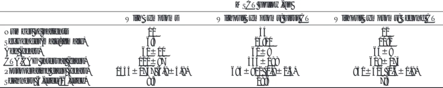
Table 1. Demographic Data for the Accuracy of MSCT Accuracy of MSCT in Evaluation of Graft Patency Immediate Long-term
Post-op Follow-up
Number of patients 13 11
Gender (male:female) 10:3 6:5
Age (years) 63±60 59±80
CTA-CAG interval (days) 1.8±2.2 2.9±2.2 Postoperative days (years) 5.8±1.7 2064±1594
(5.7±4.4) Scanners (4-slice:16-slice) 13:0 11:0
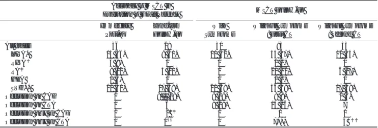
Table 2. Demographic Data of the MSCT follow-up Subset of Patients
MSCT Follow-up
With Symptoms Without Symptoms: First CT Without Symptoms: Second CT
Number of patients 11 34 12
SexGender(male:female) 6:5 24:10 10:2
Age (years) 061±10 61±80 63±80
CTA-CAG interval (days) 120±97 345±198 619±174
Postoperative days (years) 1434±1757 (3.9±4.8) 695±912 (1.9±2.5) 951±325 (2.6±0.9)
Scanners (4-slice:16-slice) 8:3 28:6 7:5
The other 34 patients (93 grafts), who did not experience any chest symptoms, were also enrolled and they re- ceived follow-up CTA. Twelve of these 34 patients (35 total grafts) underwent another CTA approximately one year later. Information about these subjects is summa- rized in Tables 2 and 3. During the follow-up period when we used CTA, a 16-slice CT was installed in the institute and this was then used to determine post-op graft patency. The hospital ethics committee approved this protocol, and all the patients gave us and informed consent.
Multi-slice CT: Graft Angiography Protocol
CTA scans were performed using a 4-slice CT scanner (LightSpeed Plus, GE Medical Systems, Milwaukee, Wisconsin, U.S.A.) or 16-slice CT scanner (Sensation 16, Siemens, Forchheim, Germany). We did not pre-med- icate any of the patients.
Protocol for 4-slice CT
The scan range was adjusted to cover all the grafts, from the uppermost implanted graft vessel at the as- cending aorta to the diaphragmatic surface. For the case of the patient with a gastroepiploic artery graft, the scan range was extended lower to include the pancreas. Prior to the CTA scan, the patients were injected with a 15-ml
bolus injection of non-ionic contrast agent (370 iodine mg/mL, Iopamiro; Bracco, Milan, Italy) in order to mea- sure the circulation time. The scan delay was set at five seconds after the time of peak enhancement of the as- cending aorta. The CTA scan was performed in a cranio- caudal manner. The gantry rotation time was 500 ms, and the tube current was 270-300 mA at 120 kV. The breath-holding time, which depended on the patient’s heart rate and the anatomic coverage, ranged from 25 to 40 seconds. Pitch also depended on the patient’s heart rate at the beginning of the scan or the heart rate that was input by the operator, which was adjusted automat- ically in increments of 0.1 according to the prepro- grammed manufacturer settings within a range of 1.0- 4.0. For imaging in a patient with a heart rate that varied from 45 to 75 beats per minute, the pitch varied from 1.2 to 1.5. The total CTA scan contrast dose ranged from 120-180 mL, and this depended on the total scan time.
The patients received the contrast agent through an 18- gauge needle in the antecubital vein, and they were in- jected at a rate of 4 mL/s for the first half of the total scan time, and at 2 mL/s for the second half.
Protocol for 16-slice CT
A real-time bolus tracking technique was used in or- der to trigger the scan. The scan delay was set at six sec-
Table 3. Number of Grafts in Each Subset of Patients
Accuracy of MSCT in
Evaluation of Graft Patency MSCT Follow-up
Immediate Long-term With Without Symptoms Without Symptoms
Post-op Follow-up Symptoms : First CT : Second CT
All grafts 36 29 31 93 35
LITA* 13 (36) 09 (31) 10 (32) 34 (37) 12 (34)
RITA 3 (8) 0 0 2 (2) 0
RA 08 (22) 03 (10) 0 20 (22) 06 (17)
GEA § 1 (3) 0 0 1 (1) 0
SVG** 11 (31) 17 (59) 21 (68) 36 (39) 17 (49)
Occlusion on CAG 0 8 (28) 9 (29) 8 (9) 2 (6)
Occlusion on CTA 0 6 9 (29) 15 (16) 7
Occlusion only on CAG 0 2 0 0 0
Occlusion only on CTA 0 1§§ 0 7*** 5
*. Left internal thoracic artery graft . Right internal thoracic artery graft . Radial artery graft
§. Gastroepiploic artery graft
**. Saphenous vein graft
. Including two grafts that were not diagnosed due to the lack of selective angiography . Two grafts that were not diagnosed due to lack of selective angiography
§§. A case with competitive flow
***. Newly developed occlusion on CTA . Newly developed occlusion on CTA
: The number in parentheses is the percentage in each subset of patients.
onds after the enhancement of the ascending aorta reached 100 HU. The CT parameters were as follows:
12×0.75 mm collimation, 0.42-seconds rotation speed, 500 effective mA at 120 kV and 2.7 mm/rotation table- feed. The amount of nonionic contrast agent (370 iodine mg/mL, Iopamiro; Bracco, Milan, Italy) ranged from 100 to 140 ml, and it was injected during a single inspiratory breath-hold (duration: 24.2±3.4 s) via an 18- or 20- gauge needle through an antecubital vein (flow rate: 4 mL/s). A multi-cyclic, segmented reconstruction algo- rithm was used. The temporal resolution was from 110 to 210 ms. Radiation dose modulation was not used be- cause the images should be reconstructed not only at a diastolic phase, but also at a systolic phase to obtain bet- ter qualities. The other, unmentioned protocol details were essentially the same as those for 4-slice CT.
Multi-slice CT: Reconstruction and Interpretation
With using the 4-slice CT scans, the axial image data sets were reconstructed with a 1.25-mm thickness and a 1.0-mm increment by using retrospective ECG gating at 40% and 70% of the cardiac cycle (the R wave-to-R wave interval). With using the 16-slice CT scans, axial image data sets were reconstructed with 1.0-mm thick- ness and a 0.5-mm increment at one or two cardiac phases, which were selected by using the preview re- construction function, where the phase had the least motion artifacts. On the basis of the reconstructed data sets, the three-dimensional volume rendered images, the thin or thick slab of maximal intensity projection im- ages and the curved multi-planar reformatted images were reconstructed for each bypass graft on an off-line workstation. Two radiologists, who were unaware of the invasive coronary arteriography outcomes, evaluat- ed all the CT data by working in consensus. The bypass grafts were considered occluded if they showed no con- trast material filling on any images, or if they showed only a short segment of contrast material in the proxi- mal stump. Otherwise, the grafts were considered patent.
Conventional Graft Angiography
Bypass graft contrast angiography was performed us- ing a digital angiographic system (Hicor, Simens, Erlangen, Germany). Every graft was selectively catheterized for performing selective angiography.
However, some composite grafts could not be directly catheterized due to an anastomosis with the distal part of another graft. In that case, indirect angiography was
completed with a proximal injection into the other graft.
In some patients with a left internal thoracic artery graft (LITA), left subclavian arteriography was performed in place of selective graft angiography. Non-visualization of the contrast agent into the grafts was considered to be an occlusion. Failure to cannulate a targeted graft from the aorta, without any evidence of contrast filling ac- cording to the aortography, was tentatively considered to be an occlusion of the graft as well.
Data Analysis
The results from CAG served as the standard of refer- ence. The bypass graft patency diagnostic accuracy of CTA was expressed as the sensitivity, specificity and the positive and negative predictive values. Any discrepan- cies between CTA and CAG were analyzed.
Results
Accuracy of MSCT
In 13 patients, no grafts were occluded in the immedi- ate postoperative period. CAG showed occlusion of eight grafts in 11 patients in the long-term follow-up group. CTA correctly diagnosed six of these occlusions, including four SVGs and two radial artery grafts. The re- maining two grafts were diagnosed as being patent on CTA. These grafts entailed a SVG connected from the aorta to the first obtuse marginal branch (Fig. 1), and a SVG connected from another graft (Fig. 2). Upon review of these cases, there was no doubt that the grafts were patent on CTA, as there was a continuous, strong opaci- fication of the lumen with contrast material. Upon ex- amining the CAG, we concluded that the two cases had been tentatively diagnosed as occluded because the grafts could not be visualized. The selective angiography failed because a surgically placed metal indicator was not present for the graft in the former case and the com- posite graft was distally connected from another graft in the latter case.
There was only one false positive case, in which the CTA showed no opacification of a LITA connected to the middle segment of the left anterior descending artery. We noticed that the graft was patent, but it had competitive flow on angiography. The sensitivity, speci- ficity, positive predictive value, negative predictive val- ue and accuracy were 75% (6/8), 98% (56/57), 86% (6/7), 97% (56/58) and 95% (63/65). Graft occlusion was deter- mined to be 9% (6/65) of the grafts, including 2 ambigu- ous cases that were tentatively diagnosed as occluded on
CAG. The sensitivity, specificity, positive predictive val- ue and negative predictive value were 100% (6/6), 98%
(58/59), 86% (6/7) and 100% (58/58), respectively.
Overall accuracy was 98% (64/65).
MSCT Follow-Up
The CTA and CAG results were concordant for 31 grafts (11 patients) in those patients with ischemic symp- toms. These results included the detection of nine oc- cluded grafts (29%). Two (20%) of the 10 LITAs and sev-
A B
C D
Fig. 2. 64-year-old male patient. On the 1-year follow-up angiograpy after surgery (A), the catheter tip is located at the proximal SVG and the contrast agent is injected. The SVG connected from another SVG is visualized only by the slow retrograde filling (arrow), which does not reach the proximal anastomosis. Seven days later, CTA (B) shows the graft patent. Two years lat- er, the follow-up curved MPR CT im- age (C) and the volume rendered im- age (D) confirm the patency of the graft. The patient had reported no symptom.
A B
Fig. 1. 59-year-old male patient. A vol- ume rendered image with a superior view (A) and an axial image (B) of the contrast-enhanced CT angiography both reveal a patent saphenous vein graft. The patient underwent surgery eleven years prior. The graft coursed posterior to the aorta, without a metal indicator at the proximal anastomosis.
The selective angiography failed, and the graft was not visualized by aortic root angiography (not shown).
en (33%) of the 21 SVGs were occluded. For 93 grafts (34 asymptomatic patients), CAG demonstrated eight occluded grafts (8.6%). The following CTA revealed sev- en more occluded grafts, which was 8.2% of the 85 patent grafts on the previous CAG. Therefore, there was a total of 15 occluded grafts detected on CTA (16%) (Table 4). In 12 patients, the second follow-up CTA was performed within 347±128 days after the first CTA and it revealed five more graft occlusions (15.2% of the 33 originally patent grafts). Newly observed occlusions were found in two of the 11 previously patent LITAs (18%), and also in three of the six previously patent radi- al artery grafts (50%). All the occluded radial artery grafts were composite grafts.
Discussion
Even though single-slice CT was not sensitive detect- ing for graft occlusion, it could detect graft patency.
However, both 4- and 16-slice CT can be used for the detection of both graft occlusion and patency (4). With the introduction of 4-detector CT into clinical practice, the sensitivity (93-100%) and specificity (97.8-100%) of CT for detecting patency or occlusion has increased (5-10). Compared to a 4-slice CT, the16-slice CT demonstrated a significantly better image quality, re- gardless of the type of graft, for all bypass segments, with the exception of distal anastomosis (11). The accu- racy of a 16-slice CT for the diagnosis of graft patency has slightly improved as compared to that of the 4-slice CT; 16 slice CT demonstrating a sensitivity of 96-100%
and a specificity of 93-100% (12-17).
In the present study, the accuracy for diagnosing graft patency and occlusion with using 4-slice CT was 98%.
This percentage is considered to be relatively high as compared to the previous studies, even though no β- blockers were used and no cases were excluded from this study. Graft patency could be determined by
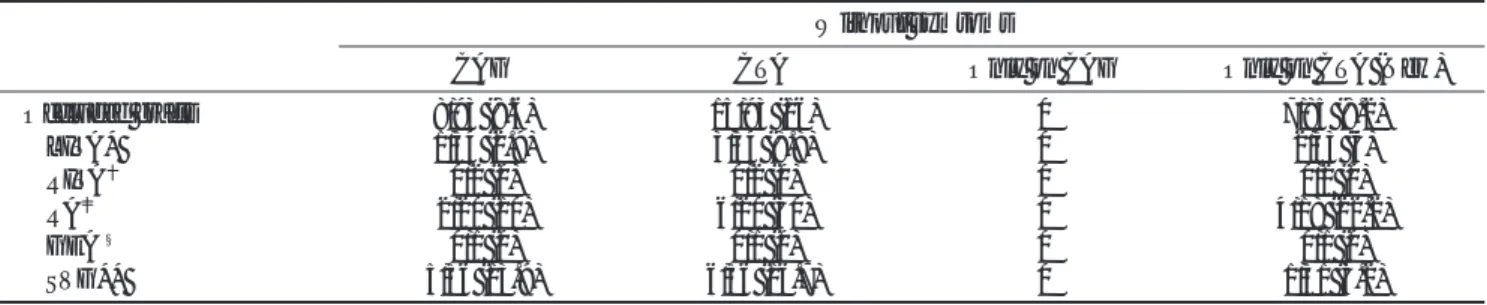
Table 4. Follow-up MSCT in the Patients Without Symptoms
Without symtoms
CAG CTA Only on CAG Only on CTA (New)
Occluded grafts 8/93 (8.6) 15/93 (16) 0 7/85 (8.2)
LITA* 1/34 (2.9) 3/34 (8.8) 0 2/33 (6)
RITA 0/2 (0) 0/2 (0) 0 0/2 (0)
RA 2/20 (10) 6/20 (30) 0 4/18 (22.2)
GEA§ 0/1 (0) 0/1 (0) 0 0/1 (0)
SVG** 5/36 (13.9) 6/36 (16.7) 0 1/31 (3.2)
*. Left internal thoracic artery graft . Right internal thoracic artery graft . Radial artery graft
§. Gastroepiploic artery graft
**. Saphenous vein graft
: The number in parentheses is the percentage in each subset.
A B
Fig. 3. 54-year-old male patient. The one week follow-up angiograpy after surgery (A) shows the left internal tho- racic artery graft and the radial aratery composite graft are both patent. The two-year follow-up CT angiography (B) shows the two grafts are atent, which is well correlated with the pre- vious angiography.
demonstrating at least two different levels of the con- trast-filled graft lumen (22). Therefore, motion artifacts or metal artifacts (i.e., that resulted from clips obscuring some segments of grafts) did not necessarily reduce the accuracy of MSCT. In this study, no case of graft occlu- sion showed any additional patent segments or a patent proximal segment that was longer than 15 mm.
Graft patency could not be determined when selective graft angiography was not performed: this was caused by the limited accessibility of complex composite grafts or by grafts without metallic markers (10). In this case, MSCT accurately demonstrated patency or occlusion, and this is an advantage of MSCT over conventional graft angiography. In the presence of competitive flow in a graft, effective lumen contrast-filling may not be possible and MSCT may incorrectly diagnose the physi- cally patent graft as being occluded. This was the only incidence of false graft patency interpretation in our study. Therefore, readers of CTA scans should be aware of this possibility when determining the presence of an occlusion.
MSCT was very effective and accurate for following up graft patency during the post-CABG period for both symptomatic and asymptomatic patients (Fig. 3).
Examining the immediate postoperative graft patency is important because early occlusion may occur within the first week to month (a 10% likelihood in SVGs, 1% in LITA, 6% in RITA and 4-10% of radial grafts) and this is mainly due to spasm (23). Determining the etiology of ischemic symptoms after bypass graft surgery is an im- portant clinical issue and it is essential for delivering proper management. The cause of recurrent chest pain varies with the time after surgery. When symptoms are first noted in the immediate postoperative period, it may be due to a technical problem with a graft or with early graft closure; these are not reliably detected with MRI (24). In our study, all grafts in the patients that were fol- lowed during the immediate postoperative period were patent on CAG and MSCT. After the first few postopera- tive months, the symptoms are usually caused by both graft problems and by the progression of atherosclerosis in the non-bypassed vessels. For the symptomatic pa- tients after bypass grafting, the angiographic graft fail- ure rate is approximately double that in the patients without symptoms, which was compatible to the high occlusion rate of the symptomatic group (29%) in this study (25). Although the time interval between CTA and CAG for the symptomatic patients was about six months, the MSCT results were the same as those for
the CAG in all cases. MSCT was very sensitive for de- tecting newly developed graft failure in the patients without symptoms. This implies that many patients with newly developed graft failure do not present with symptoms.
Off-pump coronary artery bypass grafting (OPCAB) combined with the aorta no-touch technique has been accepted as an effective procedure to avoid neurologic and aortic complications, and also to reduce the opera- tive risks (26). Using the radial artery for a composite graft in context of the no-touch aorta technique has been well documented. However, the long-term patency of a radial artery composite graft has not been fully studied.
In the present study, the high occlusion rate of the radial artery graft on the first or second follow-up CTA reflect- ed the increased use of radial artery composite grafts with the OPCAB technique at our institute. This result demonstrated that MSCT could be used as a noninva- sive modality for evaluating new bypass grafting tech- niques.
Graft stenosis was not evaluated with MSCT in this study. The image quality was not sufficient to diagnose graft stenosis, primarily because we did not use a β- blocker to control the heart rate, nor did we exclude any candidates with coexisting arrhythmia or tachycardia.
We also did not exclude any cases with severe post-scan motion artifacts. MSCT has a limitation to functionally assess graft flow. Even though the study population and the CTA protocols were heterogenous, and the time in- terval between the two modalites was variable, the re- sults of this study showed the usefulness of MSCT in daily practice for a variety of situations.
In conclusion, in terms of patency and occlusion as compared to conventional invasive angiography, 4- or a 16-slice MSCT is a very practical, highly accurate, and reproducible noninvasive tool for evaluating coronary artery grafts during both the immediate postoperative period and on the long-term follow-up, and this is re- gardless of the patient’s symptoms or heart rate.
References
1. Lipton MJ, Takaro T, Clayman J, Maley T. Veterans Administra- tion Cooperative Study of medical versus surgical treatment for stable angina—progress report. Section 10. Factors associated with long-term graft patency after coronary artery bypass surgery. Prog Cardiovasc Dis 1986;28:301-308
2. Wyman RM, Safian RD, Portway V, Skillman JJ, McKay RG, Baim DS. Current complications of diagnostic and therapeutic cardiac catheterization. J Am Coll Cardiol 1988;12:1400-1406
3. Kennedy JW. Complications associated with cardiac catheteriza-
tion and angiography. Cathet Cardiovasc Diagn 1982;8:5-11 4. Stein PD, Beemath A, Skaf E, Kayali F, Janjua M, Alesh I, et al.
Usefulness of 4-, 8-, and 16-slice computed tomography for detec- tion of graft occlusion or patency after coronary artery bypass grafting. Am J Cardiol 2005;96:1669-1673
5. Ropers D, Ulzheimer S, Wenkel E, Baum U, Giesler T, Derlien H, et al. Investigation of aortocoronary artery bypass grafts by multi- slice spiral computed tomography with electrocardiographic-gated image reconstruction. Am J Cardiol 2001;88:792-795
6. Yoo KJ, Choi D, Choi BW, Lim SH, Chang BC. The comparison of the graft patency after coronary artery bypass grafting using coro- nary angiography and multi-slice computed tomography. Eur J Cardiothorac Surg 2003;24:86-91
7. Nieman K, Pattynama PM, Rensing BJ, Van Geuns RJ, De Feyter PJ. Evaluation of patients after coronary artery bypass surgery: CT angiographic assessment of grafts and coronary arteries. Radiology 2003;229:749-756
8. Burgstahler C, Kuettner A, Kopp AF, Herdeg C, Martensen J, Claussen CD, et al. Non-invasive evaluation of coronary artery by- pass grafts using multi-slice computed tomography: initial clinical experience. Int J Cardiol 2003;90:275-280
9. Marano R, Storto ML, Maddestra N, Bonomo L. Non-invasive as- sessment of coronary artery bypass graft with retrospectively ECG-gated four-row multi-detector spiral computed tomography.
Eur Radiol 2004;14:1353-1362
10. Moore RK, Sampson C, MacDonald S, Moynahan C, Groves D, Chester MR. Coronary artery bypass graft imaging using ECG-gat- ed multislice computed tomography: comparison with catheter an- giography. Clin Radiol 2005;60:990-998
11. Khan MF, Herzog C, Landenberger K, Maataoui A, Martens S, Ackermann H, et al. Visualisation of non-invasive coronary bypass imaging: 4-row vs. 16-row multidetector computed tomography.
Eur Radiol 2005;15:118-126
12. Schlosser T, Konorza T, Hunold P, Kuhl H, Schmermund A, Barkhausen J. Noninvasive visualization of coronary artery bypass grafts using 16-detector row computed tomography. J Am Coll Cardiol 2004;44:1224-1229
13. Martuscelli E, Romagnoli A, D’Eliseo A, Tomassini M, Razzini C, Sperandio M, et al. Evaluation of venous and arterial conduit pa- tency by 16-slice spiral computed tomography. Circulation 2004;110:3234-3238
14. Burgstahler C, Beck T, Kuettner A, Drosch T, Kopp AF, Heuschmid M, et al. Non-invasive evaluation of coronary artery bypass grafts using 16-row multi-slice computed tomography with 188 ms temporal resolution. Int J Cardiol 2006;106:244-249 15. Salm LP, Bax JJ, Jukema JW, Schuijf JD, Vliegen HW, Lamb HJ, et
al. Comprehensive assessment of patients after coronary artery by- pass grafting by 16-detector-row computed tomography. Am Heart J 2005;150:775-781
16. Chiurlia E, Menozzi M, Ratti C, Romagnoli R, Modena MG.
Follow-up of coronary artery bypass graft patency by multislice computed tomography. Am J Cardiol 2005;95:1094-1097
17. Anders K, Baum U, Schmid M, Ropers D, Schmid A, Pohle K, et al. Coronary artery bypass graft (CABG) patency: assessment with high-resolution submillimeter 16-slice multidetector-row comput- ed tomography (MDCT) versus coronary angiography. Eur J Radiol 2006;57(3):336-344.
18. Dewey M, Lembcke A, Enzweiler C, Hamm B, Rogalla P.
Isotropic half-millimeter angiography of coronary artery bypass grafts with 16-slice computed tomography. Ann Thorac Surg 2004;77:800-804
19. Song MH, Ito T, Watanabe T, Nakamura H. Multidetector com- puted tomography versus coronary angiogram in evaluation of coronary artery bypass grafts. Ann Thorac Surg 2005;79:585-588 20. Dorgelo J, Willems TP, van Ooijen PM, Panday GF, Boonstra PW,
Zijlstra F, et al. A 16-slice multidetector computed tomography protocol for evaluation of the gastroepiploic artery grafts in pa- tients after coronary artery bypass surgery. Eur Radiol 2005;15:
1994-1999
21. Gurevitch J, Gaspar T, Orlov B, Amar R, Dvir D, Peled N, et al.
Noninvasive evaluation of arterial grafts with newly released mul- tidetector computed tomography. Ann Thorac Surg 2003;76:1523- 1527
22. Bateman TM, Gray RJ, Whiting JS, Sethna DH, Berman DS, Matloff JM, et al. Prospective evaluation of ultrafast cardiac com- puted tomography for determination of coronary bypass graft pa- tency. Circulation 1987;75:1018-1024
23. Tatoulis J, Royse AG, Buxton BF, Fuller JA, Skillington PD, Goldblatt JC, et al. The radial artery in coronary surgery: a 5-year experience—clinical and angiographic results. Ann Thorac Surg 2002;73:143-147
24. Kalden P, Kreitner KF, Wittlinger T, Voigtlander T, Krummenauer F, Kestel J, et al. Assessment of coronary artery bypass grafts: val- ue of different breath-hold MR imaging techniques. AJR Am J Roentgenol 1999;172:1359-1364
25. Buxton BF, Durairaj M, Hare DL, Gordon I, Moten S, Orford V, et al. Do angiographic results from symptom-directed studies reflect true graft patency? Ann Thorac Surg 2005;80:896-900
26. Cameron A, Davis KB, Green G, Schaff HV. Coronary bypass surgery with internal-thoracic-artery grafts—effects on survival over a 15-year period. N Engl J Med 1996;334:216-219
대한영상의학회지 2006;55:451-459
관상동맥 우회술후 다절편 전산화단층촬영 혈관조영술을 통한 우회도관의 개통성에 대한 비침습적 평가1
1연세의료원 신촌세브란스병원 영상의학과
2연세의료원 신촌세브란스병원 내과학교실
3연세의료원 신촌세브란스병원 흉부외과학교실
주승호・최병욱・서재승・김영진・김태훈・최규옥・최동훈2・유경종3
목적: 관상동맥 우회술 후 우회도관의 개통도 여부를 평가함에서 다절편 전산화단층촬영혈관조영술(CTA)의 유용 성에 대해 알아본다.
대상과 방법: 관상동맥 우회술을 시행 받은 24명의 환자에서 우회도관의 개통성에 대한 다절편 CTA의 정확도를 평가하기 위해 4채널 다절편 CTA와 CAG가 7일 이내의 간격을 두고 시행되었다. 증상이 있는 군(n=11)과 없는 군(n=34) 모두에서 추적 4- 또는 16-채널 다절편 CTA가 시행되었고, 고식적 관상동맥혈관조영술(CAG)의 결 과와 후향적으로 비교분석하였다.
결과과: 총 65개의 우회도관이 다절편 CTA로 평가되었고, 이중 6개는 폐쇄되었다. 다절편 CTA의 민감도, 특이도, 양 성예측도, 음성예측도는 각각 100%(6/6), 98%(58/59), 86%(6/7), 그리고 100%(58/58)이었다. CAG에서 평가 곤란한 2개의 우회도관이 다절편 CTA에서는 개통되어 평가되었다. 수술 후 다절편 CTA를 통해 증상을 동반하지 않는 우회도관의 새로운 폐쇄가 2년째에 8.2%에서, 3년째에 15.2%에서 발견되었다.
결론: 다절편 CTA는 관상동맥 우회술의 추적 검사에 있어 실용적이고, 정확한 비침습적 진단 검사법이다.