In daily practice, cases in which a unilateral hyperlu- cent hemithorax is seen at frontal chest radiography are often encountered. Because such an abnormality is re- lated to various conditions, including radiographic arti- facts, chest wall abnormalities, pulmonary vascular dis- orders with reduced perfusion, and lung parenchymal abnormalities with air trapping or compensatory over-
expansion, a radiographic finding of increased radiolu- cency involving one hemithorax may give rise to diffi- culties in differential diagnosis. The accurate radi- ographic interpretation of unilateral hyperlucency there- fore requires an awareness of the spectrum of these causes. The various conditions can be grouped on the basis of their underlying mechanisms and radiographic features, and according to our experience, an approach that reflects this is useful for their evaluation and differ- entiation.
In this pictorial essay, we present the imaging spec- trum of the conditions causing unilateral hyperlucent hemithorax, emphasizing the importance of a systemat- ic approach to the radiographic interpretation of the re-
Unilateral Hyperlucency of the Lung: A Systematic Approach to Accurate Radiographic Interpretation1
Hyung-Jun Noh, M.D., Yu-Whan Oh, M.D., Eun-Jeong Choi, M.D., Bo-Kyung Seo, M.D., Kyu-Ran Cho, M.D., Eun-Young Kang, M.D., Jung Hyuk Kim, M.D.
The radiographic appearance of a unilateral hyperlucent lung is related to various conditions, the accurate radiographic interpretation of which requires a structured ap- proach as well as an awareness of the spectrum of these entities. Firstly, it is important to determine whether a hyperlucent hemithorax is associated with artifacts resulting from rotation of the patient, grid cutoff, or the heel effect. The second step is to deter- mine whether or not a hyperlucent lung is abnormal. Lung that is in fact normal may appear hyperlucent because of diffusely increased opacity of the opposite hemithorax.
Thirdly, thoracic wall and soft tissue abnormalities such as mastectomy or Poland syn- drome may cause hyperlucency. Lastly, abnormalities of lung parenchyma may result in hyperlucency. Lung abnormalities can be divided into two groups: a) obstructive or compensatory hyperinflation; and b) reduced vascular perfusion of the lung due to congenital or acquired vascular abnormalities. In this article, we describe and illustrate the imaging spectrum of these causes and outline a structured approach to accurate ra- diographic interpretation.
Index words : Lung, abnormalities Lung, density Lung, diseases Lung, radiography Thorax, radiography
1Department of Diagnostic Radiology, College of Medicine and Medical Science Research Center, Korea University
Supported by grant 1999-309 from the Korea University Medical Science Research Center
Received November 22, 2002 ; Accepted December 4, 2002
Address reprint requests to : Yu-Whan Oh, M.D., Department of Diagnostic Radiology, Korea University Anam Hospital, 126-1, 5-Ka, Anam-dong, Sungbuk-gu, Seoul 136-705, South Korea.
Tel. 82-2-920-5657 Fax. 82-2-929-3796 E-mail: [email protected]
lated findings.
Radiographic Artifacts
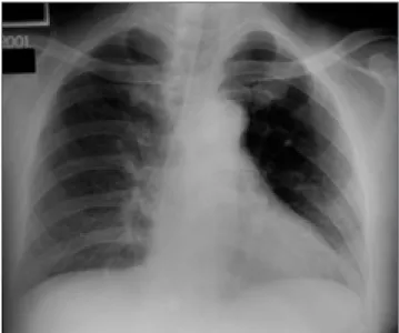
The first step in the evaluation of unilateral hyperlu- cent lung is to check for the presence of a radiographic artifact. Those that simulate unilateral hyperlucency in- clude rotation of the patient, grid cutoff, and the heel ef- fect. The radiologic technique employed may result in differential blackening of the two hemithoraces when a patient is rotated (1). The side to which the patient is ro- tated is more radiolucent because soft tissues project over one side of the chest while being rotated off the op- posite side (Figs. 1, 2). The artifact secondary to rotation is independent of the projection, which may be a pos- teroanterior or anteroposterior. This situation is particu- larly apparent in women with large breasts that are pro- jected over the lower hemithorax as additional radiopac- ity.
The radiographic grid used for removing scatter radia- tion may cause uneven attenuation of primary radiation
─grid cutoff─as a result of the mismatched geometry between the primary beam and the lead strips of the grid. Among four situations producing grid cutoff, com- bined cutoff due to lateral and focus-grid distance decen- tering causes unequal exposure, resulting in a film that
is light on one side and dark on the other (Figs. 3, 4) (2).
The intensity of film exposure on the anode side of the x-ray tube is significantly less than that on the cathode side: the angled anode target itself absorbs some of the x-ray photons emitted almost parallel to the surface of the angled target (Fig. 5) (3). This variation is known as the heel effect, normally used to obtain balanced densi- ties in radiographs of the chest with vertically different thicknesses. If two electrodes (anode and cathode) of the
Fig. 1. Changes in radiographic transmission with rotation. X- ray beam in A direction traverses a longer path through the ob- ject, and thus is more attenuated than that in B direction.
Fig. 3. Grid cutoff resulting from combined lateral and focus- grid distance decentering. There is a large loss of primary radi- ation on one side of film (C) in comparison with the other side (A).
Fig. 2. Radiographic artifact resulting from the rotation of a pa- tient. Chest radiograph taken with a patient who is slightly ro- tated to the left shows a hyperlucent left lung.
x-ray tube are horizontally oriented across the thorax, the heel effect may cause one hemithorax to appear more lucent than the other.
In cases involving these radiographic artifacts, there is
a difference in radiographic density between the two sides of the entire film, not just between the two lungs.
The reason for unilateral hyperlucency is, then, techni- cal rather than pathologic, and in order to recognize this, the relative exposure of soft tissues, especially around the shoulder girdles, should be compared.
Whether Hyperlucent or Radiodense Lung Is Abnormal
After excluding radiographic artifacts, the diagnostic approach to a unilateral hyperlucent lung requires the determination of whether the hyperlucency or radio- density observed is, in fact, abnormal. Mild diffuse in- creased opacity of one hemithorax may falsely suggest that the opposite normal lung is pathologically hyperlu- cent (Fig. 6). The best-known example is a patient in the supine position in whom a large pleural effusion extends posteriorly in layers. In such cases, other radiographic signs of pleural effusion are helpful in deciding that the relatively radiodense hemithorax is abnormal: capping of the lung apex, blunting of the costophrenic angle, loss of the sharp silhouette of the ipsilateral hemidiaphragm, a bandlike opacity separating the lateral lung margin from the chest wall, and thickening of the minor fissure (4). Occasionally, a similar situation may result from the diffuse swelling of unilateral chest wall or slight volume loss in an entire lung or lobe due to sputum plugs (Fig.
7). The latter can be observed particularly in postopera- Fig. 4. A 22-year-old man with acute lymphoblastic lym-
phoma. Portable chest radiograph shows different radiograph- ic densities between two sides of entire film. All of the scapula, ribs, soft tissue, and lung on the right side are more radiolu- cent as compared to those on the left.
Fig. 5. Heel effect. Intensity of x-ray photons emerging in B di- rection is more attenuated due to absorption of some photons by anode target itself than that in A direction.
Fig. 6. A 49-year-woman with left pleural effusion secondary to tuberculous pleurisy. Chest radiograph shows hazy opacifi- cation of left hemithorax with preserved vascular markings.
Note apical capping, blunting of costophrenic angle, loss of hemidiaphragmatic silhouette in left hemithorax.
tive patients or in those in an intensive care unit.
Chest Wall Abnormalities
The third step in the assessment of unilateral hyperlu- cent lung is to determine whether or not the thoracic wall is abnormal. Asymmetric chest wall and soft tissue abnormalities, though very obvious to the clinician, may be overlooked by a radiologist who is unaware of them.
The most common cause of such abnormalities is radi- cal mastectomy, performed because of breast cancer (Fig. 8). Rarely, a congenital defect of the pectoral mus- cles (Poland syndrome) can likewise result in a unilater- al hyperlucent lung (5). In addition, individuals involved in certain occupations, such as butchers and carpenters, may have asymmetric chest wall musculature that can lead to differences in radiographic density between the two hemithoraces. In these cases, the normal branching pulmonary vascularity of both lungs indicates that the explanation for a unilateral hyperlucent lung is asym- metry of soft tissues rather than true hyperlucency.
Abnormalities of Lung Parenchyma
When the various above-mentioned conditions are ex- cluded, the remaining causes of unilateral lung hyperlu- cency are associated with abnormalities of lung parenchyma. The radiographic density of the lung is in- fluenced by the complex interplay between air, blood,
and interstitial tissue. Two of these elements─air and blood volume─are the most important parameters with regard to the hyperlucent lung; that is, hyperlucency usually results from an increase in the volume of air or decrease in the volume of blood in the lung, or a combi- nation of both mechanisms (6).
Increase in the Volume of Pulmonary Air
The amount of air within lung parenchyma may in- crease as a result of either air trapping or compensatory hyperinflation of the lung. A unilateral hyperlucent lung
A B
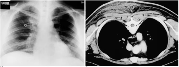
Fig. 7. A 25-year-old man with cellulitis in posterior chest wall.
A. Chest radiograph shows a diffusely increased radiopacity in right hemithorax. The left hemithorax appears relatively radiolu- cent. A safety pin is seen over right hemithorax.
B. Chest CT scan with the patient positioned prone shows a diffuse swelling in posterior chest wall, especially the right side. Ill-de- fined areas of increased attenuation are seen in thickened subcutaneous fat plane of posterior chest wall.
Fig. 8. A 56-year-old woman who underwent mastectomy for right breast cancer. Chest radiograph shows a hyperlucent right lung. Normal shadow of right breast is not present.
occurring in association with air trapping (obstructive hyperinflation) is present in various conditions: foreign body aspiration, inflammatory bronchial strictures, en- dobronchial tumors, congenital lobar emphysema, bronchial atresia, Swyer-James syndrome, unilateral bullous emphysema or cystic lung diseases, and extrin- sic bronchial compression due to mediastinal lesions (7-13). The common radiographic finding of these con- ditions is a hyperlucent lung, with spreading and nar- rowing of the pulmonary vessels (Fig. 9). The volume of the affected lung varies, and may be the equivalent of a lung which is small, of normal size, or enlarged. Where lung volume is large, as in cases of congenital lobar em- physema, bronchial atresia, and bullous emphysema, for example, the mediastinum shifts toward the con-
tralateral side (Fig. 10). The characteristic radiologic fea- ture of obstructive hyperinflation is the presence of air trapping during expiration: chest radiography reveals no appreciable change in the volume of the affected lung at full expiration, whereas the contralateral normal lung decreases in volume (Fig. 9). This radiographic finding is very valuable in distinguishing these disorders from oth- er conditions resulting in a unilateral hyperlucent lung.
When a lobe becomes atelectatic or is resected, the re- maining lobe hyperexpands by way of compensation, and at chest radiography this is marked by the presence of a small or normal-sized hyperlucent lung, spreading of the vascular markings, and displacement of the ipsi- lateral hilum (Fig. 11). If lobar atelectasis, notably of the left lower lobe, is chronic and extreme, the atelectatic
A B
C D
Fig. 9. A 44-year-old woman with Swyer-James syndrome.
A. Chest radiograph in full inspiration shows a hyperlucent left lung with markedly decreased vascular markings.
B. Expiratory radiograph shows no reduction of left lung volume in comparison with right lung.
C. Thin-section CT obtained at level of left main bronchus shows large area of low attenuation in left upper and lower lobes.
D. Pulmonary angiogram shows reduced size and number of pulmonary vessels to the left lung.
lobe may be inconspicuous because it is wedged against the mediastinum and is often hidden by the heart (14).
In contrast to obstructive hyperinflation, an expiratory radiograph depicts a normal decrease in the volume of the hyperinflated lung.
Decreased Pulmonary Blood Volume
Considerable reduced vascular perfusion in one lung may give rise to hyperlucency of that lung at chest radi- ography. The disorders involved are either congenital or acquired; the former, which cause diminished vascular perfusion in one lung, are rare and include aplasia of a pulmonary artery (its proximal interruption) and pul- monary hypoplasia (15,16). These two conditions are ra- diographically characterized by a reduction in the vol- ume of the hyperlucent lung, a small hilum, poor vascu- larization, and ipsilateral shift of the mediastinum (Fig.
12). The pulmonary vascularity of the affected lung is markedly reduced and atypical because blood supply to all or part of the lung is provided by systemic collateral vessels arising from the aorta. In addition, an anomalous pulmonary vein draining into the inferior vena cava may be seen in cases of hypoplasia of the right lung─
the scimitar sign in hypogenetic lung syndrome. The ra- diologic findings of proximal pulmonary artery interrup- tion may mimic those of Swyer-James syndrome.
However, a chest radiograph obtained at full expiration usually serves to differentiate between the two disor- ders. In contrast to Swyer-James syndrome, air trapping is absent where the proximal pulmonary artery is inter-
rupted.
Acquired disorders include pulmonary thromboem- bolism, fibrosing mediastinitis, sequelae of mediastinal irradiation, and mediastinal or hilar tumors (17-19). In these cases, either intrinsic or extrinsic pulmonary arter- ial obstruction may result in marked diminution of arte-
A B
Fig. 10. A 21-year-old man with congenital bronchial atresia.
A. Chest radiograph shows a hyperlucent left lung with increased lung volume. Soft tissue opacity (arrow) is seen adjacent to the left hilum. The mediastinum is shifted to the right side.
B. Thin-section CT shows mucoid impaction (arrow) in anterior segmental bronchus of left upper lobe that is seen as soft tissue opacity on plain radiograph. Large areas of left upper lobe are overinflated and hypovascular.
Fig. 11. A 60-year-old woman who has endobronchial tubercu- losis causing left lower lobe atelectasis. Chest radiograph shows collapsed left lower lobe (black arrow) behind the heart and downward displacement of left hilum. The hyperlucency of left lung is caused by compensatory expansion of left upper lobe that results in spreading of the pulmonary vessels so that there are fewer vessels per unit volume. Calcified small granu- lomas (white arrow) are noted in left lower lung zone.
rial blood perfusion. The affected lung is manifested by a radiographically abnormal unilateral hyperlucent lung, with no evidence of air trapping on both inspirato- ry and expiratory radiographs. In contrast to congenital disorders, the affected lung in acquired vascular disor- ders is of normal size and the ipsilateral hilum is either of normal size or enlarged (Fig. 13).
Summary
Various conditions including radiographic artifacts,
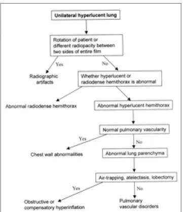
chest wall abnormalities, and pulmonary vascular and parenchymal disorders are associated with the radi- ographic appearance of a unilateral hyperlucent lung. In patients in whom this is observed, accurate radiographic interpretation requires both a structured approach and awareness of the radiographic spectrum of these condi- tions (Fig. 14).
A B
Fig. 12. A 20-year-old man with aplasia of right pulmonary artery.
A. Chest radiograph shows reduced volume of right lung with a small hilum. Note decreased and atypical pulmonary vascularity of right lung.
B. Pulmonary angiogram shows complete absence of right pulmonary artery.
A B
Fig. 13. A 35-year-old man with pulmonary thromboembolism.
A. Chest radiograph shows a hyperlucent right lung and abrupt tapering of right lower lobar pulmonary artery (arrow).
B. Contrast-enhanced CT scan shows thromboembolism in bilateral pulmonary arteries with right one involved more severely.
References
1. Joseph AE, de Lacey GJ, Bryant TH, Stoker DJ, Ayr G. The hyper- transradiant hemithorax: the importance of lateral decentering, and the explanation for its appearance due to rotation. Clin Radiol 1978;29:125-131
2. Curry TS, Dowdey JS, Murry RC. Christensen’s physics of diagnostic radiology, 4th ed. Malvern: Lea & Febiger, 1990:99-117
3. Wolbarst AB. Physics of radiology. Norwalk: Appleton & Lange, 1993: 94-102
4. Wilson AG. Pleura and pleural disorders. In: Armstrong P, Wilson AG, Dee P, Hansell DM, eds. Imaging of diseases of the chest. 2nd ed. St. Luois: Mosby, 1995: 641-716
5. Pearl M, Chow TF, Friedman E. Poland’s syndrome. Radiology 1971; 101: 619-623
6. Remy-Jardin M, Remy J, Giraud F, Wattinne L, Gosselin B.
Computed tomography assessment of ground-glass opacity: semi- ology and significance. J Thorac Imaging 1993; 8: 249-64
7. Blazer S, Naveh Y, Friedman A. Foreign body in the airway. A re- view of 200 cases. Am J Dis Child 1980;134:68-71
8. Spitzer SA, Segal I, Lubin E, Nili M, Levy M. Unilateral increased transradiancy of the lung caused by bronchial carcinoid tumour.
Thorax 1980;35:739-744
9. Wegener WA, Velchik MG. Ventilation-perfusion scintigraphy in an adult with congenital unilateral hyperlucent lung. Clin Nucl Med 1990;15:683-7
10. Genereux GP. Bronchial atresia: a rare cause of unilateral lung hy- pertranslucency. J Can Assoc Radiol 1971 Mar;22:71-82
11. Swyer PR, James GCW. A case of unilateral pulmonary emphyse- ma. Thorax 1953; 8: 133-136
12. Angstadt JD, Cohn HE, Steiner RM. Unilateral hyperlucent lung due to bullous disease. Chest 1986;90:437-8
13. Fukumoto T, Uyama T, Sakiyama S, Kondo K, Monden Y.
Mediastinal esophageal cyst causing unilateral hyperlucent lung.
Jpn J Thorac Cardiovasc Surg 1999;47:141-3
14. Felson B. Chest roentgenology. Philadelphia: WB Saunders, 1973:
92-133
15. Grum CM, Yarnal JR, Cook SA, Cordasco EM, Tomashefski JF.
Unilateral hyperlucent lung. Non-invasive diagnosis of pulmonary artery agenesis. Angiology 1981;32:194-207
16. Schawohl P, Hennig K, Thomas E. Bronchologic aspects of unilat- eral hypertransparency of the lung caused by hypoplasia of the pulmonary artery. Bronches 1970 Nov-Dec;20(6):404-15
17. Schlozman DL, Kerby GR, Ruth WE. Chronic pulmonary artery thrombosis with features of unilateral hyperlucent lung syndrome.
Am J Med 1971;50:547-51
18. Wieder S, White TJ 3rd, Salazar J, Gold RE, Moinuddin M, Tonkin I. Pulmonary artery occlusion due to histoplasmosis. AJR Am J Roentgenol 1982 Feb;138(2):243-251
19. Wencel ML, Sitrin RG. Unilateral lung hyperlucency after medi- astinal irradiation. Am Rev Respir Dis 1988;137:955-7
Fig. 14. Algorithm depicting radiologic approach to unilateral hyperlucent lung.
대한방사선의학회지 2002;47:615-623
일측성 폐의 음영감소:
정확한 방사선사진 판독을 위한 체계적 접근11고려대학교 의과대학 의과학연구원 진단방사선과학교실
고대안암병원 진단방사선과
노형준・오유환・최은정・서보경・조규란・강은영・김정혁
흉부X선사진상 일측폐의 음영감소 소견은 여러 가지 질환을 포함한 다양한 상황에서 관찰된다. 따라서 일측폐의 음 영감소에 대한 정확한 방사선학적 진단을 하기위해서는 이들 원인에 대한 다양성을 잘 인식하여야 하며 이와 더불어 영상소견에 대하여 체계적인 접근방법이 필요하다. 첫째, 음영이 감소된 편측흉부가 환자 자세의 회전, 격자 절단 (gird cutoff), 힐효과 (heel effect) 등의 인공물에 의한 것인 지를 우선 판단하여야 한다. 다음 단계는 음영 감소된 편측흉부 가 비정상인 지를 결정하여야 한다. 왜냐하면 반대쪽 편측흉부가 비정상적으로 음영이 약간 증가하면 정상 편측흉부가 음영이 감소된 것으로 오진할 수가 있기 때문이다. 세번째는 유방절제술 혹은 폴란드 증후군 (Poland syndrome) 등에 서와 같이 흉벽 이상으로 인해 편측흉부의 음영이 감소된 것인 지를 구분하여야 한다. 마지막으로 폐실질의 이상에 의 해 음영감소가 발생할 수가 있다. 폐실질 이상에는 크게 두 종류로 나눌 수 있으며 여기에는 폐쇄성 혹은 보상성 과팽창 과 혈관질환에 의한 폐순환의 감소가 포함된다. 이 임상화보에서는 편측흉부의 음영감소와 관련된 여러 가지 원인과 정확한 판독을 위한 체계적 접근방법에 대한 기술과 예들을 보여주고자 한다.