2013, Vol. 57, No. 1
Printed in the Republic of Korea http://dx.doi.org/10.5012/jkcs.2013.57.1.63
Vanadyl Binary Schiff Base Complexes Containing N
2O
2Coordination Sphere:
Synthesis, Ab Initio Calculations and Thermodynamic Properties
Mozaffar Asadi*, Mohammad Hadi Ghatee, Susan Torabi, Khosro Mohammadi†, and Fatemeh Moosavi‡ Chemistry Department, College of Sciences, Shiraz University, Shiraz 71454, I.R. Iran.
*E-mail: [email protected]; [email protected]
†Chemistry Department, Faculty of Sciences, Persian Gulf University, Bushehr 75169, I.R. Iran
‡Department of Chemistry, Ferdowsi University of Mashhad, Mashhad 91779, Iran (Received October 17, 2012; Accepted December 5, 2012)
ABSTRACT. Some vanadyl complexes were synthesized by treating a methanolic solution of the appropriate Schiff base ligand and one equivalent of VO(SO4)2 to yield [(VOL21–14)](L=Salicylaldehyde’s derivatives, Schemes 1, 2). These oxovana- dium (IV) complexes were characterized based on their FT-IR, UV-Vis spectroscopy and elemental analysis. The IR spectra suggest that coordination takes place through azomethine nitrogen and phenolate oxygen. In addition, the formation constants of the oxovanadium (IV) binary complexes were determined in methanolic medium. The ab initio calculations were also carried out to determine the structural and the geometrical properties of one of the complexes and its calculated vibrational frequencies were investigated.
Key words: Oxovanadium (IV), Formation constants, Thermodynamics, Ab initio calculations
INTRODUCTION
Schiff base ligands are considered “privileged ligands”
because they are easily prepared by condensation between aldehydes and primary amines. Schiff base ligands are able to coordinate with many different metals,1−5 and to stabilize them in various oxidation states. The Schiff base complexes have been used in catalytic reactions6 and as models for biological systems.7,8 It has been reported that the structure of the substituent bonded to the imino nitro- gen affects the coordination geometry of the complex.
During the past two decades, considerable attention has been paid to the chemistry of the metal complexes of Schiff bases containing nitrogen and other donors.9,10 The interest in the coordination chemistry of vanadium com- plexes has grown enormously over the last few decades due to the role of vanadium in several biological processes, such as haloperoxidation,11 phosphorylation,12 glycogen metabolism,13,14 and insulin mimicking.15,16 More recently, vanadium(IV) coordination compounds have been shown to catalyze selective oxidation of alkenes by molecular oxygen.17−21
In view of recent interest in the energetics of the metal ligand bonding in metal chelates involving N, O donor ligands,22−30 we tried to synthesize Schiff base complexes derived from ligands involving N2O2 donor sphere. So, the aim of the present work was to support and evaluate
the chelation behavior of this class of ligands having C=N and OH groups, towards VO(IV) metal ion and to evaluate their thermodynamic properties. Also, the conformational stability of the bis(5-bromo-salicylideneanilinato) oxova- nadium (IV) molecule was investigated through quantum mechanical calculations. Geometry optimizations of cis and trans conformers were performed, and the corresponding relative energies were compared. A complete vibrational analysis of the complex was performed by infrared data with quantum mechanical calculations. Infrared spectros- copy is among the traditional methods of analysis, and particularly powerful for nondestructive characterization of substances including living and drug materials.31 The calculated vibrational spectra were analyzed based on each vibrational mode, which allowed us to obtain a quan- titative as well as qualitative interpretation of the infrared spectrum.
EXPERIMENTAL Materials
All solvents and chemicals were purchased from Merck, Fluka or Aldrich and used without further purification.
Apparatus and Techniques
The infrared spectra of all ligands and their complexes were recorded in the range 4000−400 cm−1 using a Shi-
madzu FTIR–8300 spectrophotometer applying the KBr disc technique. The UV-Visible absorption spectra were recorded using Perkin-Elmer Lambda 2 spectrophotom- eter at room temperature. The Elemental analysis was car- ried out by Thermo Finnigan-Flash-1200. The NMR spectra were recorded by a Bruker Avance DPX 250 MHz spec- trometer.
Synthesis of the Ligands
The Schiff bases were prepared by mixing equimolec- ular amounts of 5-Br-salicylaldehyde or 5-MeO-salicy- laldehyde and aniline or substituted anilines in the 3- and 4-positions by methoxy, hydroxy, chloro, bromo, nitro and cyano groups in 10 mL absolute ethanol in a round bottomed flask equipped with a condenser. The mixture was brought into reflux for 4 h. The products obtained after cooling were filtered off and crystallized from abso- lute ethanol. The product’s solids were dried under vac- uum and kept dry in a desiccator over anhydrous calcium chloride. Melting points were measured and elemental anal- ysis for the prepared Schiff bases was done. The results obtained were in good agreement with the calculated values.
The prepared Schiff bases have the structural formula shown in Scheme 1.
Synthesis of the Complexes
A methanolic solution (10 mL) of vanadyl sulfate VO(SO4)2
·nH2O (0.25 mmol) was added dropwise to a stirred solu- tion of the Schiff base (0.5 mmol) in a mixture of degassed MeOH (10 mL) and NEt3 (1 mmol). The reaction mixture was refluxed for 2 h under N2, and then cooled to room temperature (RT), affording a greenish-grey solid product which was isolated by vacuum filtration, washed with cold methanol, and then dried overnight in vacuo at RT.32 The prepared Schiff base complexes have the structural formula shown in Scheme 2.
Scheme 1. The structural formula of the Schiff bases.
Scheme 2. The structural formula of the Schiff base complexes.
Schiff bases R X
[VO(L1)2]a Bis(5-bromo-salicylideneanilinato)oxovanadium(IV) H Br
[VO(L2)2]a Bis(5-bromo-salicylidene-4-methoxyanilinato)oxovanadium(IV) 4-OCH3 Br [VO(L3)2]a Bis(5-bromo-salicylidene-4-bromoanilinato)oxovanadium(IV) 4-Br Br [VO(L4)2]a Bis(5-bromo-salicylidene-3-chloroanilinato)oxovanadium(IV) 3-Cl Br [VO(L5)2]a Bis(5-bromo-salicylidene-4-chloroanilinato)oxovanadium(IV) 4-Cl Br [VO(L6)2]a Bis(5-bromo-salicylidene-3-cyanoanilinato)oxovanadium(IV) 3-CN Br [VO(L7)2]a Bis(5-bromo-salicylidene-4-cyanoanilinato)oxovanadium(IV) 4-CN Br [VO(L8)2]a Bis(5-bromo-salicylidene-3-nitroanilinato)oxovanadium(IV) 3-NO2 Br [VO(L9)2]a Bis(5-methoxy-salicylidene-4-methoxyanilinato)oxovanadium(IV) 4-OCH3 OCH3 [VO(L10)2]a Bis(5-methoxy-salicylidene-4-hydroxyanilinato)oxovanadium(IV) 4-OH OCH3
[VO(L11)2]a Bis(5-methoxy-salicylidene-4-bromoanilinato)oxovanadium(IV) 4-Br OCH3
[VO(L12)2]a Bis(5-methoxy-salicylidene-4-chloroanilinato)oxovanadium (IV) 4-Cl OCH3 [VO(L13)2]a Bis(5-methoxy-salicylidene-4-cyanoanilinato)oxovanadium(IV) 4- CN OCH3 [VO(L14)2]b Bis(5-bromo-salicylidenebenzylaminato)oxovanadium(IV) H Br
an=0, bn=1
Thermodynamic Studies of Complex Formation The formation constants, Kf, of the VO(IV) complexes were determined by spectrophotometric titration of a fixed concentration of the ligands (5×10⁻⁵ M) with various con- centrations of the metal sulfate (1×10⁻⁵−1.7×10⁻⁴ M) at 25oC and at constant ionic strength (0.1 M NaClO4). The inter- action of NaClO4 with the ligands was negligible. In a typ- ical titration, 2.5 mL of the ligand solution was transferred into the thermostated cell compartment of the UV-Visible instrument, which was kept at constant temperature (±0.1oC) by circulating water, and was titrated by the metal ion solution.
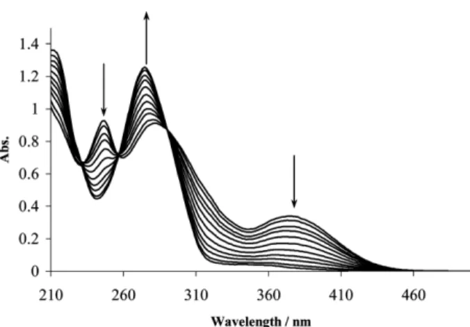
The titration was performed by adding aliquots of the metal ion with a Hamilton µL syringe to the ligand. The absorption measurements were carried out at various wavelengths where the difference in absorption was the maximum after equilibrium. The formed complex shows different absorption from the free ligand, while the metal ion solution shows no absorption at those wavelengths. As an example, the variation of the electronic spectra for
H2L13, titrated with various concentrations of VO (SO4)
·nH2O at 25oC in MeOH is shown in Fig. 1. The same procedure was followed for all other systems. The elec- tronic spectra of the formed complexes at the end of titration were the same as the electronic spectra of the separately synthesized complexes.
RESULTS AND DISCUSSION
Physico-chemical Characterizations and Geometri- cal Configuration of the Complexes
VO(IV) salt reacts with Schiff base ligands in 1:2 molar ratio in alcoholic medium to afford greenish-grey com- plexes. The ligand and its complexes are stable at room temperature and are nonhygroscopic. The synthesized ligands and their complexes were characterized by spectral tech- niques and elemental analysis. Apart from this, thermo- dynamic properties of the complexes were studied and the optimized geometry of one of the newly synthesized com- pounds has been elucidated by ab initio calculations.
IR analysis
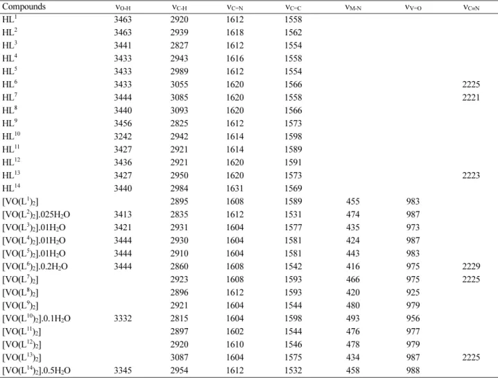
The IR spectra provide valuable information regarding the nature of functional groups attached to the metal atom.
The ligands and the metal complexes were characterized mainly using the azomethine band. The main infrared bands and their assignments are listed in Table 2. The vanadyl complex shows a band at ~940 cm−1 attributed to V=O fre- quency.33 In addition the spectra of the ligands show –C=N band in the region 1608−1620 cm−1, which is shifted to lower frequencies (1606−1612 cm−1) through complex forma- tion indicating the involvement of the –C=N nitrogen in the metal ion coordination.34,35 Assignment of the proposed coordination sites is further supported by the appearance Table 1. The physical properties of the prepared compounds
Compounds mp (oC) Color
HL1 125 Orange
HL2 160 Yellow
HL3 180 Yellow
HL4 130 Yellow orange
HL5 160 Yellow
HL6 164 Orange
HL7 190 Orange
HL8 190 Pale yellow
HL9 160 Yellow
HL10 167 Orange
HL11 130 Orange
HL12 110 Orange
HL13 160 Yellow
HL14 90 Yellow
[VO(L1)2] >250 Olive green
[VO(L2)2] 250 Olive green
[VO(L3)2] 250 Green
[VO(L4)2] >250 Light green
[VO(L5)2] >250 Green
[VO(L6)2] >250 Light green
[VO(L7)2] >250 Light green
[VO(L8)2] >250 Pale green
[VO(L9)2] >250 Green
[VO(L10)2] 250 Pale green
[VO(L11)2] >250 Olive green
[VO(L12)2] >250 Pale green
[VO(L13)2] >250 Green
[VO(L14)2] >250 Green
Fig. 1. The variation of the electronic spectra of H2L13titrared with VO(SO4).nH2O at 25oC in 96% methanol.
of medium bands at 400−450 cm−1 and 450−500 cm−1 which could be attributed to υ(M-O) and υ(M-N) respectively.36,37 Thus the oxovanadium (IV) complexes have the general structure which were shown in Scheme 2.
Elemental Analysis
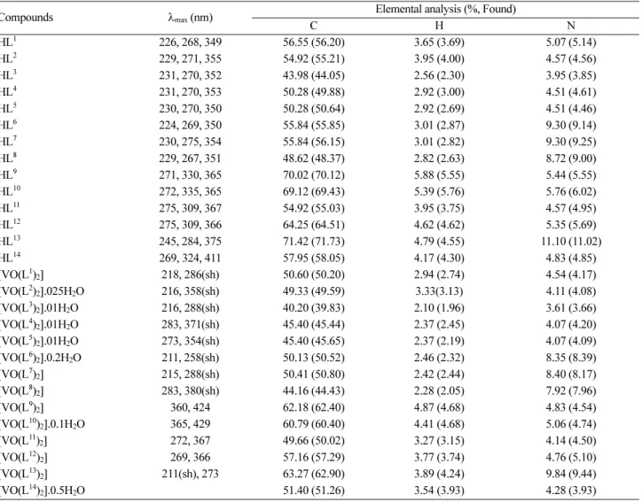
The stoichiometry of the ligands and vanadyl complexes were confirmed by their elemental analysis. The metal/
ligand ratio was found to be 1:2 has been arrived at by esti- mating the carbon, hydrogen, and nitrogen contents of the complexes. Elemental analysis of ligands and their VO (IV) complexes show good agreement with the proposed struc- tures of the ligands and their complexes (Table 3).
UV-Vis Analysis
The ligands show two absorption bands at UV-Visible region. A n-π* transition band at 326−410 nm and a π-π*
transition band at 240−297 nm are shown in the ligands.
These absorption bands show a slight shift to higher energy
in the complexes that is evident for unalteration of the structure of ligands upon complexation (Table 3).
All the vanadyl (IV) complexes have a band at 340−470 nm in methanol corresponding to a d-d transition band.
This band is not always observed, being often buried beneath a high intensity charge transfer band (or more accurately the low energy tail of that band), and when it is observed it is generally a shoulder (Table 3). UV-Vis spectra of HL13 and its oxovanadium (IV) are shown in Fig. 2.
1H NMR
In the 1H NMR spectral data of the salicylideneaniline ligands, the hydroxy proton is in the range 10−13 ppm. The spectral data of the ligands show a singlet (1H) signal at
~8.5 ppm which can be assigned to the azomethine proton group. The signals of the hydrogens of the phenyl group are appeared at δ = 6.3−8 ppm. The signal appeared as a singlet at 4.45 ppm. The protons of the methoxy groups show a signal at ~3.8 ppm.38,39
Table 2. IR Spectral data (cm−1) of the compounds
Compounds νO-H νC-H νC=N νC=C νM-N νV=O νC≡N
HL1 3463 2920 1612 1558
HL2 3463 2939 1618 1562
HL3 3441 2827 1612 1554
HL4 3433 2943 1616 1558
HL5 3433 2989 1612 1554
HL6 3433 3055 1620 1566 2225
HL7 3444 3085 1620 1558 2221
HL8 3440 3093 1620 1566
HL9 3456 2825 1612 1573
HL10 3242 2942 1614 1598
HL11 3427 2921 1614 1589
HL12 3436 2921 1620 1591
HL13 3427 2950 1620 1573 2223
HL14 3440 2984 1631 1569
[VO(L1)2] 2895 1608 1589 455 983
[VO(L2)2].025H2O 3413 2835 1612 1531 474 987
[VO(L3)2].01H2O 3421 2931 1604 1577 435 973
[VO(L4)2].01H2O 3444 2930 1604 1581 424 987
[VO(L5)2].01H2O 3444 2910 1604 1581 443 983
[VO(L6)2].0.2H2O 3444 2860 1608 1542 416 975 2229
[VO(L7)2] 2923 1608 1593 466 975 2225
[VO(L8)2] 2896 1612 1593 420 925
[VO(L9)2] 2921 1604 1544 480 979
[VO(L10)2].0.1H2O 3332 2815 1604 1598 493 956
[VO(L11)2] 2897 1602 1544 476 977
[VO(L12)2] 2920 1610 1546 478 979
[VO(L13)2] 3087 1604 1575 434 987 2225
[VO(L14)2].0.5H2O 3345 2954 1612 1532 458 988
Table 3. UV-Visa and elemental analysis data of the compounds
Compounds λmax (nm) Elemental analysis (%, Found)
C H N
HL1 226, 268, 349 56.55 (56.20) 3.65 (3.69) 5.07 (5.14)
HL2 229, 271, 355 54.92 (55.21) 3.95 (4.00) 4.57 (4.56)
HL3 231, 270, 352 43.98 (44.05) 2.56 (2.30) 3.95 (3.85)
HL4 231, 270, 353 50.28 (49.88) 2.92 (3.00) 4.51 (4.61)
HL5 230, 270, 350 50.28 (50.64) 2.92 (2.69) 4.51 (4.46)
HL6 224, 269, 350 55.84 (55.85) 3.01 (2.87) 9.30 (9.14)
HL7 230, 275, 354 55.84 (56.15) 3.01 (2.82) 9.30 (9.25)
HL8 229, 267, 351 48.62 (48.37) 2.82 (2.63) 8.72 (9.00)
HL9 271, 330, 365 70.02 (70.12) 5.88 (5.55) 5.44 (5.55)
HL10 272, 335, 365 69.12 (69.43) 5.39 (5.76) 5.76 (6.02)
HL11 275, 309, 367 54.92 (55.03) 3.95 (3.75) 4.57 (4.95)
HL12 275, 309, 366 64.25 (64.51) 4.62 (4.62) 5.35 (5.69)
HL13 245, 284, 375 71.42 (71.73) 4.79 (4.55) 11.10 (11.02)
HL14 269, 324, 411 57.95 (58.05) 4.17 (4.30) 4.83 (4.85)
[VO(L1)2] 218, 286(sh) 50.60 (50.20) 2.94 (2.74) 4.54 (4.17)
[VO(L2)2].025H2O 216, 358(sh) 49.33 (49.59) 3.33(3.13) 4.11 (4.08)
[VO(L3)2].01H2O 216, 288(sh) 40.20 (39.83) 2.10 (1.96) 3.61 (3.66)
[VO(L4)2].01H2O 283, 371(sh) 45.40 (45.44) 2.37 (2.45) 4.07 (4.20)
[VO(L5)2].01H2O 273, 354(sh) 45.40 (45.65) 2.37 (2.19) 4.07 (4.09)
[VO(L6)2].0.2H2O 211, 258(sh) 50.13 (50.52) 2.46 (2.32) 8.35 (8.39)
[VO(L7)2] 215, 288(sh) 50.41 (50.80) 2.42 (2.44) 8.40 (8.17)
[VO(L8)2] 283, 380(sh) 44.16 (44.43) 2.28 (2.05) 7.92 (7.96)
[VO(L9)2] 360, 424 62.18 (62.40) 4.87 (4.68) 4.83 (4.54)
[VO(L10)2].0.1H2O 365, 429 60.79 (60.40) 4.41 (4.68) 5.06 (4.74)
[VO(L11)2] 272, 367 49.66 (50.02) 3.27 (3.15) 4.14 (4.50)
[VO(L12)2] 269, 366 57.16 (57.29) 3.77 (3.74) 4.76 (5.10)
[VO(L13)2] 211(sh), 273 63.27 (62.90) 3.89 (4.24) 9.84 (9.44)
[VO(L14)2].0.5H2O 51.40 (51.26) 3.54 (3.93) 4.28 (3.93)
aIn methanol.
Table 4. 1H NMR spectroscopic dataof the compounds (δ in ppm) Compounds H−C=N Ar−H OH −CH2− OCH3 aHL1 8.56 6.91−7.52 13.28
aHL2 8.53 6.89−7.49 13.44 3.81
aHL3 8.54 6.69−7.67 10.94
aHL4 8.54 6.93−7.67 10.93
aHL5 8.53 6.69−7.61 10.94
aHL6 8.54 6.93−7.65 10.93
aHL7 8.53 6.90−7.62 10.94
aHL8 8.53 6.69−7.61 10.94
aHL9 8.57 6.87−7.29 10.42 3.51, 4.12
bHL10 8.83 6.79−7.29 12.75 3.72
aHL11 8.56 6.90−7.56 10.65 3.83
aHL12 8.58 6.86−7.46 10.65 3.86
aHL13 8.56 6.90−7.74 10.65 3.83
aHL14 8.41 6.32−7.74 11.39 4.45
aIn CDCl3. bIn d6-DMSO.
Fig. 2. UV–Vis spectra of the HL13 ligand (––), VO(L13)2 com- plex separately synthesized (...), and the product at the end of titration (---).
Thermodynamic Studies
To study the effect of the steric and the electronic param- eters of the ligands on the formation constants and the thermodynamic free energy of complexation, the interac- tion of the ligands as donors and VO (IV) as acceptor was carried out. The formation constants, Kf, were calculated using SQUAD computer program,40,41 designed to calcu- late the best values for the formation constants of the pro- posed reaction model (reaction 1) by employing a non- linear, least-squares approach. The free energy change
∆Go values of the formed complexes were calculated from
∆Go=−RT lnKf at 25oC (See Table 5).
2 HLx+ VO(SO4)·nH2O
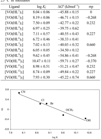
F [VO(Lx)2]·nH2O + SO42−+ 2 H+ (1) As the results show, in the para substituted Schiff base ligands, the formation constants (logKf) varies as can be expected from the electronic effects of the substituents at positions 4,4. Thus, the formation constants decrease accord- ing to the sequence OH > OCH3> H > Br > Cl > CN. In fact,
for the selected Schiff bases, Hammett type relationships were found between the logKf values and σp, the para-sub- stituent constant.42 Such correlations are shown in Fig. 3.
COMPUTATIONAL DETAILS
The electronic structure and the optimized geometries of the stable conformers of the complex bis (5-bromo-sal- icylideneanilinato)oxovanadium (IV) were computed by the Hartree-Fock (HF) method using the Gaussian 03 program43 employing 6-311G basis set. The infrared absorption inten- sity was calculated in the harmonic approximation, at the same level of theory as in the geometry optimization to verify the adequacy of method and the basis set.
Optimized Geometries and Energies
Geometry optimization was performed by HF/6-311G method on the two conformers of [VO(L1)2·H2O] com- plexes. Comparison of the energies shows that the trans conformer is the more stable molecule. Careful exami- nation of the conformer’s structure indicates that the tar- get complex does not involve water molecule coordinated to vanadium. This is evident by the fact that distances between the V and O atoms of the cis (2.354 Å) and trans (4.196 Å) conformers are larger than a V−O single bond (1.791 Å).44 Therefore, it is adequate to continue the investi- gation on the stable trans conformer without water coor- dinated as it has been already confirmed experimentally.45 The equilibrium geometry of the complex has been deter- mined by the energy minimization with the same basis set, i.e., 6-311G. The relative energies, dipole moments, HOMO, LUMO, and the energy gaps for the two conformers are given in Table 6. The relative energies show that the trans conformer, shown in Fig. 4, is the stable structure of the target complex. The energy difference between conform- ers is 7.88 kJ/mol, which is larger by about three times of the thermal energy kT (at room temperature where k is Bolt- zmann constant). As a result, there is no possibility of coex- istence of these two conformers at room temperature.
From the other side of view, it can be found that (Table 6) the cis conformer having higher dipole moment and less stable HOMO electronic state is not favorable as the trans conformer.
The calculated electron density on the rings containing Br indicates that the electron orbital of HOMO is local- ized over the rings having Br, while of the LUMO is dis- tributed over the rings. As a result, the HOMO-LUMO energy gap of the trans complex increases. The stability of the trans conformer can be in part accounted by consid- Table 5. The formation constants, log Kf, for the complexes at
25oC in methanol
Ligand log Kf ∆Go (kJmol−1) σp [VO(HL1)2] 8.04 ± 0.06 −45.88 ± 0.15 0 [VO(HL2)2] 8.19 ± 0.06 −46.71 ± 0.15 −0.268 [VO(HL3)2] 7.50 ± 0.09 −42.77 ± 0.22 0.232 [VO(HL4)2] 6.97 ± 0.25 −39.75 ± 0.62
[VO(HL5)2] 7.11 ± 0.57 −40.55 ± 0.43 0.227 [VO(HL6)2] 6.72 ± 0.46 −38.33 ± 0.41
[VO(HL7)2] 7.02 ± 0.13 −40.03 ± 0.32 0.660 [VO(HL8)2] 6.05 ± 0.05 −34.50 ± 0.12
[VO(HL9)2] 9.62 ± 0.45 −54.86 ± 0.41 −0.268 [VO(HL10)2] 10.47 ± 0.11 −59.71 ± 0.27 −0.370 [VO(HL11)2] 8.98 ± 0.51 −51.21 ± 0.47 0.232 [VO(HL12)2] 8.74 ± 0.09 −49.84 ± 0.22 0.227 [VO(HL13)2] 7.93 ± 0.30 −45.22 ± 0.74 0.660
Fig. 3. Linear correlation between the para substituted constants, σp, and logKf for the substituted salicylideneaniline Schiff bases with VO(SO4).nH2O in methanol at 25 ºC.
ering that its electric dipole moment is less than that of the cis. Fig. 5 shows the distribution of the HOMO and LUMO orbitals over the stable conformer.
Vibrational Assignment
For the coformer [VO(L1)2] shown in Fig. 4, the num- ber of normal mode is 150 [(3N-6), where N is the num- ber of atoms N=52]. The conformation obtained from the geometry optimization exhibits no special molecular sym- metries, and hence the molecule belongs to the C1 point group. Consequently, all the 150 fundamental vibrations of the gas phase molecule belong to the A irreducible rep- resentation and are both IR and Raman active.
Vibrational Frequencies
Comparison of the calculated frequencies at HF/6-311G level with experimental values (Table 2) reveals an over- estimation of the wavenumber of the vibrational modes, which can be attributed to the neglect of the anharmo- nicity present in a real system. The calculated infrared absorption spectra are shown in Fig. 6.
The spectrum predicted by HF method shows the fin- Table 6. Calculated properties for cis and trans conformers of the
[VO(L1)2] complex at HF/6-311G level of theory
Trans Cis
EHF (a. u.) −7415.950 −7415.947
µ (Debye) 1.471 3.0217
point group C1 C1
HOMO (a.u.) −0.110 −0.108
LUMO (a.u.) 0.049 0.049
HOMO-LUMO gap (eV) 4.318 4.272
Fig. 4. The structure of the trans conformer of VO(L1)2.
Fig. 5. HOMO (left) and LUMO (right) orbitals of the trans conformer of VO(L1)2 in vacuo.
Fig. 6. Calculated infrared absorption spectra of the [VO(L1)2] complex versus frequency (cm−1) in vacuo.
gerprint of the complex in the range of 400−1820 cm−1 and the C−H stretching can be seen around 1820 cm−1.
C−X vibrations, X=O, C, H, N, Br
The predicted stretching modes at 1165 and 1451.71 cm−1 correspond to bands of the IR spectra. The C−O bond in this complex is located near the center of the molecule (vanadium). C=C stretching bands can be seen at 1253.93, 1783, 1779.59, and 1181.37 cm−1. The C=C bond near the Br group vibrates at a lower frequency compared to the vibration of C=C bond on the ring in the farrest position to Br. The C=C stretching mode in the benzene ring is 1783 cm−1, which occurs at a higher frequency relative to the C=C bond in the ring involving Br (1779.59 cm−1).
The bending vibration of C−H groups connected to the rings occurs at 1631 cm−1. The C−H modes also depend upon the location of the bond. Near the electron acceptor bromide, it can be observed at 959.9, 1056.57, and 1260.52 cm−1, while far from this electron acceptor, it can be found in a wide range from 970 cm−1 to 1300 cm−1. In addition, the C−H bond near the nitrogen atom of the complex vibrates at 1570 cm−1. The stretching mode of C=N bond is calculated to be at 1811.97 and 1820.38 cm−1. The smaller wavenumbers at 96.95 and 165.95 cm−1 rep- resent the torsional mode of C=N. The stretching vibra- tion of CN group on the ring has the strongest band in the IR spectrum. The normal vibrational mode of C−Br in the bromobenzene ring was predicted to be at 1179.85 cm−1.
V−Y vibrations, Y=O, N
The four modes of V=O vibrations can be identified by the bands at 593.04, 727.35, 732.69, and 1164 cm−1. The first one corresponds to the bending motion and the sec- ond and the third ones to the symmetric and asymetric stretchings, respectively. The forth one could hardly be identified. The vibrational modes of the V−N bond are seen at 65.85, 378.52, and 5477.96 cm−1 of the IR spectra.
Lattice Vibrations
The lattice vibration can not be associated with any vibrational mode of the single molecule. Interestingly, the lattice vibrations are usually observed below 200 cm−1. The lattice modes associated with the translations and librations of the whole molecule can be only observed by far infrared, dispersive Raman, or tetrahertz spectroscopy.46 Since HF method overestimates these modes, the lattice vibrational mode in trans conformer of [VO(L1)2] are observed at frequencies below 350 cm−1.
Interaction Energy Between Complex and Water To provide more insights into the nature of the inter- action between [VO(L1)2] complex and water molecule, a systematic approach was taken into account. The inter- action energy of complex is defined as the difference between the energy of the complex with water (E[VO(L1)2·H2O]) and the sum of the energies of the pure H2O(EH2O) and [VO(L1)2] (E[VO(L1)2]) species:
(2) For this purpose each isolated [VO(L1)2.H2O] and its corresponding species ([VO(L1)2] and H2O) were opti- mized at HF/6-311g level of theory as mentioned above.
The calculated interaction energies are −25.420 and
−23.883 kJ/mol for the trans and the cis conformers, respectively. Since the interaction energy for the trans (−25.420 kJ/mol) and for the cis (−23.883 kJ/mol) con- formers is small, it can be confirmed again that this com- plex does C contain water molecule coordinated to the vanadium.
Charge Distribution on the Complex
The atomic charges calculated by ab initio method at HF/6-311g level of theory by Natural Bond Orbital (NBO) for two conformers indicate that the most positive charge, +2.0216 C, is on the center of the trans complex (vana- dium atom). This charge value may be one of the effective factors in determining the stability of trans conformer over the cis conformer.
CONCLUSIONS
The structural, geometrical and the thermodynamic properties of the oxovanadium(IV) complexes have been investigated. Geometry optimization on the cis and trans conformers shows that the cis conformer is less stable and the energy difference between these two conformers is 7.88 kJ/mol. Thus, calculations using HF mathod with 6- 311g basis set show that the energy difference between the conformers is much larger than kT, such that there is no possibility of coexistence of conformers.
In conformer trans, there does not exist the possibility of intramolecular bond formation between V−O (the oxy- gen of the water) as indicated by the large distance between V and O atoms, e.g., 4.196 Å. This means that no water is coordinated to vanadium as confirmed by the calculated
E kJmol( –1) =
2625.50 E[ [VO L( )12⋅H2O](a.u.) E– H2O(a.u.)–E[VO L( )12](a.u.)]
interaction energy. Vibrational spectroscopy and compu- tational chemistry have been applied for investigating the most stable conformer of bis(5-bromo-salicylideneanili- nato) oxovanadium (IV). Infrared spectrum was recorded, and vibrational bands were assigned based on the HF cal- culations. In general, the calculated modes observed were overestimated. Using the elaborated method, which includes the correlations like DFT, shall improve the results.
A striking feature of the present work is the study of the interaction energy, which reveals small interaction between H2O and [VO(L1)2] in [VO(L1)2.H2O], e.g., −25.420 kJ/
mol. This energy lies below a V−O bond energy and it can be concluded that bis(5-bromo-salicylideneanilinato)oxo- vanadium (IV) does not have a coordinated water. Accord- ing to the thermodynamic studies, the formation constant of the complexes depends upon the steric and the elec- tronic characteristic of the ligands. Moreover, the molecular electronic structure of each complex plays an important role on its thermodynamic properties. It is evident that there is a close relationship between these various prop- erties.
Acknowledgments. We are grateful to Shiraz University Research Council for their financial support.
REFFERENCES 1. Khalil, S. M. E. Chem. Papers 2000, 54, 12.
2. Osman, A. H. Transition. Met. Chem. 2006, 31, 35.
3. Sallam, S. A. Transition. Met. Chem. 2006, 31, 46.
4. Cindri´c, M.; Strukan, N.; Vrdoljak, V.; Kajfez, T.; Kame- nar, B. Croatica. Chim. Acta 2003, 76, 257.
5. Sousa, C.; Freire, C.; Castro, B.; Molecules 2003, 8, 894.
6. Hamilton, D. E.; Drago, R. S.; Zombeck, A. J. Am. Chem.
Soc. 1987, 109, 374.
7. Chen, D.; Martel, A. E.; Inorg. Chem. 1987, 26, 1026.
8. Costamagna, J.; Vargas, J.; Latorre, R.; Alvarado, A.;
Mena, G. Coord. Chem. Rev. 1992, 119, 67.
9. Chatterjee, D.; Mitra, A. J. Coord. Chem. 2004, 57, 175.
10. Minu, G.; Bhowon, H.; Li Kam Wah, A.; Dosieah Ridana, M. O. Ramalingum Lacour, D.; Synth. React. Inorg. Metal- Org. Chem. 2004, 34, 1.
11. Macedo-Ribeiro, S.; Hemrika, W.; Renirie, R.; Wever, R.;
Messerschimdt, A. J. Biol. Inorg. Chem. 1999, 4, 209.
12. Cornman, C. R.; Zovinka, E. P.; Meixner, M. H. Inorg.
Chem. 1995, 34, 5099.
13. Crans, D. C.; Bunch, R. L.; Theisen, L. A. J. Am. Chem.
Soc. 1989, 111, 7597.
14. Nour-Elden, A. F.; Craig, M. M.; Gresser, M. J. J. Biol.
Chem. 1985, 260, 6836.
15. Thompson, K. H.; McNeill, J. H.; Orvig, C. Chem. Rev.
1999, 99, 2561.
16. Tasiopoulos, A. J.; Troganis, A. N.; Evangelou, A.; Rap- topoulou, C. P.; Terzis, A.; Deligiannakis, Y.; Kabanos, T.
A. Chem. Eur. J. 1999, 5, 910.
17. Boghaei, D. M.; Behzad, M. Synth. Reac. Inorg. Met-Org Chem. 2005, 35, 261.
18. Boghaei, D. M.; Bezaatpour, A.; Behzad, M. J. Mol.
Catal. A 2006, 245, 12.
19. Mohebbi, S.; Boghaei, D. M.; Sarvestani, A. H.; Salimi, A. Appl. Catal. A 2005, 278, 263.
20. Mohebbi, S.; Nikpour, F.; Rayati, S. J. Mol. Catal. A 2006, 256, 265.
21. Mohebbi, S.; Sarvestani, A. H. Transition Met. Chem. 2006, 31, 749.
22. Asadi, M.; Sarvestani, A. H.; Asadi, Z. Setoodehkhah, M.
Metal Org. Chem. 2005, 35, 639.
23. Asadi, M.; Mohammadi, Kh.; Kiyanfar, A. H. J. Iran.
Chem. Soc. 2006, 3, 247.
24. Asadi, M.; Kianfar, A. H.; Torabi, S.; Mohammadi, Kh.;
J. Chem. Thermodynamics 2008, 40, 523.
25. Asadi, M.; Kianfar, A. H.; Abbasi, M. J. Chem. Res. 2007, 56.
26. Asadi, M.; Asadi, Z. J. Coord. Chem. 2008, 61, 640.
27. Asadi, M.; Sarvestani, A. H. Can. J. Chem. 2001, 79, 1360.
28. Asadi, M.; Sarvestani, A. H. J. Chem. Res. 2002, 520.
29. Asadi’ M.; Sarvestani, A. H.; Ahmadi, M. B.; Moham- madi, Kh.; Asadi, Z. J. Chem. Thermodynamics 2004, 36, 141.
30. Absalan, Gh.; Asadi, M.; Kamran, S.; Torabi, S.; Sheikhian, L. Sensors and Actuators 2010, 147B, 31.
31. Chalmers, J. M.; Griffiths, P. R. Handbook of Vibrational Spectroscopy, Eds.; Wiley New York, 2002.
32. Mohammadi, Kh.; Thompson, K. H.; Patrick, B. O.; Storr, T.; Martins, C.; Polishchuk, E.; Yuen, V. G.; McNeill, J.
H.; Orvig, C. J. Inorg. Biochem. 2005, 99, 2217.
33. Xiu, R. B.; Mintz, F. L.; You, X. Z.; Wang, R. X.; Yue, Q.; Meng, Q. J.; Lu, Y. J.; Derveer, D. V. Polyhedron 1996, 15, 4585.
34. Iskander, M. R.; Ei-Syed, L.; Ismail, K. Z. Trans. Met.
Chem. 1979, 4, 225.
35. Thankamony, M.; Mohanan, K. Ind. J. Chem. 2007, 46A, 247.
36. West, D. X.; Nassar, A. A. Trans. Metal. Chem. 1999, 24, 617.
37. Tumer, M.; Koksal, H.; Sener, M. K.; Serin, S. Trans. Metal.
Chem. 1999, 24, 13.
38. Kumar, D. N.; Garg, B. S. Spectrochim. Acta, Part A 2006, 64, 141.
39. Asadi, M.; Mohammadikish, M.; Mohammadi, Kh. Cent.
Eur. J. Chem. 2010, 8, 291.
40. Leggett, D. L. Computational Methods for the Determi- nation of Formation Constant; Plenum Press: New York, 1985.
41. Dey, D. K.; Saha, M. K.; Gielen, M.; Kemmer, M.; Biese- mans, M.; Willem, R.; Gramlich, V.; Mitra, S. Organomet.
Chem. 1999, 590, 88.
42. Gordon, A. J.; Ford, R. A. The Chemist’s Companion: A Handbook of Practical Data, Techniques, and References, Vol. 145; Wiley: New York, 1972.
43. Frisch, M. J.; et al. Gaussian 03, Revision B.03; Gaussian Inc., Pittsburgh PA 2003.
44. Hardcastlet, F. D.; Wachs, I. E. J. Phys. Chem. 1991, 95, 5031.
45. Asadi, M.; Ghatee, M. H.; Torabi, S.; Mohammadi, Kh.;
Moosavi, F. Journal of Iranian Chemical Society 2010, 7, 1021.
46. Mishra, S.; Chaturvedi, D.; Tandon, P.; Gupta, V. P.; Ayala, A. P.; Honorato, S. B.; Siesler, H. W. J. Phys. Chem. A 2009, 113, 273.
![Fig. 6. Calculated infrared absorption spectra of the [VO(L 1 ) 2 ] complex versus frequency (cm −1 ) in vacuo.](https://thumb-ap.123doks.com/thumbv2/123dokinfo/5299087.154365/7.892.104.786.821.1079/calculated-infrared-absorption-spectra-complex-versus-frequency-vacuo.webp)
