INTRODUCTION
Bronchogenic cysts are congenital malformations thought to develop from abnormal budding of the tracheobronchial tree (1). In normal development, the tracheal bud arises from the embryonic foregut around the fourth week of gestation, and undergoes further branching. If abnormal budding occurs dur- ing this process, a blind-ended pouch lined with pseudostrati- fied columnar respiratory epithelial cells develops and forms a bronchogenic cyst. The location of a bronchogenic cyst de- pends on when the abnormal budding occurred. The cysts are most commonly seen in the mediastinum, especially in the middle and the posterior mediastinum, and can be located in lung parenchyma. The attenuation value is dependent on the contents of the cyst. Although the name is bronchogenic ‘cyst’, the attenuation value can be more than 100 HU if the cyst con- tains a large amount of protein or calcium. Due to variability in
location and internal density, it is often difficult to diagnose bronchogenic cysts on CT scan.
With this background, Cardinale et al. (2) proposed diagnostic criteria based on those two factors, the location and the CT den- sity of the lesion. According to these criteria, if the mass was lo- cated in a typical location (the middle mediastinum) and showed typical attenuation (fluid attenuation, 0-20 HU), a diagnosis of bronchogenic cyst was considered. However, if the mass was lo- cated in an atypical location for bronchogenic cysts and showed soft-tissue attenuation, other diagnoses were considered.
In our study, we encountered 3 cases of anterior mediastinal well-circumscribed masses with soft-tissue attenuation. Be- cause of their location and attenuation characteristics these le- sions were diagnosed as thymoma preoperatively. However, they were later identified to be bronchogenic cysts on histopa- thology. As mentioned above, anterior mediastinal broncho- genic cysts are extremely rare. Furthermore, there have been a
J Korean Soc Radiol 2012;66(4):347-351
Received September 22, 2011; Accepted March 2, 2012 Corresponding author: Soo-Youn Ham, MD Department of Radiology, Korea University Anam Hospital, Korea University College of Medicine, 73 Inchon-ro, Seongbuk-gu, Seoul 136-705, Korea.
Tel. 82-2-920-5578 Fax. 82-2-929-3796 E-mail: [email protected]
Copyrights © 2012 The Korean Society of Radiology
Bronchogenic cysts are congenital malformations that are thought to develop from abnormal budding of the embryonic foregut. They are most commonly seen in the mediastinum, especially in the middle and the posterior mediastinum, and they typ- ically show water-attenuation. Anterior mediastinal bronchogenic cyst is extremely rare. Anterior mediastinal bronchogenic cysts with soft-tissue attenuation have not been reported in the English-language literature up until now. We discovered 3 cas- es of anterior mediastinal well-circumscribed lesions showing soft-tissue attenua- tion which were diagnosed as thymoma preoperatively, but were later identified as bronchogenic cysts on pathology.
Index terms Bronchogenic Cyst Anterior Mediastinum Soft-Tissue Attenuation
Anterior Mediastinal Bronchogenic Cysts Manifesting as a Solid Mass: A Report of Three Cases
1연조직 음영의 앞종격동 기관지낭 3예1
Sung-Hye Yoo, MD
1, Soo-Youn Ham, MD
1, Eun Jeong Choi, MD
1, Yu-Whan Oh, MD
1, Kwang Taik Kim, MD
2, Cheol Whan Kim, MD
3Departments of 1Radiology, 2Thoracic and Cardiovascular Surgery, 3Pathology, Korea University Anam Hospital, Korea University College of Medicine, Seoul, Korea
with an incidentally-discovered anterior mediastinal mass. She did not complain of any symptoms, and there was no past his- tory of illness. Chest radiography revealed no significant ab- normality in the chest. Transverse pre-contrast enhanced CT scan (mediastinal window settings) (Fig. 2) showed a 3.2 cm well-circumscribed soft-tissue attenuated (ROI = 25 HU, SD = 3.91) lesion in the anterior mediastinum. This lesion showed homogenous internal attenuation which was less than that of soft tissue such as chest wall muscle. After injection of contrast material, there was slight thin-wall enhancement. Internal en- hancement was not seen. We tentatively diagnosed thymic cyst or thymoma based on the location and density of the lesion.
However, on pathology, this lesion was confirmed to be a bron- chogenic cyst.
Case 3
A 65-year-old female patient was admitted to our cardiology center with chest pain. EKG showed no significant abnormality, and serum cardiac enzymes were in the normal range. We could not detect any abnormality in the chest radiography. The cardiologist recommended a chest CT scan be performed. A transverse pre-contrast enhanced CT scan (mediastinal win- dow settings) (Fig. 3A) showed a 2-cm well-circumscribed soft-tissue attenuated (ROI = 57 HU, SD = 45.60) lesion in the anterior mediastinum. This lesion showed homogenous attenua- tion similar to that of soft tissue. In contrast-enhanced CT scan (Fig. 3B), there was no enhancement of the lesion (57 HU → 47 HU). According to the classification proposed by Cardinale et few cases of anterior mediastinal bronchogenic cysts showing
water-attenuation and therefore mimicking thymic cysts that have been reported on previously (3). However, anterior medi- astinal bronchogenic cysts with soft-tissue attenuation have not been reported in the English literature up until now. The cases in our study indicate the possibility of an anterior mediastinal location for bronchogenic cysts, and the potential for these cysts to show soft-tissue attenuation.
CASE REPORT
Case 1
A 43-year-old male patient presented to our outpatient clinic with an incidentally-discovered mass in the anterior mediasti- um. His medical history was unremarkable except for a history of mitral valve insufficiency but no significant abnormality was noted on physical examination or in the laboratory tests. Chest radiography showed no abnormal finding except for bilateral pulmonary edema. Non-contrast enhanced chest CT scan (Fig.
1A) showed a well-circumscribed soft-tissue homogenously-at- tenuated (ROI = 35 HU, SD = 19.15) mass in the anterior me- diastinum. Because the location and density of this mass were compatible with thymoma, this lesion was diagnosed as being a thymoma preoperatively, but it was later identified as a bron- chogenic cyst on pathology (Fig. 1B).
Case 2
A 46-year-old female patient was admitted to our hospital
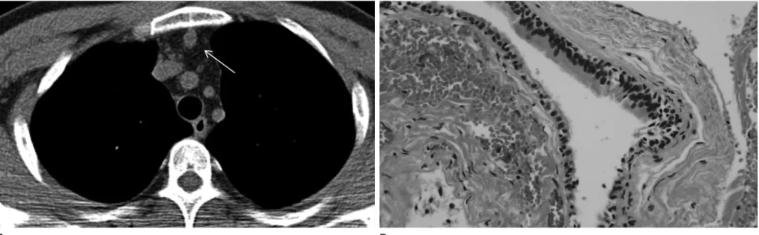
Fig. 1. Bronchogenic cyst in a 43-year-old asymptomatic man. Transverse view of non-contrast enhanced chest CT scan (mediastinal window settings) (A) shows a well-circumscribed homogenous soft-tissue attenuated (ROI = 35 HU, SD = 19.15) lesion (arrow) in the anterior mediasti- num. On microscopy (H&E, × 400) (B), the cyst is lined with pseudostratified ciliated columnar epithelium and is without cartilage and smooth muscle components.
A B
Fig. 2. Bronchogenic cyst in a 46-year-old asymptomatic woman. Transverse pre-contrast enhanced CT scan (mediastinal window settings) shows a 3.2 cm sized well-circumscribed soft-tissue attenuated (ROI = 23 HU) lesion (arrow) in the anterior mediastinum (A). This lesion shows homogenous attenuation which is less than that for soft tissue. After contrast enhancement, slight thin-wall enhancement is noted. Internal en- hancement is not significant (arrow) (B).
A B
Fig. 3. Bronchogenic cyst in a 65-year-old woman with chest pain. Transverse pre-contrast enhanced CT scan (mediastinal window settings) (A) shows a 2-cm well-circumscribed soft-tissue attenuated (ROI = 57 HU, SD = 45.60) lesion (arrow) in the anterior mediastinum. This lesion shows homogenous attenuation similar to that of soft tissue. In contrast-enhanced CT scan, internal enhancement is not seen (arrow) (B). On microscopy [H&E, × 40 (C) and × 400 (D)], the cyst is lined with pseudostratified ciliated columnar epithelium, and is without cartilage and smooth muscle components.
C D
A B
Cardinale et al. (2) ENREF 3 have proposed diagnostic crite- ria based on the typical location and the typical CT density of bronchogenic cysts. They defined the typical location as being the subcarinal area and the typical density as water density (be- tween 0-20 HU). If the lesion showed both typical findings, it was classified as Level 1, likely diagnosis. If the lesion displayed only one of the typical findings, it was classified as Level 2, pos- sible diagnosis. If the lesion did not demonstrate any of the typ- ical findings, it was classified as Level 3, unlikely diagnosis. In that study of 21 bronchogenic cysts, there was no cyst classified as Level 3.
In our study, all of the three anterior mediastinal lesions showed soft-tissue attenuation (above 20 HU). Under Cardina- le’s classification system, these lesions would have been classi- fied as Level 3, unlikely diagnosis. Although contrast enhance- ment was not demonstrated in our cases, we misdiagnosed these lesions as thymoma due to their location and density. The cysts in our study are the first cases of anterior mediastinal bronchogenic cyst showing soft-tissue attenuation on imaging that have been reported in the medical literature.
REFERENCES
1. O'Rahilly R, Müller F. Chevalier Jackson lecture. Respiratory and alimentary relations in staged human embryos. New embryological data and congenital anomalies. Ann Otol Rhinol Laryngol 1984;93(5 Pt 1):421-429
2. Cardinale L, Ardissone F, Cataldi A, Gned D, Prato A, Solitro F, et al. Bronchogenic cysts in the adult: diagnostic criteria derived from the correct use of standard radiography and computed tomography. Radiol Med 2008;113:385-394 3. Takasuna K, Yamanda T, Makiuchi A, Kondoh R, Numanami
H, Machida E, et al. [Two cases of the bronchial cyst locat- ed in the anterior mediastinum]. Kyobu Geka 1999;52:
959-961
4. Nuchtern JG, Harberg FJ. Congenital lung cysts. Semin Pe- diatr Surg 1994;3:233-243
5. McAdams HP, Kirejczyk WM, Rosado-de-Christenson ML, Matsumoto S. Bronchogenic cyst: imaging features with clinical and histopathologic correlation. Radiology 2000;
217:441-446
6. Lyon RD, McAdams HP. Mediastinal bronchogenic cyst:
al. (2), a diagnosis of bronchogenic cyst could not even be con- sidered. Because it was thought to be a thymoma, the patient was operated on and the lesion was sent for pathologic exami- nation (Fig. 3C, D), which showed that the lesion was a bron- chogenic cyst.
DISCUSSION
Bronchogenic cysts are a congenital anomaly arising from abnormal budding of the ventral foregut, a development which occurs during the first 16 weeks of gestation (4). Instead of de- veloping into a bronchus, the abnormal ventral bud differenti- ates into a fluid-filled blind-ended pouch (5). Bronchogenic cysts typically manifest as well-circumscribed non-enhancing middle mediastinal masses with water-attenuation. Despite bronchogenic cysts not being an uncommon finding, two fac- tors cause radiologists to misdiagnose them. The first is the variable density of bronchogenic cysts, and the second is their variable location. We need to know the location of atypical sites and we need to know the full range of CT density of broncho- genic cysts, and diagnosis should take into account these two factors.
The fluid in the bronchogenic cysts contains water and pro- tein, therefore bronchogenic cysts show a wide variation in CT- density according to the proportion of protein contained in the cyst (6). Although mediastinal bronchogenic cysts typically show a density that is similar to that of water, one study on CT attenuation in 58 pathologically-confirmed bronchogenic cysts revealed that 43% of cases showed soft-tissue attenuation (above 20 HU) (5). Because of this, bronchogenic cysts can mimick other masses, such as lymphomas or neurogenic tu- mors.
Bronchogenic cysts may occur in any part of the mediasti- num (70-85%) (7, 8). Other less common sites are lung paren- chyma (15-30%) (5, 7, 9, 10), pleura and diaphragm (5). As mentioned above, bonchogenic cysts are typically located in the middle mediastinum, especially in the subcarinal area, and it is not rare to find them in the posterior mediastinum. However, only two cases of anterior mediastinal bronchogenic cysts have been reported in the English literature up until now (3). These anterior mediastinal bronchogenic cysts showed cystic attenua- tion so they were diagnosed as thymic cysts preoperatively.
adult. Chest 1994;106:79-85
9. St-Georges R, Deslauriers J, Duranceau A, Vaillancourt R, Deschamps C, Beauchamp G, et al. Clinical spectrum of bronchogenic cysts of the mediastinum and lung in the adult. Ann Thorac Surg 1991;52:6-13
10. Procacci C, Graziani R, Pelosi G, Politi L, Guarise A, Main- ardi P, et al. [Imaging of bronchogenic cysts]. Radiol Med 1996;92:41-46
demonstration of a fluid-fluid level at MR imaging. Radi- ology 1993;186:427-428
7. Suen HC, Mathisen DJ, Grillo HC, LeBlanc J, McLoud TC, Moncure AC, et al. Surgical management and radiological characteristics of bronchogenic cysts. Ann Thorac Surg 1993;55:476-481
8. Patel SR, Meeker DP, Biscotti CV, Kirby TJ, Rice TW. Presen- tation and management of bronchogenic cysts in the
연조직 음영의 앞종격동 기관지낭 3예1
유성혜
1· 함수연
1· 최은정
1· 오유환
1· 김광택
2· 김철환
3기관지낭(bronchogenic cyst)은 선천성 기형으로 배아 앞창자(embryonic foregut)의 비정상적 발아(budding)로 인해 발 생하는 것으로 알려져 있다. 이들의 대부분은 종격동, 특히 중간과 후종격동에서 발견되며 전산화단층촬영상에서 물과 유사한 음영(water attenuation)을 보이게 된다. 기관지낭이 종종 연조직 음영을 보일 수는 있으나 연조직 음영을 보이면서 앞종격동에 위치하였던 예는 현재까지 보고된 바가 없다. 이에 저자들은 비전형적 위치에 비전형적 음영을 보여 영상학적 으로 흉선종으로 오인되었던 기관지낭 3증례를 보고하는 바이다.
고려대학교 의과대학 안암병원 1영상의학과학교실, 2흉부외과학교실, 3병리과학교실