ABSTRACT
Purpose: Perforation of choledochal cyst (CC) is a relatively rare clinical presentation in pediatric populations and difficult to predict preoperatively. We assess the clinical implications by comparing clinical parameters based on a single-center experience between perforated and nonperforated CC to facilitate the appropriate management for future interventions.
Methods: A total of 92 cases of CC in pediatric patients (aged <18 years) who received surgical management between January 2003 and December 2018 at a Pusan National University Children's Hospital were reviewed. After screening the clinical features of perforated cases, we compared the demographic findings, clinical characteristics, and some laboratory results between the perforated and nonperforated groups.
Results: Perforated CC was identified in 8 patients (8.7%), and nonperforated CC in 84 patients (91.3%). Perforation can be classified into three categories: free perforation of cyst (3 cases), pinpoint perforation of cyst (2 cases), and necrotic change of cyst (3 cases). CC perforation occurred significantly more commonly in patients aged <24 months. Clinically, the perforated group showed significantly higher frequency of fever and higher C-reactive protein (CRP) level during the initial visit.
Conclusion: Perforation is more likely to be suspected in patients aged <24 months presenting together with fever and high CRP level in the initial visit. It is also necessary to keep in mind that it indicates not only a possibility of complicated disease status regardless of its association with stones but also a difficulty of applying a minimal invasive procedure and relatively increased length of hospital stay.
Keywords: Perforated; Choledochal cyst; Fever; C-reactive protein; Initial; Visit
INTRODUCTION
Pediatric patients with choledochal cyst (CC) typically present with classic symptoms of abdominal pain in the right upper quadrant, mass, and jaundice. However, some cases are asymptomatic with incidental findings or unusually complicated situations [1,2].
Furthermore, spontaneous perforation of CC is a relatively rare clinical presentation in pediatric populations and difficult to predict preoperatively. It could present as surgical abdomen secondary to bile peritonitis or delayed pseudocyst, which may affect clinical
Original Article
Received: Nov 22, 2019 1st Revised: Jan 14, 2020 2nd Revised: Feb 3, 2020 Accepted: Feb 19, 2020 Correspondence to Yong-Hoon Cho
Department of Surgery, Pusan National University School of Medicine, 49 Busandaehak-ro, Mulgeum-eup, Yangsan 50612, Korea.
E-mail: [email protected]
Copyright © 2020 by The Korean Society of Pediatric Gastroenterology, Hepatology and Nutrition
This is an open-access article distributed under the terms of the Creative Commons Attribution Non-Commercial License (https://
creativecommons.org/licenses/by-nc/4.0/) which permits unrestricted non-commercial use, distribution, and reproduction in any medium, provided the original work is properly cited.
ORCID iDs Soo-Hong Kim
https://orcid.org/0000-0001-7085-5969 Yong-Hoon Cho
https://orcid.org/0000-0003-0170-9997 Hae-Young Kim
https://orcid.org/0000-0002-2316-5815 Conflict of Interest
The authors have no financial conflicts of interest.
Soo-Hong Kim , Yong-Hoon Cho , and Hae-Young Kim
Department of Pediatric Surgery, Pusan National University Children's Hospital, Yangsan, Korea
Perforated Choledochal Cyst: Its Clinical
Implications in Pediatric Patient
progress and require an alternative management strategy. Previous studies have suggested several possible causes of perforation, however, a prominent cause remains unclear [3-9].
In this study, we evaluated the clinical characteristics of perforated CC, including its
manifestations, basic laboratory findings, and other results related to biliary tract disease and radiologic and operative findings in pediatric patients. Furthermore, this study attempted to assess clinical implications by comparing some clinical parameters between perforated and nonperforated CC to facilitate appropriate management for future interventions.
MATERIALS AND METHODS
Patient selection
We reviewed cases of CC in pediatric patients (age, <18 years) who received surgical management between January 2003 and December 2018 at a Pusan National University Children's Hospital. There were 92 patients with CC during this period, who were classified into two clinical groups: perforated and nonperforated CC. This study was approved by the Institutional Review Board (IRB No. 05-2019-159), and the data were managed with personal information protection.
Evaluation of clinical characteristics and comparison
The current study was retrospectively conducted by reviewing patients' clinical data. We analyzed basic demographic characteristics, including sex, age, and body weight during the surgery, and clinical characteristics including signs and symptoms, laboratory markers, and radiologic findings at the initial visit. After screening the clinical features of perforated cases, we compared the demographic characteristics; clinical findings, such as a presenting symptoms and sign; presence of fever and associated choledochalithiasis; start of diet; and hospital stay between the two groups. Moreover, some laboratory results, including a white blood cell (WBC) count, C-reactive protein (CRP) level, and other parameters associated with biliary tract disease were analyzed.
Statistical analysis
Analysis was performed using IBM SPSS Statistics for Windows, Version 25.0 (IBM Co., Armonk, NY, USA). Fisher's exact test for comparison of the frequencies of variables and Mann-Whitney U-test for measured laboratory values were performed. A p-value <0.05 was considered statistically significant.
RESULTS
Of 92 patients, 8 (8.7%) had perforated CC, and 84 (91.3%) had nonperforated CC.
Clinical features of perforated CC
The clinical presentation was usually abdominal pain, localized right upper quadrant pain or generalized rigidity, and fever. Surgical management intervals varied from a few hours to 15 days. Radiologic findings, usually via computed tomography, showed massive ascites or localized fluid collection around the subhepatic space in all cases. Additionally, it showed dilatation of the common bile duct in 7 cases and collapsed gallbladder in 4 cases. Combined choledocholithiasis was found in only one case. According to operative findings, perforation
can be classified into three categories: free perforation of cyst (3 cases), pinpoint perforation of cyst (2 cases), and necrotic change of cyst (3 cases). While perforation occurred at any site on the cyst, we could not find any clinical differences according to perforation type. All patients underwent a one-stage operation, cyst excision with hepaticojejunostomy, and had a good clinical course without major complications (Table 1).
Comparison between the two groups
In basic demographic findings, there was no significant difference in sex and body weight.
Patients were younger in the perforated group than the nonperforated group and a
significantly high rate of perforation was seen in patients aged <24 months (p<0.05) (Table 2).
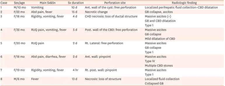
Table 1. Clinical features of perforated choledochal cyst
Case Sex/age Main Sx&Sn Sx duration Perforation site Radiologic finding
1 M/10 mo Vomiting 10 d Ant. wall of the cyst: free perforation Localized perihepatic fluid collection+CBD dilatation
2 F/22 mo Abd pain, fever 15 d Necrotic change GB collapse, ascites
3 F/18 mo Rigidity, vomiting, fever 4 d CHD necrosis: loss of ductal structure Massive ascites (+) GB and CBD dilatation Type 1
4 F/32 mo RUQ pain, vomiting, fever 5 d Post. wall of the CBD: free perforation Massive ascites GB collapse Mild dilatation of CBD
5 F/20 mo RUQ pain 2 d Rt. Lateral: free perforation Massive ascites
GB collapse Type 1
6 F/18 mo Abd pain, diarrhea, fever 2 d Ant. wall: pinpoint Massive ascites
Type IV
Multiple CBD stones 7 F/19 mo Rigidity, vomiting, fever 4 hr Rt. post. wall: pinpoint Massive ascites
Type 1
8 M/6 mo Fever 13 d Necrosis: loss of structure Localized fluid collection
Collapsed GB
Sx: symptom, Sn: sign, M: male, F: female, Abd: abdominal, RUQ: right upper quadrant, Ant.: anterior, CHD: common hepatic duct, Post.: posterior, CBD:
common bile duct, Rt.: right, GB: gallbladder.
Table 2. Demographic findings
Variable Perforated group (n=8) Nonperforated group (n=84) p-value
Composition 8.7 91.3 -
Sex
Male 2 (25.0) 13 (15.5) 0.612
Female 6 (75.0) 71 (84.5)
Age (mo) 19.5±8.7 (6.0–32) 54.2±52.5 (0–192) 0.161
Age group
Neonate - 2 (2.4)
0.005
Infant (−12 mo) 2 (25.0) 21 (25.0)
Childhood
−24 mo 5 (62.5) 10 (11.9)
>24 mo 1 (12.5) 51 (60.7)
Perforation rate
Neonate 0/2 (0.0)
0.005
Infant (−12 mo) 2/23 (8.7)
Childhood
−24 mo 5/15 (33.3)
>24 mo 1/52 (1.9)
Body weight (kg) 10.6±1.5 (5.0–12.1) 19.2±14.8 (2.4–52.1) 0.307
Values are presented as number (%) or mean±standard deviation (range).
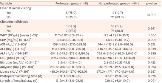
Clinically, the perforated group had a significantly higher incidence of fever during the initial visit (75.0% vs. 10.7%, p<0.05). There were 11 CC cases combined with choledocholithiasis in this study: 1 in the perforated group and 10 in the nonperforated group. However, there was no significant difference between the two groups (12.5% vs. 11.9%, p=0.591). The WBC count and CRP level were higher in the nonperforated group than in the perforated group; however, only the CRP level showed a significant difference. In the perforated group, leukocytosis was not severe. Several laboratory markers related with biliary tract disease, such as aspartate aminotransferase, alanine aminotransferase, alkaline phosphatase, gamma-glutamyl transferase, bilirubin, amylase, and lipase, were compared to determine any association with CC perforation. All these laboratory markers were elevated in the nonperforated group, despite the lack of significance between the two groups. Postoperatively, there was no significant difference in postoperative fasting time, but the length of hospital stay was significantly longer in the nonperforated group (12.2 days vs. 7.8 days, p<0.05) (Table 3).
By comparing clinical and laboratory parameters, such as age <24 months, presence of fever at the initial visit, and combined choledocholithiasis, we examined the risk ratio for developing CC perforation in both groups (Table 4). The results showed a significantly high risk ratio in cases that presented with fever at the initial visit.
Table 3. Comparison of clinical characteristics between the perforated and nonperforated groups
Variable Perforated group (n=8) Nonperforated group (n=84) p-value
Fever at initial visiting
Yes 6 (75.0) 9 (10.7) <0.001
No 2 (25.0) 75 (89.3)
Choledocholithiasis
Yes 1 (12.5) 10 (11.9) 0.961
No 7 (87.5) 74 (88.1)
WBC (103/μL) (mean 6–12)* 11.7±6.07 (6.2–23.4) 11.2±4.7 (5.6–25.7) 1.000
CRP (mg/dL) (0–0.5)* 3.4±2.6 (0.36–6.4) 1.1±1.8 (0.01–9.2) 0.006
AST (IU/L) (15–60)* 109.1±95.5 (27.0–289.0) 144.4±133.9 (18.0–626.0) 0.533 ALT (IU/L) (13–45)* 140.3±119.7 (8.0–296.0) 146.4±135.6 (5.0–566.0) 0.949 ALP (IU/L) (124–341)* 654.8±345.3 (279.0–1,191.0) 769.4±610.0 (186.0–3,042.0) 0.889 GGT (IU/L) (9–64)* 282.7±109.7 (204.0–408.0) 460.6±396.5 (10.0–1,256.0) 0.516 Bilirubin (mg/dL) (0.3–1.2)* 2.3±1.4 (0.9–4.2) 2.8±3.5 (0.25–15.1) 0.422 Amylase (IU/L) (22–80)* 205.5±206.3 (6.0–583.0) 371.7±674.1 (2.5–3,492.0) 0.924 Lipase (IU/L) (7–39)* 438.0±340.6 (127.0–923.0) 977.3±1,474.1 (7.0–5,944.0) 0.927 Postoperative fasting time (d) 3.9±1.3 (2.0–8.0) 3.5±1.1 (2.0–6.0) 0.876 Length of hospital stay (d) 12.2±4.6 (8.0–22.0) 7.8±1.2 (6.0–10.0) 0.001 Values are presented as number (%) or mean±standard deviation.
WBC: white blood cell, CRP: C-reactive protein, AST: aspartate aminotransferase, ALT: alanine aminotransferase, ALP: alkaline phosphatase, GGT: gamma-glutamyl transferase.
*Reference value.
Table 4. Predictable value of perforated choledochal cyst
Variable Perforated group (n=8) Nonperforated group (n=84) Odds ratio 95% Confidence interval Age group
<24 mo 7 33 9.8 1.153–83.271
>24 mo 1 51
Fever
Yes 6 9 25.0 4.374–142.9
No 2 75
Stone
Yes 1 10 1.057 0.118–9.51
No 7 74
DISCUSSION
CC perforation with subsequent bile peritonitis is a rare clinical presentation in pediatric patients. Its incidence has been known to range between 1.8% and 12.2% in the reported literature [3,4,10-12]. The present study showed 8 cases of perforation (8.7%) that could be classified into three different types without a predilection site: free perforation of cyst (3 cases), pinpoint perforation of cyst (2 cases), and necrotic change of cyst (3 cases). However, there was no significant clinical difference in the location or type of perforation, which may be attributed to the small number of available cases. Therefore, it would be necessary to increase the sample size and prolong the duration of such a study to better determine the nature of CC perforation prevalence in pediatric patients. Nonetheless, it has been generally accepted that CC is more prevalent in teenage girls, aligning with our findings, which also revealed greater number of female patients in both the perforated and nonperforated groups.
There were no significant differences in age between the two groups (p=0.161) although patients in the nonperforated group were older (54.2±52.5 vs. 19.5±8.7 months). Additionally, there was no significant difference in body weight at the time of the surgery. In this study the oldest patient in the perforated group was aged 32 months. When considering the age of the patients in this study, we could identify a significantly high perforation rate in patients aged
<24 months and a rare occurrence of perforation in older patients (p=0.005).
To date, risks or predisposing factors causing CC perforation have not been well established.
We presume that there are three main possible causes related to perforation. First, the impacted plug in the bile duct and consequent intraluminal high pressure may result in perforation of the fragile cystic wall [3-5]. Considering the physiological characteristics of CC, multiple stones in the distal portion of the cyst and fibrosis or inflammation can be predisposing factors of perforation. The authors investigated the association of stone formation in all cases but could not find a significant relationship between perforation and presence of stone in this study (p=0.591). Moreover, CC perforation may be further characterized using the Todani classification, which we were unable to do because we could not primarily determine the type of perforation. The second possible risk factor for perforation is pancreaticobiliary malunion (PBM), which may exacerbate the development of CC [3,6,7]. Although we believed that the relationship between PBM and perforation is significant, we could not present such results in this study group due to its retrospective design, rendering the comparison between the two patient groups unfeasible. The third risk factor for perforation is related to the anterior wall of the common bile duct, which is more susceptible to an ischemic event due to anatomical impairment leading to poor blood supply, only from the marginal artery. In the event of hypoperfusion, it may cause focal ischemia of the common bile duct wall and perforation in severe situations [8,9].
Clinically, a perforated CC usually manifests as a form of bile peritonitis, in which most of our cases showed signs and symptoms of peritonitis, such as generalized abdominal pain and fever. These clinical manifestations are nonspecific, so it is difficult to confidently attribute them to this specific disease. However, there were significantly more patients with fever at the initial visit in the perforated group, which together with younger age may be clinically important. When fever and leukocytosis are simultaneously present, it is usually considered a result of an inflammatory reaction, but the WBC count was not greatly increased to signify inflammation, despite perforation. Thus, a significantly higher CRP level seems to suggest its clinical implication in the perforated group. Besides, laboratory results for biliary tract disease markers did not show significant differences between the two groups in this study,
although most results showed higher levels of the respective biomarkers in the nonperforated group. This might be a result of a transient change due to improvement of bile flow after perforation. In the management of both groups, all patients showed a favorable course after primary one-stage operation irrespective of disease status, as reported in previous studies [13,14]. However, the length of hospital stay was significantly longer in the perforated group in the present study. Because we managed the perforated group by open surgical procedure, it implies the poor likelihood of applying a minimal invasive surgical procedure for CC perforation and might affect the length of hospital stay.
This study was limited by the small number of perforated cases in a single institution and the comparison of limited data due to its retrospective design. A greater number of cases to further validate the clinical implications of our results are necessary in the future.
In conclusion, CC perforation is more likely to be suspected in younger patients, especially those aged <24 months, who present with fever and high CRP level during the initial visit. Furthermore, considering the management for, it is necessary to keep in mind that perforated CC not only implies a complicated disease status regardless of its association with stones but also poses the challenge of using minimal invasive procedure to reduce the associated length of hospital stay.
ACKNOWLEDGEMENTS
This work was supported by a 2-year research grant of Pusan National University.
REFERENCES
1. de Vries JS, de Vries S, Aronson DC, Bosman DK, Rauws EA, Bosma A, et al. Choledochal cysts: age of presentation, symptoms, and late complications related to Todani's classification. J Pediatr Surg 2002;37:1568-73.
PUBMED | CROSSREF
2. Saing H, Han H, Chan KL, Lam W, Chan FL, Cheng W, et al. Early and late results of excision of choledochal cysts. J Pediatr Surg 1997;32:1563-6.
PUBMED | CROSSREF
3. Ando H, Ito T, Watanabe Y, Seo T, Kaneko K, Nagaya M. Spontaneous perforation of choledochal cyst. J Am Coll Surg 1995;181:125-8.
PUBMED
4. Ando K, Miyano T, Kohno S, Takamizawa S, Lane G. Spontaneous perforation of choledochal cyst: a study of 13 cases. Eur J Pediatr Surg 1998;8:23-5.
PUBMED | CROSSREF
5. Yamoto M, Urushihara N, Fukumoto K, Miyano G, Nouso H, Morita K, et al. Usefulness of laparoscopic cholecystostomy in children with complicated choledochal cyst. Asian J Endosc Surg 2015;8:153-7.
PUBMED | CROSSREF
6. Ohkawa H, Takahashi H, Maie M. A malformation of the pancreatico-biliary system as a cause of perforation of the biliary tract in childhood. J Pediatr Surg 1977;12:541-6.
PUBMED | CROSSREF
7. Ng WT, Cheung CH, Chan S. Is spontaneous perforation of the bile duct in children due solely to pancreatico-biliary maljunction? Pediatr Surg Int 2002;18:565-6.
PUBMED | CROSSREF
8. Northover JM, Terblanche J. A new look at the arterial supply of the bile duct in man and its surgical implications. Br J Surg 1979;66:379-84.
PUBMED | CROSSREF
9. Lloyd JR. The etiology of gastrointestinal perforations in the newborn. J Pediatr Surg 1969;4:77-84.
PUBMED | CROSSREF
10. Arda IS, Tuzun M, Aliefendioglu D, Hicsonmez A. Spontaneous rupture of extrahepatic choledochal cyst:
two pediatric cases and literature review. Eur J Pediatr Surg 2005;15:361-3.
PUBMED | CROSSREF
11. Yamaguchi M. Congenital choledochal cyst. Analysis of 1,433 patients in the Japanese literature. Am J Surg 1980;140:653-7.
PUBMED | CROSSREF
12. Tan KC, Howard ER. Choledochal cyst: a 14-year surgical experience with 36 patients. Br J Surg 1988;75:892-5.
PUBMED | CROSSREF
13. Ngoc Son T, Thanh Liem N, Manh Hoan V. One-staged or two-staged surgery for perforated choledochal cyst with bile peritonitis in children? A single center experience with 27 cases. Pediatr Surg Int
2014;30:287-90.
PUBMED | CROSSREF
14. Ohba G, Yamamoto H, Nakayama M, Honda S, Taketomi A. Single-stage operation for perforated choledochal cyst. J Pediatr Surg 2018;53:653-5.
PUBMED | CROSSREF