ABSTRACT
Purpose: Due to the difficulty of the hygienic care and sanitary management of abutment teeth and subpontic areas associated with fixed dental prostheses (FDPs), intrabony defects occur and accelerate due to the accumulation of plaque and calculus. This study aimed to evaluate the efficacy of regenerative periodontal surgery for intrabony defects associated with FDPs.
Methods: The study inclusion criteria were met by 60 patients who underwent regenerative treatment between 2016 and 2018, involving a total of 82 intrabony defects associated with FDPs. Periodontal osseous lesions were classified as 1-, 2-, and 3-wall intrabony defects and were treated with an enamel matrix derivative in combination with bone graft material. The changes in clinical (pocket probing depth [PPD] and clinical attachment level [CAL]) and radiographic (defect depth and width) outcomes were measured at baseline and at 6, 12, and 24 months.
Results: Six months after regenerative treatment, a significant reduction was observed in the PPD of 1-wall (P<0.001), 2-wall (P<0.001), and 3-wall (P<0.001) defects, as well as a significant reduction in the CAL of 2-wall (P<0.001) and 3-wall (P<0.001) intrabony defects.
However, there was a significant increase in the CAL of 1-wall intrabony defects (P=0.003).
Radiographically, a significant reduction in the depth of the 3-wall (P<0.001) defects and a significant reduction in the width of 2-wall (P=0.008) and 3-wall (P<0.001) defects were observed. The depth decreased in 1-wall defects; however, this change was not statistically significant (P=0.066).
Conclusions: Within the limitations of the current study, regenerative treatment of 2- and 3-wall intrabony defects associated with FDPs improved clinical and radiological outcomes. Additional prospective studies are necessary to confirm our findings and to assess long-term outcomes.
Keywords: Enamel matrix proteins; Fixed partial denture; Periodontal guided tissue regeneration; Periodontitis
Research Article
Received: Sep 29, 2020 Revised: Feb 6, 2021 Accepted: Mar 10, 2021
*Correspondence:
Jae-Hong Lee
Department of Periodontology, Daejeon Dental Hospital, Institute of Wonkwang Dental Research, Wonkwang University College of Dentistry, 77 Dunsan-ro, Seo-gu, Daejeon 35233, Korea.
E-mail: [email protected] Tel: +82-42-366-1114 Fax: +82-42-366-1115
Copyright © 2021. Korean Academy of Periodontology
This is an Open Access article distributed under the terms of the Creative Commons Attribution Non-Commercial License (https://
creativecommons.org/licenses/by-nc/4.0/).
ORCID iDs Yeon-Tae Kim
https://orcid.org/0000-0001-7209-3208 Seong-Nyum Jeong
https://orcid.org/0000-0003-4890-989X Jae-Hong Lee
https://orcid.org/0000-0002-2375-0141 Funding
This research was supported by Wonkwang University in 2021.
Author Contributions
Conceptualization: Yeon-Tae Kim, Jae-Hong Lee, Seong-Nyum Jeong; Formal analysis:
Yeon-Tae Kim, Jae-Hong Lee, Seong-Nyum Jeong; Investigation: Yeon-Tae Kim, Jae-Hong Lee, Seong-Nyum Jeong; Methodology:
Yeon-Tae Kim, Jae-Hong Lee, Seong-Nyum
Yeon-Tae Kim 1, Seong-Nyum Jeong 2, Jae-Hong Lee 2,*
1 Daejeon Dental Care Center for Persons with Special Needs, Daejeon Dental Hospital, Institute of Wonkwang Dental Research, Wonkwang University College of Dentistry, Daejeon, Korea
2 Department of Periodontology, Daejeon Dental Hospital, Institute of Wonkwang Dental Research, Wonkwang University College of Dentistry, Daejeon, Korea
Effectiveness of porcine-derived xenograft with enamel matrix derivative for periodontal
regenerative treatment of intrabony defects associated with a fixed
dental prosthesis: a 2-year follow-up retrospective study
Periodontal Science
Jeong; Project administration: Yeon-Tae Kim, Jae-Hong Lee, Seong-Nyum Jeong; Writing - original draft: Yeon-Tae Kim, Jae-Hong Lee, Seong-Nyum Jeong; Writing - review & editing:
Yeon-Tae Kim, Jae-Hong Lee, Seong-Nyum Jeong.
Conflict of Interest
No potential conflict of interest relevant to this article was reported.
INTRODUCTION
In the past and present decades, fixed dental prostheses (FDPs) have served as an efficient and predictable treatment modality for partially edentulous patients. According to a systematic review and meta-analysis, conventional FDPs show a 10-year long-term survival rate of 89.1%
(95% confidence interval [CI], 81.0%–93.8%) and a success rate of 71.1% (95% CI, 47.7%–
85.2%); these rates reflect inevitable biological and mechanical complications such as dental caries, periodontitis, loss of retention, abutment fractures, and prosthetic fractures [1].
Most studies have confirmed that FDPs directly or indirectly affect the health of periodontal tissue associated with FDPs [2-5]. Fayyad and al-Rafee [3,4] reported that the probability of recurrence of periodontitis after 5 years of functional loading was 12%. Periodontitis was the predominant cause of FDP failure, accounting for 36.6% of cases in which FDPs failed [4].
The difficulty of the maintenance care of the abutment teeth and subpontic area of FDPs causes alveolar bone loss to develop and accelerate due to the accumulation of plaque and calculus [6]. Specifically, subgingival margin preparation, poor or irregular restoration margins, and over- or under-contoured restorations lead to localized periodontal breakdown [7-9]. However, since there is a limit to the reliability and accuracy of clinical periodontal measurements compared to natural teeth, current evidence is still insufficient to determine whether FDPs directly intensify or exaggerate alveolar bone loss [5].
Oral pathological bacteria-induced inflammatory pathways cause persistent destruction of the supporting bone and periodontal tissues [10]. In particular, when periodontal osseous lesions occur adjacent to FDPs, bone defects become wider and deeper; therefore, an appropriate treatment strategy is needed to improve clinical outcomes. Although multiple studies have explored various treatment modalities for periodontitis-induced intrabony defects, no studies have yet focused on regenerative periodontal surgery for the treatment of intrabony defects associated with FDPs. Therefore, this study aimed to evaluate the efficacy of regenerative treatment of intrabony defects associated with FDPs.
MATERIALS AND METHODS
Data collection
The study protocol was approved by the Institutional Review Board of Daejeon Dental Hospital, Wonkwang University (approval No. W2009/001-001). This observational study was conducted in accordance with the STROBE guidelines. Data were collected from the clinical and periapical radiographic records of all included patients who underwent regenerative periodontal surgery at the Department of Periodontology, Daejeon Dental Hospital, Wonkwang University, between September 2016 and August 2018.
The following inclusion criteria were applied: 1) age ≥19 years old, 2) the presence of an intrabony defect associated with FDP (≥3 mm clinical attachment level [CAL] at the site of greatest loss) in the premolar or molar regions, 3) having completed conventional pre- surgical treatment (including scaling and root planing) and stable clinical periodontal status (full-mouth plaque score and full-mouth bleeding score on probing<25%), and 4) healthy or controlled systemic diseases (including diabetes mellitus and cardiovascular diseases) or conditions. Patients with the following conditions were excluded: 1) previously surgically
treated at the same surgical site, 2) current heavy smokers (≥10 cigarettes per day), and 3) patients who received no or irregular supportive periodontal treatment.
Surgical procedure
All regenerative treatments were performed by 1 board-certified periodontal specialist (JHL).
After local infiltration anesthesia (2% lidocaine, 1:100,000 epinephrine), an intrasulcular incision of the intrabony defect associated with FDP was performed without an additional vertical incision. A full-thickness flap was minimally elevated to extend to the subpontic area in order to expose the intrabony defect using the Orban knife and #12, #15, and #15c blades. The remaining plaque, calculus, and granulation tissues were carefully removed using manual curettes (Standard and mini Gracey curettes; Hu-Friedy, Chicago, IL, USA) and an ultrasonic scaler (SONICflex air scaler; KaVo, Biberach, Germany). Additional bleeding control, root conditioning, and decontamination of the intrabony defect were performed with tetracycline HCl at a concentration of 50 mg/mL for 2 minutes, and then the intrabony defect was thoroughly rinsed with sterile saline. Subsequently, the intrabony defect was compactly filled with a mixture of demineralized porcine bone matrix (the Graft 0.25 g; Purgo Biologics, Seongnam, Korea) and enamel matrix derivative (EMD, Straumann® Emdogain 0.3 mL; Straumann, Basel, Switzerland). No periosteal releasing incision was performed, and tension-free primary closure was achieved using modified horizontal mattress and interrupted sutures with a non-absorbable polytetrafluorethylene monofilament (Biotex®;
Purgo Biologics) (Figure 1).
Post-surgical procedure
The treated patients were prescribed postoperative medication (antibiotics [amoxicillin 500 mg thrice daily], analgesics [ibuprofen 200 mg thrice daily]) for 5 to 7 days. In addition, all patients were instructed to rinse their mouths with 15 mL of 0.12% chlorhexidine digluconate for 1 minute, twice a day for 2 weeks. Two weeks after periodontal surgery, the sutures were removed, and the surgical site was thoroughly and gently cleansed with sterile saline. For hygiene in the subpontic area, we educated patients on how to use superfloss. Clinical and radiographic examinations with professional tooth cleaning were scheduled and performed at 6, 12, and 24 months, respectively.
Clinical and radiographic parameters
During regenerative treatment, periodontal osseous lesions were classified as 1-, 2-, and 3-wall intrabony defects. [11] Changes in the clinical (pocket probing depth [PPD] and CAL) and radiographic (defect depth and width) parameters of intrabony defects were measured at baseline (before surgery) and at 6, 12, and 24 months on periapical radiographs. Defect depth was calculated as the distance between the crest of the alveolar bone and the bottom of the bone defect. Defect width was measured as the horizontal distance between the alveolar crest at the bone defect in the direction perpendicular to the long axis of the tooth. All clinical parameters were measured by the operator who performed the periodontal surgery (JHL) using a periodontal probe (CP 15 UNC; Hu-Friedy). All radiographic measurements were conducted by a single calibrated examiner not involved with the periodontal treatment, using a medical image viewer (Osirix X 11 64-bit version; Pixmeo SARL, Geneva, Switzerland).
Statistical analysis
Descriptive statistics of baseline characteristics are presented, including the frequency, proportion, mean, and standard deviation of 82 intrabony defects in 60 patients. The Kolmogorov–Smirnov test was conducted to assess normality distribution. The t-test was
performed to identify significant differences in all clinical and radiographic outcomes at baseline, 6, 12, and 24 months for the 82 intrabony defects. Before the study, the examiner was calibrated to minimize intra-examiner variability. All radiographic parameters were measured thrice in 10 intrabony defects. The intraclass correlation coefficient scores were over 0.9 for all measures of reliability. All statistical calculations were conducted using statistical software (SPSS Statistics version 26.0; IBM Corp., Armonk, NY, USA), and the significance level was set at 5%.
Baseline Regenerative periodontal surgery
6 months
Follow-up 12 months
Follow-up 24 months
Follow-up A
B G H I
J
C D E F
Figure 1. Two-year follow-up after regenerative treatment of an intrabony defect associated with a FDP. (A) Periapical radiograph before periodontal surgery, (B) periapical radiograph after periodontal surgery with a bone graft, (C) a deep intrabony defect associated with an FDP was detected by a periodontal probe, (D) minimal elevation of a full-thickness flap to expose the intrabony defect, (E) the bone defect was compactly filled with a mixture of demineralized porcine bone matrix and enamel matrix derivative, (F) tension-free primary closure was achieved using interrupted and modified horizontal mattress sutures, (G, H) periapical radiograph at 6 and 12 months, respectively, after periodontal surgery, (I, J) periapical radiograph and clinical photo at 24 months after periodontal surgery.
FDP: fixed dental prosthesis.
RESULTS
Baseline characteristics
Sixty patients with 82 intrabony defects associated with FDP were included, comprising 35 (58.3%) men and 25 (41.7%) women with a mean age of 53.8±11.2 years (range, 28–76 years).
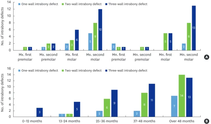
There were 14 (23.3%) patients with diabetes, 42 (70.0%) nonsmokers, and 18 (30.0%) smokers, all of whom smoked less than 10 cigarettes per day, (Table 1). The intrabony defects were distributed in the posterior jaw as follows: maxillary first premolar, n=2 (2.4%); second premolar, n=5 (6.1%); first molar, n=11 (13.4%); second molar, n=25 (30.5%); mandibular first premolar, n=4 (4.9%); second premolar, n=2 (2.4%), first molar, n=10 (12.2%); and second molar, n=23 (28.0%). The mean period of functional loading before regenerative treatment was 60.2±39.3 months (range, 9–212 months) (Figure 2).
Clinical and radiographic outcomes
Table 2 provides the clinical and radiographic outcomes at baseline and at 6, 12, and 24 months after regenerative treatment. Clinically, at 6 months after regenerative treatment, a significant reduction in PPD was observed, from 7.2±1.1 mm to 5.1±1.6 mm (P<0.001), as well as a significant reduction in CAL from 7.5±1.1 mm to 5.8±2.2 mm (P<0.001).
Radiographically, at 6 months after regenerative treatment, a significant reduction in defect depth, from 5.0±1.4 mm to 3.8±1.8 mm (P<0.001), was observed, as well as a significant reduction in defect width from 1.5±0.7 mm to 1.2±0.7 mm (P=0.004).
Figure 3 presents a comparison of changes in the clinical and radiographic outcomes based on the classification of intrabony defects associated with FDP. Clinically, at 6 months after regenerative treatment, a significant reduction was found in the PPD of 1-wall (P<0.001), 2-wall (P<0.001), and 3-wall (P<0.001) intrabony defects, as well as a significant reduction in the CAL of 2-wall (P<0.001) and 3-wall (P<0.001) intrabony defects. Radiographically, at 6 months after regenerative treatment, a significant reduction was found in the depth of the 3-wall (P<0.001) defects, as well as a significant reduction in the width of the 2-wall (P=0.008) and 3-wall (P<0.001) defects.
Table 1. Baseline characteristics of included patients and intrabony defects associated with fixed dental prostheses
Variables Values
Demographic factors 60 (100.0)
Sex
Male 35 (58.3)
Female 25 (41.7)
Age (yr) 53.8±11.2
Diabetes mellitus 14 (23.3)
Smoking status
Non-smoker 42 (70.0)
Current smoker (<10 cigarettes per day) 18 (30.0)
Intrabony defects 82 (100.0)
Defect morphology
One-wall 12 (14.6)
Two-wall 29 (35.4)
Three-wall 41 (50.0)
Period of functional loading (mon) 60.2±39.3
Data are shown as mean±standard deviation or number (%).
DISCUSSION
The characteristics of bone defect morphology, including depth, width, angulation, and number of defect walls, have been found to be closely related to the prognosis of periodontal Table 2. Clinical and radiographic outcomes at baseline, 6, 12, and 24 months after regenerative treatment of intrabony defects associated with fixed dental prostheses
Parameters (mm) Baseline 6-month follow-up 12-month follow-up 24-month follow-up
Mean±SD (95% CI) Mean±SD (95% CI) P valuea) Mean±SD (95% CI) P valueb) Mean±SD (95% CI) P valuec) Clinical outcomes
PPD
One-wall 7.6±0.7 (7.2–7.9) 6.7±1.0 (6.3–7.1) <0.001 6.4±1.3 (5.8–6.9) 0.342 6.3±1.3 (5.8–6.9) 0.892 Two-wall 7.2±1.1 (6.7–7.6) 5.2±1.2 (4.7–5.7) <0.001 4.9±1.1 (4.4–5.3) 0.330 5.0±1.2 (4.5–5.5) 0.732 Three-wall 6.9±1.3 (6.4–7.4) 3.5±0.4 (3.3–3.7) <0.001 3.8±1.2 (3.3–4.3) 0.171 3.8±1.1 (3.3–4.3) 0.906 CAL
One-wall 8.0±0.8 (7.7–8.3) 8.7±0.7 (8.3–9.0) 0.003 8.6±0.7 (8.3–8.9) 0.698 8.5±0.7 (8.2–8.8) 0.780 Two-wall 7.4±1.0 (7.0–7.8) 5.5±1.3 (4.9–6.0) <0.001 5.1±1.3 (4.6–5.7) 0.336 5.2±1.3 (4.6–5.7) 0.938 Three-wall 7.1±1.3 (6.5–7.6) 3.7±0.6 (3.4–3.9) <0.001 3.9±1.2 (3.4–4.4) 0.462 3.9±1.1 (3.4–4.4) 0.895 Radiographic outcomes
Defect depth
One-wall 5.1±1.6 (4.5–5.8) 4.3±1.7 (3.6–5.0) 0.066 4.9±2.0 (4.1–5.7) 0.216 4.8±1.9 (4.0–5.6) 0.859 Two-wall 5.0±1.4 (4.4–5.6) 4.4±1.6 (3.7–5.1) 0.116 4.4±1.6 (3.7–5.0) 0.968 4.5±1.7 (3.8–5.2) 0.722 Three-wall 4.8±1.1 (4.3–5.2) 2.5±1.4 (1.9–3.1) <0.001 2.6±1.6 (1.9–3.3) 0.868 2.6±1.9 (1.8–3.4) 0.943 Defect width
One-wall 1.8±0.6 (1.6–2.1) 1.9±0.7 (1.7–2.2) 0.549 1.9±0.7 (1.6–2.2) 0.827 1.9±0.7 (1.7–2.2) 0.828 Two-wall 1.5±0.7 (1.3–1.8) 1.2±0.4 (1.0–1.3) 0.008 1.1±0.3 (0.1–1.2) 0.254 1.0±0.4 (0.8–1.1) 0.293 Three-wall 1.3±0.8 (0.9–1.6) 0.6±0.3 (0.4–0.7) <0.001 0.6±0.4 (0.4–0.7) 0.968 0.7±0.3 (0.5–0.8) 0.498 Boldface indicates P values below the significance level of 0.05.
PPD: probing pocket depth, CAL: clinical attachment loss, SD: standard deviation, CI: confidence interval.
P values for comparisons between a)baseline vs. 6 months, b)6 months vs. 12 months, and c)12 months vs. 24 months, respectively.
One-wall intrabony defect Two-wall intrabony defect Three-wall intrabony defect
One-wall intrabony defect Two-wall intrabony defect Three-wall intrabony defect
B
0–12 months 13–24 months 25–36 months 37–48 months Over 48 months
1 2 2
7 1
6 8
14
3 5
9 11 13
0 2 4 6 8 10 12 14 16
No. of intrabony defects
Mx. first A
premolar Mx. second
premolar Mn. first
premolar Mn. second premolar Mx. first
molar Mx. second
molar Mn. first
molar Mn. second molar
1 2
5
2 2
1 2 3
8
1 1
5
8
1 2
6
12
1 1
5
13
0 2 4 6 8 10 12 14
No. of intrabony defects
Figure 2. (A) Frequency distribution of intrabony defects according to their position in the jaw, (B) period of functional loading before regenerative treatment.
regeneration in intrabony defects [12,13]. In the present study, during 24 months of observation after regenerative periodontal surgery for intrabony defects associated with FDPs, the clinical and radiographic outcomes, including CAL, PPD, and defect depth and width, significantly improved, except for 1-wall intrabony defects. This is because a larger number of defect walls provide a stable environment for grafting materials, clot formation, and bridging vascular and cellular elements from the periodontal ligament and osseous walls [13]. These findings are consistent with systematic reviews and meta-analyses of randomized controlled clinical trials assessing the efficacy of regenerative treatment for periodontal intrabony defects [14,15].
A previous review indicated that abutment teeth associated with FDPs showed more plaque and food retention and gingival inflammation than the non-abutment teeth [5]. In our previous study of 184 periodontally compromised patients with 2- and 3-wall intrabony defects, the baseline PPD was found to be 6.3±1.7 mm and the CAL was 7.1±1.6 mm [16].
When the patients of this study were limited to 2- and 3-wall intrabony defects, the baseline PPD was 7.0±1.2 mm and CAL was 7.3±1.1 mm, respectively. In a recent randomized controlled clinical trial, which was only limited to 1-wall intrabony defects, the baseline PPD was reported to be 7.3±0.6 mm and CAL was 7.8±0.6 mm [17]. When the patients of this study were likewise limited to 1-wall intrabony defects, the baseline PPD was 7.6±0.7 mm and CAL was 8.0±0.8 mm, respectively. In light of these results, we carefully suggest that the intrabony defects associated with FDPs show more severe bone loss than conventional intrabony defects associated with chronic periodontitis.
Baseline 6 months 12 months 24 months A
−4.0
−3.2
−2.4
−1.6
−0.8 0
Changes of probing pocket depth (mm)
−5 B
−4
−3
−2
−1 0 1 2
Changes of clinical attachment level (mm)
Baseline 6 months 12 months 24 months
−1.0 D
−0.5 0 0.5
Changes of defect width (mm)
Baseline 6 months 12 months 24 months
−3 C
−2
−1 0 1
Changes of defect depth (mm)
Baseline 6 months 12 months 24 months
One-wall intrabony defect Two-wall intrabony defect Three-wall intrabony defect Total
−0.9 −1.2 −1.2
−2.1 −2.3
−2.0 −2.2 −2.2
−2.2
−3.4 −3.1 −3.1
−0.6
−0.6 −0.5
−0.9
−0.2 −0.2
−1.2 −1.0 −1.0
−2.2 −2.2 −2.1
0.1 0.1 0.1
−0.3 −0.4 −0.4
−0.4 −0.5
−0.7 −0.7 −0.6
−0.6
0.7 0.6 0.5
−1.6 −1.7 −1.7
−2.0 −2.3 −2.3
−3.4 −3.2 −3.2
Figure 3. Comparison of changes of clinical and radiographic outcomes according to the classification of intrabony defects associated with fixed dental prostheses. (A) Probing pocket depth at 6, 12, and 24 months, (B) clinical attachment loss at 6, 12, and 24 months, (C) defect depth at 6, 12, and 24 months, (D) defect width at 6, 12, and 24 months.
Many clinical studies in the past decades have demonstrated that guided tissue regeneration is an effective and successful treatment modality for regenerative surgery of intrabony defects [18,19]. However, guided tissue regeneration has major drawbacks, such as an additional cost burden, technique sensitivity, and serious postoperative complications (barrier membrane exposure, wound dehiscence and fenestration, and infection) [20]. Therefore, in recent years, various alternative regeneration techniques and materials without the use of a barrier membrane, particularly a non-resorbable barrier membrane, have been introduced [21].
EMD is expected not only to enhance periodontal regeneration through new cementum formation and connective tissue attachment but also to promote soft tissue wound healing and reduce patients' subjective discomfort [16,17,22]. In an attempt to achieve periodontal regeneration without the adjunctive use of barrier membranes, EMD has become an acceptable treatment option in most periodontal practices [23,24]. Nemoto et al. [25]
reported that the use of EMD with bone graft material resulted in similar improvements in periodontal regeneration with or without a barrier membrane. Another split-mouth clinical trial also showed that PPD, CAL, and the filled bone rate significantly improved regardless of the use of a barrier membrane [26].
Previous studies showed that the combination of bone grafting material and EMD had no additional benefit when compared to EMD alone [27,28]. However, a recent systematic review and meta-analysis reported that the adjunctive use of bone grafting materials in combination with EMD may result in an additional clinical benefit in terms of PPD reduction of 4.22±1.20 mm (95% CI, 3.96–4.24 mm) and CAL gain of 3.76±1.07 mm (95% CI, 3.51–3.75 mm) compared with EMD without bone grafts [24]. In our study, at 24 months after regenerative treatment, the intrabony defects associated with FDPs also showed a mean PPD reduction of 4.1±1.7 mm and a CAL gain of 3.4±1.5 mm (P<0.001).
This study included all types of intrabony defect morphology in premolar and molar positions, but the lack of a sufficient number of tooth positions and defect types is considered as a major limitation. In particular, only 13 cases of maxillary and mandibular premolars and 12 cases of 1-wall intrabony defects were included. Therefore, more cases associated with FDPs and careful interpretation are needed to prevent selection bias.
In conclusion, within the limitations of the current study, the effectiveness of porcine-derived xenograft with EMD for periodontal regenerative treatment of 2- and 3-wall intrabony defects associated with FDPs improved clinical and radiological parameters. Further high-quality prospective studies on long-term outcomes and a greater number of cases are necessary to confirm our findings.
REFERENCES
1. Tan K, Pjetursson BE, Lang NP, Chan ES. A systematic review of the survival and complication rates of fixed partial dentures (FPDs) after an observation period of at least 5 years. Clin Oral Implants Res 2004;15:654-66.
PUBMED | CROSSREF
2. Hochman N, Yaffe A, Ehrlich J. Splinting: a retrospective 17-year follow-up study. J Prosthet Dent 1992;67:600-2.
PUBMED | CROSSREF
3. Fayyad MA, al-Rafee MA. Failure of dental bridges: III--effect of some technical factors. J Oral Rehabil 1996;23:675-8.
PUBMED
4. Fayyad MA, al-Rafee MA. Failure of dental bridges. II. Prevalence of failure and its relation to place of construction. J Oral Rehabil 1996;23:438-40.
PUBMED | CROSSREF
5. Knoernschild KL, Campbell SD. Periodontal tissue responses after insertion of artificial crowns and fixed partial dentures. J Prosthet Dent 2000;84:492-8.
PUBMED | CROSSREF
6. Freilich MA, Niekrash CE, Katz RV, Simonsen RJ. Periodontal effects of fixed partial denture retainer margins: configuration and location. J Prosthet Dent 1992;67:184-90.
PUBMED | CROSSREF
7. Valderhaug J, Ellingsen JE, Jokstad A. Oral hygiene, periodontal conditions and carious lesions in patients treated with dental bridges. A 15-year clinical and radiographic follow-up study. J Clin Periodontol 1993;20:482-9.
PUBMED | CROSSREF
8. Müller HP. The effect of artificial crown margins at the gingival margin on the periodontal conditions in a group of periodontally supervised patients treated with fixed bridges. J Clin Periodontol 1986;13:97-102.
PUBMED | CROSSREF
9. Valderhaug J, Birkeland JM. Periodontal conditions in patients 5 years following insertion of fixed prostheses. Pocket depth and loss of attachment. J Oral Rehabil 1976;3:237-43.
PUBMED | CROSSREF
10. Meyle J, Chapple I. Molecular aspects of the pathogenesis of periodontitis. Periodontol 2000 2015;69:7-17.
PUBMED | CROSSREF
11. Papapanou PN, Tonetti MS. Diagnosis and epidemiology of periodontal osseous lesions. Periodontol 2000 2000;22:8-21.
PUBMED | CROSSREF
12. Reynolds MA, Kao RT, Camargo PM, Caton JG, Clem DS, Fiorellini JP, et al. Periodontal regeneration - intrabony defects: a consensus report from the AAP regeneration workshop. J Periodontol 2015;86:S105-7.
PUBMED | CROSSREF
13. Blumenthal NM, Alves ME, Al-Huwais S, Hofbauer AM, Koperski RD. Defect-determined regenerative options for treating periodontal intrabony defects in baboons. J Periodontol 2003;74:10-24.
PUBMED | CROSSREF
14. Díaz-Faes L, Fernández-Somoano A, Magán-Fernández A, Mesa F. Efficacy of regenerative therapy in aggressive periodontitis: a systematic review and meta-analysis of randomised controlled clinical trials.
Clin Oral Investig 2020;24:1369-78.
PUBMED | CROSSREF
15. Barbato L, Selvaggi F, Kalemaj Z, Buti J, Bendinelli E, Marca M, et al. Clinical efficacy of minimally invasive surgical (MIS) and non-surgical (MINST) treatments of periodontal intra-bony defect. A systematic review and network meta-analysis of RCT's. Clin Oral Investig 2020;24:1125-35.
PUBMED | CROSSREF
16. Lee JH, Park YS, Kim YT, Kim DH, Jeong SN. Assessment of early discomfort and wound healing outcomes after periodontal surgery with and without enamel matrix derivative: an observational retrospective case-control study. Clin Oral Investig 2020;24:229-37.
PUBMED | CROSSREF
17. Lee JH, Kim DH, Jeong SN. Adjunctive use of enamel matrix derivatives to porcine-derived xenograft for the treatment of one-wall intrabony defects: two-year longitudinal results of a randomized controlled clinical trial. J Periodontol 2020;91:880-9.
PUBMED | CROSSREF
18. Needleman I, Tucker R, Giedrys-Leeper E, Worthington H. A systematic review of guided tissue regeneration for periodontal infrabony defects. J Periodontal Res 2002;37:380-8.
PUBMED | CROSSREF
19. Murphy KG, Gunsolley JC. Guided tissue regeneration for the treatment of periodontal intrabony and furcation defects. A systematic review. Ann Periodontol 2003;8:266-302.
PUBMED | CROSSREF
20. Needleman IG, Worthington HV, Giedrys-Leeper E, Tucker RJ. Guided tissue regeneration for periodontal infra-bony defects. Cochrane Database Syst Rev 2006:CD001724.
PUBMED | CROSSREF
21. Reynolds MA, Aichelmann-Reidy ME, Branch-Mays GL, Gunsolley JC. The efficacy of bone replacement grafts in the treatment of periodontal osseous defects. A systematic review. Ann Periodontol 2003;8:227-65.
PUBMED | CROSSREF
22. Lee JH, Kim DH, Jeong SN. Comparative assessment of anterior maxillary alveolar ridge preservation with and without adjunctive use of enamel matrix derivative: A randomized clinical trial. Clin Oral Implants Res 2020;31:1-9.
PUBMED | CROSSREF
23. Li W, Xiao L, Hu J. The use of enamel matrix derivative alone versus in combination with bone grafts to treat patients with periodontal intrabony defects: a meta-analysis. J Am Dent Assoc 2012;143:e46-56.
PUBMED | CROSSREF
24. Matarasso M, Iorio-Siciliano V, Blasi A, Ramaglia L, Salvi GE, Sculean A. Enamel matrix derivative and bone grafts for periodontal regeneration of intrabony defects. A systematic review and meta-analysis.
Clin Oral Investig 2015;19:1581-93.
PUBMED | CROSSREF
25. Nemoto Y, Kubota T, Nohno K, Nezu A, Morozumi T, Yoshie H. Clinical and CBCT evaluation of combined periodontal regenerative therapies using enamel matrix derivative and deproteinized bovine bone mineral with or without collagen membrane. Int J Periodontics Restorative Dent 2018;38:373-81.
PUBMED | CROSSREF
26. Kubota T, Nemoto Y, Nohno K, Nezu A, Morozumi T, Yoshie H. A comparable study of combinational regenerative therapies comprising enamel matrix derivative plus deproteinized bovine bone mineral with or without collagen membrane in periodontitis patients with intrabony defects. Open J Stomatol 2018;8:277-86.
27. Lekovic V, Camargo PM, Weinlaender M, Nedic M, Aleksic Z, Kenney EB. A comparison between enamel matrix proteins used alone or in combination with bovine porous bone mineral in the treatment of intrabony periodontal defects in humans. J Periodontol 2000;71:1110-6.
PUBMED | CROSSREF
28. Zucchelli G, Amore C, Montebugnoli L, De Sanctis M. Enamel matrix proteins and bovine porous bone mineral in the treatment of intrabony defects: a comparative controlled clinical trial. J Periodontol 2003;74:1725-35.
PUBMED | CROSSREF