| Abstract |
Purpose: The purpose of this study was to investigate the differences in muscle thickness and ground reaction force of the vastus medialis oblique and vastus lateral oblique muscles during squats at ankle angles of toe 0°, toe in 10°, and toe out 10°.
Methods: In this study, 9 male and 17 female students in their 20s participated in a randomized controlled trial and were compared according to the ankle angles of toe 0°, toe in 10°, and toe out 10°. To determine the reliability and measurement of muscle thickness according to ankle angle using ultrasound equipment and muscle thickness, the participants’ ankle angles-toe 0°, toe in 10°, and toe out 10°-were measured three times at the vastus medialis oblique and vastus lateralis oblique muscles during squats. At the same time, the maximum vertical ground reaction force was measured with a force plate. A total of three measurements were taken and averaged, and two minutes of squat movements were assessed between ankle angles to prevent target action.
Results: The results of this study illustrated that the reliability of the vastus medialis oblique muscles and vastus lateralis oblique muscles in ankle angle was high. The difference in muscle thickness was significantly greater in comparing the toe out10° angle with the toe 0° angle than between toe in 10° and toe out 10° in vastus medialis oblique and vastus lateralis oblique (p < 0.05).
There was no statistically significant difference between the ankle angle of toe 0° and toe in 10° (p > 0.05). The maximum vertical ground reaction force was significantly greater at toe out 10° than at the ankle angle of toe 0° and toe out 10° and between toe in 10° and toe out 10° (p < 0.05). There was no statistically significant difference in the comparison between toe 0° and toe in
†Corresponding Author :Su-Kyong Lee ([email protected])
Original Article Open Access
발목각도 Toe 0°, Toe in 10°, Toe out 10°에 따른 스쿼트 운동이 안쪽넓은근과 가쪽넓은근의 근두께와 지면반발력에 미치는 영향
안수홍⋅이수경1†1)
동의대학교 보건의과학대학원, 1동의대학교 물리치료학과, 건강기능성 소재연구소
The Effect of Squat Exercise According to Ankle Angle-Toe 0°, Toe In 10°, Toe Out 10°-on Muscle Thickness and Ground Reaction Force of Vastus Medialis Oblique and Vastus Lateralis Oblique Muscles
Su-Hong Ahn, P.T., M.S⋅Su-Kyong Lee, P.T., Ph.D1†
Department of Biomedical Health Science, Graduate School, Dong-Eui University
1Department of Physical Therapy, College of Nersing and Healthcare Sciences, Dong-Eui University, The Research Institute Health for Functional Material
Received: December 20, 2019 / Revised: January 14, 2020 / Accepted: January 22, 2020
ⓒ 2020 Journal of Korea Proprioceptive Neuromuscular Facilitation Association
This is an Open Access article distributed under the terms of the Creative Commons Attribution Non-Commercial License (http://creativecommons.org/licenses/by-nc/3.0) which permits unrestricted non-commercial use, distribution, and reproduction in any medium, provided the original work is properly cited.
10° (p > 0.05).
Conclusion: Squatting at an ankle angle of toe out 10° increases the dorsi flexion; thus, the stability of the ankle and the thickness of both oblique muscles increased to perform more effective squats. In addition, as the base of support widens, it is thought that the stability of the posture increases so that squat training can be performed safely.
Key Words: Squat, Ankle angle, Muscle thickness, Ground reaction force
Ⅰ. 서 론
최근 문명의 발달과 생활수준의 향상으로 인해 일 상 속에서 쇼핑이나 은행 업무 등을 컴퓨터 및 스마트 폰으로 대체하여 장시간 의자에 앉아있거나 신체를 많이 움직이지 않고도 일이 해결되는 추세이다. 이로 인하여 신체활동과 운동량이 감소하여 고혈압, 당뇨 병 등 각종 성인병과 질병 발병률이 증가하여 사회적 인 문제로 야기되고 있다(Nam, 2017). 따라서, 요즘 현대인들은 건강증진에 대한 관심과 욕구가 증가함에 따라 운동에 대한 실천이 커지고 있으며, 그 중에서 가장 손쉽게 접할 수 있는 운동으로 웨이트 트레이닝 이 있다(Oh, 2014).
웨이트 트레이닝은 근력을 증가시킬 수 있는 안전 하고 효과적인 운동방법이며(Peterson et al., 2010), 자 신의 체중 및 중량기구 등을 사용하여 다양한 방법으 로 운동이 가능하다(Kwon et al., 2012). 운동종류로는 스쿼트(squat), 런지(lunge), 벤치 프레스(bench press) 등이 있으며(Choi, 2015), 이러한 운동들은 여러부위의 근육들이 복합적으로 사용하는 큰 근육 운동으로써 짧은 시간내에 근육을 활성화시켜 근력 향상에 효과 적이다(Kim, 2010). 특히, 스쿼트는 대표적인 닫힌사 슬운동(closed kinetic chain exercise, CKCE)으로써 신 체의 먼 쪽 부위인 양 발을 지면에 고정한 상태에서 다리의 굽힘과 폄의 동작을 수행하며, 몸통의 중심부 와 중량 부하가 위아래 방향으로 움직이는 운동이다 (Lim et al., 2018). 이러한 스쿼트는 넙다리네갈래근에 많은 영향을 미치는데 그 중에서도 안쪽넓은근(vastus medialis oblique)과 가쪽넓은근(vastus lateralis oblique)
에 많은 영향을 미치며(Consitt et al., 2002), 체중지지를 통하여 안쪽넓은근, 가쪽넓은근의 근력을 강화시키고 보다 많은 관절을 움직여, 기능적인 근동원 패턴을 촉진시키고 고유수용성 감각을 자극시킨다(Selseth et al., 2000). 더 나아가 여러 근육군들이 협응 동작을 만들고 엉덩관절, 무릎관절, 발목관절들이 서로 상호 작용을 하여 움직임으로써(Labella, 2004), 양쪽 다리 에 균등하게 힘이 가해지기 때문에 다른 다리 운동 동작들보다 큰 힘을 발휘하고 안정감이 있다(Choi et al., 2015). 또한, 스쿼트는 상황에 따라서 다양한 방법 으로 응용하여 실시할 수 있는 장점이 있는데(Bompa, 2018) 예를 들면 무릎관절 굽힘 각도와 종아리의 돌림 각도, 양발의 벌림 정도 및 한 다리 서기 스쿼트 등 과 같이 몸의 내부에 변화를 주어 수행하는 방법이 있으며, 벽에 기댄 상태에서 스쿼트 및 짐볼을 이용한 스쿼트 등 과 같은 외부환경을 이용하여 수행하는 스 쿼트 방법이 있다(Oh, 2013). 이처럼 다양한 방법에 따른 스쿼트는 관절들이 유기적인 움직임을 통해 서 로 다른 모멘트 값이 형성되며, 무릎과 발의 각도 및 근육의 작용에 따라서 운동 효과가 서로 다르게 나타 나기 때문에 중요한 선행조건이 된다고 하였다(Lee, 2008; Fry, 2003). 특히, 발의 위치와 회전각도의 변화는 전반적으로 스쿼트 동작에 많은 영향을 미쳐 변화할 수 있는 요인이며, 그 중에서도 발목의 toe in과 toe out에 따라서 신체에 미치는 영향이 크기 때문에 지속 적인 연구가 필요하다(Jun, 2018). 그러나 발의 과도한 회전은 다리의 관절 모멘트에 부정적인 영향을 미친 다고 하였으며, 발목의 toe의 각도가 10°일 때 효과적 인 스쿼트를 시행할 수 있다고 하였다(Almosnino et
al., 2013; Schoenfeld, 2010; Song, 2019). 이러한 선행연 구를 토대로 스쿼트 시 발목 각도에 따라서 신체에 제공되는 긍정적인 효과를 알아보고자 하였으며, 다 양한 운동 효과를 극대화시키는 기초적인 자료로 제 시할 필요가 있다(Yoo et al., 2004). 또한, 발목 각도에 따른 스쿼트 운동 시 대부분의 선행연구들은 근활성 도에만 초점을 맞추었으며, 근두께의 변화에 대한 연 구는 미비한 실정이다. 따라서 본 연구에서는 스쿼트 시 3가지 발목각도 toe 0°, toe in 10°, toe out 10°에 따른 안쪽넓은근과 가쪽넓은근의 근두께와 동작 수행 시 관절의 힘과 토크 값에 대한 정보를 정확하게 분석 하기 위해 힘판(force plate)을 이용하여 지면반발력 (ground reaction force)을 함께 알아보고자 한다(Min
& Kim, 2015).
Ⅱ. 연구 방법
1. 연구 대상
본 연구는 부산광역시 D대학교에 재학하고 있는 건강한 성인 남자 9명 여자 17명 총 26명을 대상으로 진행하였으며, 다리와 몸통과 관련된 질환 및 통증과 관절가동범위의 제한이 없는 자, 자세조절이나 보행 에 불편함과 통증이 없는 자, 스쿼트 수행이 가능한 자, 본 연구의 내용을 충분히 이해하고 자발적인 참여
의사를 보인 자를 선정하여 연구를 진행하였다.
2. 측정 방법 및 도구
1) 발목각도에 따른 스쿼트
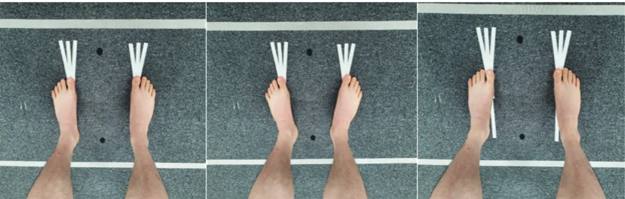
본 연구는 3가지 발목각도 toe 0°, toe in 10°, toe out 10°에 따라서 스쿼트를 실시하였다(Jun, 2018)(Fig.
1). 무작위 배정(randomized controlled trial)을 통하여 측정 순서를 결정하였고, 몸은 똑바로 세운 상태에서 양 발을 대상자의 어깨넓이의 120%로 벌리고 양팔은 팔짱을 낀 상태에서 수행하였다(Earl et al., 2001; Nam, 2008). 시선은 정면을 바라보게 하였고 무릎관절 각도 는 90°를 유지하도록 하였다. 실험 전, 스쿼트 자세에 대한 교육을 충분히 하였으며, 각 발목각도에 따라서 3회 반복 측정하였다. 반복된 측정으로 발생할 수 있는 근 피로 및 대상작용을 최소화하기 위해서 Kendall 등(2005)은 각 운동 측정 간 2분의 휴식을 취하는 것이 효율적이라고 하였다. 따라서 본 연구에서 각 발목각 도에 따른 스쿼트 동작 간 2분간의 휴식을 취하도록 하였다(Jun, 2018).
2) 초음파 측정장비
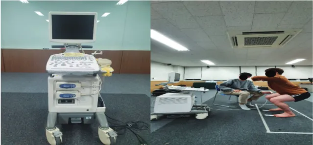
발목각도에 따른 스쿼트 시 진단용 초음파 영상장 치(Aloka, SSD-3500X, Aloka Co. Ltd, Japan)를 사용하
Fig. 1. Squat according to the ankle angle.
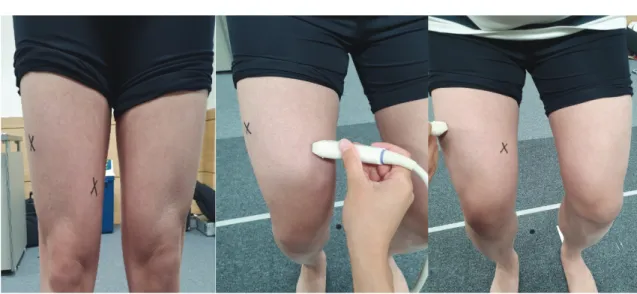
여 근두께를 측정하였다(Fig. 2)(Fig. 3). 탐촉자는 근육 전용인 10MHz의 직선형 탐촉자를 사용하였으며, 오 른쪽 안쪽넓은근과 가쪽넓은근 2가지 근육을 측정하 였다. 초음파 장비를 이용한 근두께의 측정자 내 신뢰 도는 연구대상자의 발목각도 toe 0°, toe in 10°, toe out 10°에서 스쿼트 시 안쪽넓은근 ICC (1, 3)와 가쪽넓 은근 ICC (1, 3)를 3회 반복 측정하여 급내 상관계수 (intraclass correlation coefficient, ICC)를 산출하였다.
초음파 장비에 대한 측정결과 값의 일관성을 얻기 위 해 사전에 사용방법을 충분히 익힌 1명이 실시하였다
(Kim & Kwon, 2017). 연구 대상자를 바로 서게 한 후 초음파 겔을 피부와 직선 탐촉자에 충분히 바른 후 초음파 영상과 해부학적 지식에 근거하여 시진 및 촉진을 하였으며, 측정부위와 직선 탐촉자가 수직이 되게 위치하여 중앙에 닿도록 위치시켰다. 안쪽넓은 근의 측정 부위는 넙다리뼈 중간 1/2 지점에서 무릎뼈 위쪽까지 스캔하여, 근두께가 가장 두꺼운 지점을 측 정하였다. 수평면에서 안쪽넓은근이 넙다리뼈의 안쪽 가장자리와 만나는 점에서 넙다리뼈를 가로지르지 않 고 안쪽넓은근의 표면에서 가장 멀리 뻗어 나갈 수 있는 지점을 가상의 법선으로 설정하였고, 그 법선을 90° 회전시킨 선 즉, 안쪽넓은근의 안쪽 가장자리와 만나는 점까지의 거리로 정의하였다(Yong, 2018)(Fig.
4). 가쪽넓은근의 측정부위는 근육의 최대근수축을 유 도하게 하여 근두께가 가장 두꺼운 부분을 육안으로 확인한 후, 넙다리뼈의 바깥부위 위앞위엉덩뼈가시와 무릎뼈의 상극점 사이 2/3 지점에서 측정하였다(Park, 2016)(Fig. 4). 측정자의 주관적인 요소가 영향을 미칠 수 있다고 판단하여 한 사람이 측정하였으며, 정확한 측정을 위해 실험용 펜으로 측정부위에 표시해 두었 다(Kim et al., 2011). 각 근육을 3회씩 반복 측정한 후 평균값을 산출하였다.
Fig. 2. Ultrasound imaging.
Fig. 3. Ultrasound measuring equipment.
3) 힘판
본 연구에서는 발목각도 toe 0°, toe in 10°, toe out 10°에 따른 스쿼트 시 최대 수직 지면반발력(Fz) 의 변화를 측정하기 위하여 힘판(AMTI, Newton, USA)을 사용하였다(Fig. 5). A/D 카드(AMTI, Newton, USA)를 통해 최대 수직 지면반발력의 데이터를 수집하였고, 표본 주파수의 신호는 1초당 100개의 데이터를 수집 하기 위해 200Hz로 설정하였으며, A/D 컨버터 (VSAD-102-3C)를 통해 지면반발력의 아날로그 데이 터를 디지털로 변환하여 자료를 분석하였다(Chung, 2016). 수집된 데이터 자료의 신뢰도를 높이기 위해
실험 전에 각 대상자는 힘판 위에 똑바로 서게 한 후 영점세팅을 한 후 실험을 진행하였다. 발목각도에 따 른 최대 수직 지면반발력(Fz)의 변화량은 모니터로 실시간으로 제공되었으며, Bio analysis 프로그램을 통 해 뉴턴(Newton) 값으로 반환하여 사용하였다(Chung, 2016). 최대 수직 지면반발력(Fz)은 3회씩 반복 측정한 후 평균값을 산출하였다.
3. 연구 절차
본 실험에 앞서 연구대상자에게 실험방법에 대하여 충분히 설명하였고, 무작위 배정(randomized controlled Fig. 4. Muscle thickness measurement site.
Fig. 5. Force plate.
trial)비교를 통해 발목각도 toe 0°, toe in 10°, toe out 10°에 따른 스쿼트를 실시하였다. 스쿼트 시 안쪽넓은 근과 가쪽넓은근의 근두께를 측정하기 위하여 초음파 장비(Aloka, SSD-3500X, Aloka Co. Ltd, Japan)를 사용 하였고 동시에 안쪽넓은근 ICC (1, 3)와 가쪽넓은근 ICC (1, 3)의 근두께에 따른 측정자 내 신뢰도를 측정하 였다. 또한, 힘판(AMTI, Newton, USA)을 통해 최대 수직 지면반발력(Fz)을 측정하였다. 총 3회 반복 측정 하여 평균화였고 대상작용을 방지하기 위해서 각 발 목각도에 따른 스쿼트 동작 간 2분간의 휴식을 취하도 록 하였다.
4. 자료 분석
본 연구는 SPSS 18.0 for Windows 프로그램을 이용 하여 발목각도 toe 0°, toe in 10°, toe out 10°에 따른 스쿼트 시 근두께 및 최대 수직 지면반발력(Fz)의 차이 를 알아보기 위해 반복측정 분산분석(repeated measure ANOVA)을 사용하여 분석하였고, 개체 내 대비검정 을 실시하였다. 통계처리에 대한 유의수준(α)은 0.05 로 하였다. 또한, 발목각도에 따른 스쿼트 시 초음파 진단장치의 측정자내 신뢰도를 분석하기 위해서 급내 상관계수(intraclass correlation coefficient, ICC)를 분석 하였다.
Ⅲ. 연구 결과
1. 연구 대상자의 일반적인 특성
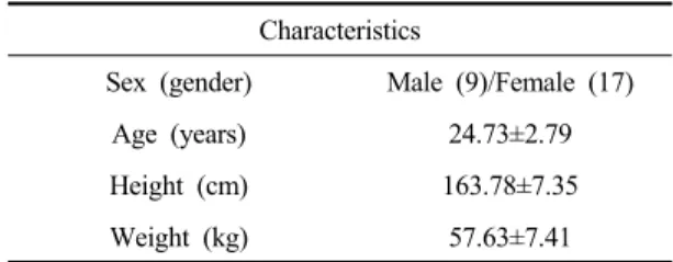
본 연구는 정상 성인 남녀 26명을 대상으로 실험을 실시하였으며, 연구 대상자의 평균 연령은 24.73±2.79 세였으며, 평균 신장은 163.78±7.35㎝, 평균 체중은 57.63±7.41㎏이었다(Table 1).
Characteristics
Sex (gender) Male (9)/Female (17)
Age (years) 24.73±2.79
Height (cm) 163.78±7.35
Weight (kg) 57.63±7.41
Table 1. General characteristics of subjects (n=26)
2. 발목각도에 따른 스쿼트 시 초음파 진단장치 측정의 신뢰도
진단용 초음파를 사용한 휴식 시 서 있는 자세와 발목각도에 따른 스쿼트에서 안쪽넓은근과 가쪽넓은 근의 근두께 측정자내 신뢰도 분석 결과를 Table 2에 나타내었다. 안쪽넓은근의 근두께 신뢰도 ICC (1, 3)는 발목각도 toe 0°에서는 0.95, toe in 10°에서는 0.94, toe out 10°에서도 0.94였다(Table 2). 가쪽넓은근의 근두께 신뢰도 ICC(1, 3)는 발목각도 0°에서는 0.92, toe in 10°
에서는 0.90, toe out 10°에서는 0.89였다. 따라서 발목 각도에 따른 스쿼트 시 안쪽넓은근과 가쪽넓은근의 측정자내 신뢰도는 높다고 볼 수 있다.
Ankle angle Muscle ICC (1, 3) 95% CI
neutral 0° VMO 0.95 0.91∼0.98
VLO 0.92 0.85∼0.96
toe out 10° VMO 0.94 0.89∼0.97
VLO 0.89 0.81∼0.95
toe in 10° VMO 0.94 0.88∼0.97
VLO 0.90 0.83∼0.95
VMO: vastus medialis oblique VLO: vastus lateralis oblique ICC: intraclass correlation coefficient CI: confidence interval
Table 2. Reliability of ultrasound diagnosis equipment measurement of vastus medialis oblique muscles and vastus lateralis oblique muscles during in squats according to ankle angle
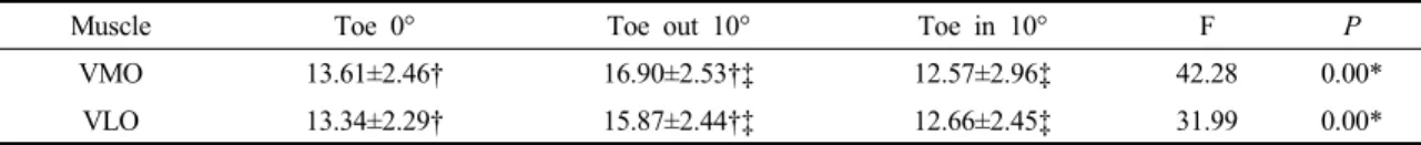
3. 발목각도에 따른 스쿼트 시 안쪽넓은근과 가쪽 넓은근의 근두께 변화 차이의 비교
발목각도에 따른 스쿼트 시 초음파를 통한 오른쪽 안쪽넓은근과 가쪽넓은근의 근두께는 발목각도 toe 0°와 toe out 10°간 비교와 toe in 10°와 toe out 10°간 비교에서 통계적으로 toe out 10°에서 유의한 증가가 있었고(p<0.05), 발목각도 toe 0°와 toe in 10°간 비교 에서는 통계적으로 유의한 차이가 없었다(p>0.05) (Table 3).
4. 발목각도에 따른 스쿼트 시 최대 수직 지면반발력 변화 차이의 비교
발목각도에 따른 스쿼트 시 힘판을 통한 오른쪽 최대 수직 지면반발력은 발목각도 toe 0°와 toe out 10°
간 비교와 toe in 10°와 toe out 10°간 비교에서 통계적으 로 toe out 10°에서 유의한 증가가 있었고(p<0.05), 발목 각도 toe 0°와 toe in 10°간 비교에서는 통계적으로 유의 한 차이가 없었다(p>0.05)(Table 4).
Ⅳ. 고 찰
본 연구는 발목각도 toe 0°, toe out 10°, toe in 10°에 따른 스쿼트 시 안쪽넓은근과 가쪽넓은근의 근두께와 지면반발력의 차이를 알아보고자 하였다. Na (2013)는 발목각도에 따른 스쿼트 시 발목각도 toe 0°보다 toe out을 한 상태에서 안쪽넓은근과 가쪽넓은근의 근활 성도는 더 증가한다고 하였고 Song과 So (2019) 또한 발목을 toe out 10°를 한 상태에서 스쿼트 동작 시 안쪽 넓은근과 가쪽넓은근의 근활성도는 증가한다고 하였 으며, Son (2007)도 무릎을 90°굽힘 스쿼트 동작 수행 시 가쪽넓은근의 근활성도는 발목각도 0°보다 toe out 10°에서 증가한다고 하였다. 본 연구에서는 발목각도 0°보다 발목 toe out 10°에서 스쿼트 시 안쪽넓은근과 가쪽넓은근의 근두께가 증가하였는데 근육이 작용함 에 따라 근두께는 커지며(Shi et al., 2007), 근활성도가 증가함에 따라 근두께도 커진다(Kim, 2011). 따라서 선행연구의 결과와 같이 발목 toe out 시 안쪽넓은근과 가쪽넓은근의 근활성도가 증가하여 근두께가 증가했 다고 볼 수 있다. 이는 발목 toe out의 자세가 엉덩관절
Muscle Toe 0° Toe out 10° Toe in 10° F P
VMO 13.61±2.46† 16.90±2.53†‡ 12.57±2.96‡ 42.28 0.00*
VLO 13.34±2.29† 15.87±2.44†‡ 12.66±2.45‡ 31.99 0.00*
VMO: vastus medialis oblique, VLO: vastus lateralis oblique
*p<0.05, Mean±SD
†significant difference between Toe 0°and Toe out 10°(p<0.05)
‡significant difference between Toe out 10°and Toe in 10°(p<0.05)
Table 3. Comparison of muscle thickness of the vastus medialis oblique muscle and vastus lateralis oblique muscle
during squat according to ankle angle (unit: mm)
GRF Toe 0° Toe out 10° Toe in 10° F P
F (z) 64.82±24.50† 82.54±31.84†‡ 68.12±25.90‡ 5.30 0.01*
GRF: ground reaction force, F (z): vertical ground reaction force
*p<0.05 Mean±SD
†significant difference between Toe 0°and Toe out 10°(p<0.05)
‡significant difference between Toe out 10°and Toe in 10°(p<0.05)
Table 4. Maximum vertical ground reaction force during squats with ankle angle (unit: newton)
모음에 저항을 주는 자세이므로 스쿼트 동작동안 엉 덩관절과 무릎관절이 바깥돌림 됨에 따라 힘의 방향 도 바깥쪽으로 작용하게 된다. 그 힘에 대항하기 위하 여 엉덩관절 모음 시 안쪽 방향으로 힘을 작용하는 안쪽넓은근이 수축함에 따라 근활성도가 증가하여 근 두께가 증가했다고 볼 수 있다(Song & So, 2019). 가쪽 넓은근은 발목 toe out 시 엉덩관절과 발의 위치가 변함 에 따라 넙다리네갈래근의 각도가 증가하여 근육의 기시와 정지의 거리가 감소하게 된다(Yoo et al., 2004).
즉, 가쪽넓은근의 당김선이 짧아짐에 따라 근수축이 일어나게 되어 수축하지 않았을 때 보다 더 많이 사용 되어지기 때문에 근 두께가 증가했다고 볼 수 있다 (Blake et al., 1981).
또한, 발목 toe in 10°보다 toe out 10°에서도 스쿼트 시 안쪽넓은근과 가쪽넓은근의 근두께가 증가하였다. Yoo 등(2004)은 스쿼트 동작 시 toe out 자세가 toe in 자세보다 안쪽넓은근과 가쪽넓은근의 근활성도가 증 가한다고 하였고 Park (2010)은 무릎을 굽히는 런지 동작 시 toe out 자세가 toe in 자세보다 안쪽넓은근과 가쪽넓은근의 근활성도가 더 증가한다고 하였다. 발 목 toe in 10° 자세는 발목이 안쪽번짐과 발바닥굽힘의 증가하는 역학적 구조로써(Abdel-aziem & Draz, 2014), 발목 바깥쪽의 구조물들에 스트레스가 가해져 안정성 이 감소하게 되어 발목불안정성이 나타나게 된다 (Fong et al., 2007; Safran et al., 1999). Palmitier 등(1991) 은 올바른 스쿼트 동작이 발목관절의 굽힘을 증가시 킨다고 하였고, Kathiresan 등(2010)은 발목관절의 굽 힘이 감소가 되면 정확한 동작의 스쿼트 자세가 가능 하지 않다고 하였다. 따라서 본 연구에서도 발목 toe in 10°에서 스쿼트 시 발목이 안쪽번짐 및 발바닥 굽힘 이 되면서 발등굽힘이 상대적으로 감소하여 발목이 불안정성이 증가하였으며, 발목의 불안정성은 다리의 근력과 안정성에 상관관계가 있으므로 근두께가 감소 하였다고 생각된다(Lee, 2014).
또한, 지면반발력에서도 발목 toe out 10°에서 스쿼 트 시 발목각도 0°에서 보다 더 크게 나와 유의한 차이 가 있었다. Song (2019)은 스쿼트 경험이 있는 자들을
대상으로 발목각도에 따른 스쿼트 동작 시 발목 toe out 20°에서 지면반발력의 크기가 증가한다고 하였고, Woo (2019)는 발목 toe out의 자세는 발의 안쪽의 지면 반발력이 더 높다고 하였다. 또한, Lee (2019)는 발을 toe out 시킨 동작은 기저면의 넓이가 커짐에 따라 자세 의 안정성이 증가할 것이라고 예상하였다. 따라서 본 연구에서도 발목 toe out은 발의 접지형태가 바깥쪽으 로 향하기 때문에 이를 보상하기 위하여 다리의 안쪽 으로 체중이 가해지면서 안쪽에 지면반발력이 커짐에 따라 지면반발력의 평균 값들이 증가하였다고 볼 수 있으며(Jung, 2004), 기저면의 면적이 넓어짐에 따라 자세 안정성도 커져 지면반발력이 증가했다고 볼 수 있다.
또한, 발목 toe in 10°보다 toe out 10°에서도 스쿼트 시 지면반발력이 증가하였는데, Hertel 등(2002)은 발 목 toe in 자세는 정상적인 발보다 중심압력이 불안정 하다고 하였고, Wang 등(2006)은 발목 toe in 자세는 지지면과의 접촉면이 감소되어 발목관절의 운동성이 감소한다고 하였다. 이는 발목 toe in 자세는 발목의 불안정성을 증가시켰으며, 발바닥과 지면의 접촉 부 위가 감소하여 체중이 발바닥의 일정부위에만 집중되 어 압력분배가 적절하게 일어나지 못해서 지면반발력 이 감소했다고 생각된다(Woo, 2019). 따라서 발목 toe out 10°에서 스쿼트 시 발등굽힘이 증가하여 발목의 안정성과 더불어 안쪽넓은근과 가쪽넓은근의 근두께 가 증가하여 더 효과적인 스쿼트를 수행할 수 있으며, 기저면 또한 넓어짐에 따라 자세의 안정성이 증가하여 보다 안전하게 스쿼트 훈련을 할 수 있다고 판단된다.
Ⅴ. 결 론
본 연구는 건강한 성인 남자 9명, 여자17명을 대상 으로 발목각도 toe 0°, toe out 10°, toe in 10°에 따른 스쿼트 시 안쪽넓은근과 가쪽넓은근의 근두께와 지면 반발력의 차이를 알아보고자 하였다. 본 연구결과 발 목각도 toe 0°와 toe in 10°에서보다 toe out 10°에서
안쪽넓은근과 가쪽넓은근의 근두께가 증가하였고 지 면반발력 또한 증가하여 유의한 차이가 있었다. 이러 한 결과는 발목 toe out 10°에서 스쿼트를 시행하는 것이 안쪽넓은근과 가쪽넓은근의 근두께를 증가시켜 훈련효과를 증대시킬 수 있고 기저면 또한 증가함에 따라 안전하게 스쿼트 훈련을 할 수 있기 때문에 임상 적 중재로 널리 활용해 볼 필요가 있다.
References
Abdel-aziem AA, Draz AH. Chronic ankle instability alters eccentric eversion/inversion and dorsiflexion/
plantarflexion ratio. Journal of Back and Musculoskeletal Rehabilitation. 2014;27(1):47-53.
Almosnino S, Kingston D, Graham RB. Three-dimensional knee joint moments during performance of the bodyweight squat: effects of stance width and foot rotation. Journal of Applied Biomechanics. 2013;
29(1):33-43.
Blake RL, Burns DP, Colson JP. Etiology of atraumatic medial knee pain. Journal of the American Podiatric Medical Association. 1981;71(10):580-583.
Bompa TO, Buzzichelli CA. Theory and methodology of training, 6th ed. Champaign. Human Kinetics. 2018.
Choi NY, Jang HS, Shin YA. The effect on muscle activation in trunk and low-limbs during squat exercise on various instability surface. Korean Journal of Physical Education. 2015;54(1):505-514.
Choi WI. An analysis of core muscle activity differences by knee joint angles in squat exercise on unstable surfaces.
Korea National Sport University. Dissertation of Master’s Degree. 2015.
Chung JS. A study on change in muscles activity of low extremity and GRF and trunk movement during golf swing according to pelvic tilt. Daegu Catholic University.
Dissertation of Doctorate Degree. 2016.
Consitt LA, Copeland JL, Tremblay MS. Endogenous anabolic responses to endurance versus resistance exercise and training in woman. Sports Medicine. 2002;32(1):1-22.
Earl J, Schmitz R, Arnold B. Activation of the VMO and VL during dynamic mini-squat exercises with and without isometric hip adduction. Journal of Electromyography and Kinesiology. 2001;11(6):
381-386.
Fong DT, Hong Y, Chan LK, et al. A systematic review on ankle injury and ankle sprain in sports. Sports Medicine. 2007;37(1):73-94.
Fry AC, Smith JC, Schili BK. Effect of knee position on hip and knee torques during the barbell squat. Journal of Strength and Conditioning Research. 2003;17(4):
692-693.
Hertel J, Gay MR, Denegar CR. Differences in postural control during single-leg stance among healthy individuals with different foot types. Journal of Athletic Training.
2002;37(2):129-132.
Jun YW. Comparative study on lower limb muscle activities depending on toe angles in squat movement. Bukyong University. Dissertation of Master’s Degree. 2018.
Jung BC. Kinetic analysis of walking toe angle in walking.
Inje University. Dissertation of Master’s Degree. 2004.
Kathiresan GJN, Rayhan AN, Azila AN, et al. The relationship between ankle joint flexibility and squatting knee flexion posture in young Malaysian men. World Journal of Sport Sciences. 2010;3(1):226-230.
Kendall FP, McCreary EK, Provance PG, et al. Muscles testing and function with posture and pain, 5th ed. Baltimore.
Lippincott Williams & Wilkins. 2005.
Kim BI. During vaginal pressure change comparative analysis abdominis thickness and activity in healthy women.
Daegu University. Dissertation of Master’s Degree.
2011.
Kim BJ, Kwon HY. Correlation between peak expiratory flow and expiratory muscle thickness in reliability of the
muscle thickness measurements using ultrasonography.
Asia-pacific Journal of Multimedia Services Convergent with Art, Humanities, and Sociology.
2017;7(7):297-308.
Kim MU, Byeon SJ, Lee GE, et al. Effects of position of the back of a chair to muscle activity during computer work. Journal of Rehabilitation Science. 2011;29(1):
55-68.
Kim YH. Analysis of muscle activity with weight increase during squat movement. Busan University of Foreign Studies. Dissertation of Master’s Degree. 2010.
Kwon YJ, Park SJ, Kim K. The effect of open and closed chain exercise on lower extremity muscle activity in adults. Journal of the Korean Society of Physical Medicine. 2012;7(2):173-182.
Labella C. Patellofemoral pain syndrome: evaluation and treatment. Primary Care Clinics in Office Practice.
2004;31(4):977-1003.
Lee JH. The effect of ballet program in functional ankle instability of women’s university students.
Sookmyung Women’s University. Dissertation of Doctorate Degree. 2014.
Lee JW. The effect of foot external rotation on postural stability factors during deep squat. Konkuk University.
Dissertation of Master’s Degree. 2019.
Lee SW. The kinematic differences and distribution of joint loads according to squat type. Seoul National University. Dissertation of Master’s Degree. 2008.
Lim YT, Lee JW, Park JS, et al. Effects of kinesio taping position of shank and gender on the range of motion and moment of ankle joint during deep squat. The Korean Journal of physical education. 2018;57(1):
471-480.
Min SJ, Kim J. Inertial motion sensing-based estimation of ground reaction forces during squat motion. Journal of the Korean Society for Precision Engineering.
2015;32(4):377-386.
Na YC. Muscle activity analysis of erector spinae and rectus femoris depending on toe out angles in squat movement. Chungnam National University. Dissertation of Master’s Degree. 2013.
Nam DH. Effects of squats using visual feedback on the balance ability and thicknesses of quadriceps femoris and transversus abdominis muscle of college students in their 20s. Daegu University. Dissertation of Master’s Degree. 2017.
Nam KS. Effect of the resistance direction by an elastic band on the VMO/VL electromyographic activity ratio during dynamic Squat exercise. The Journal of Korean Society of Physical Therapy. 2008;20(3):29-34.
Oh JH. The effects of angle-whole body vibration on squat exercise of muscles of lower limb. Chonbuk National University. Dissertation of Master’s Degree. 2014.
Oh TY. The effects of squatting exercise with gym ball and wall on lower extremity muscles activation. The Korean Society of Physical Medicine. 2013;8(4):
647-653.
Palmitier RA, An KN, Scott SG, et al. Kinetic chain exercise in knee rehabilitation. Sports Medicine. 1991;60(12):
1624-1632.
Park HS. Comparative analysis on muscle activities of lower limb depending on the types and load of squat exercise.
Hanyang University. Dissertation of Master’s Degree.
2016.
Park SR. Comparison of electromyographic activity during lunge according to ankle positions in ssierum players with patellofemoral pain syndrome. Kyung-hee University. Dissertation of Master’s Degree. 2010.
Peterson MD, Rhea MR, Sen A, et al. Resistance exercise for muscular strength in older adults: a meta-analysis.
Ageing Research Reviews. 2010;9(3):226-237.
Safran MR, Benedetti RS, Bartolozzi AR, et al. Lateral ankle sprains: a comprehensive review. Part 1: etiology, pathoanatomy, histopathogenesis, and diagnosis.
Medicine and Science in Sports and Exercise.
1999;31(7):429-437.
Schoenfeld BJ. Squatting kinematics and kinetics and their application to exercise performance. Journal of Strength and Conditioning Research. 2010;24(12):
3497-3506.
Selseth A, Dayton M, Cordova ML. Quadriceps concentric EMG activity is greater than eccentric EMG activity during the lateral step-up exercise. Journal of Sport Rehabilitation. 2000;9(2):124-134.
Shi J, Zheng YP, Chen X, et al. Assessment of muscle fatigue using sonomyography: muscle thickness change detected from ultrasound images. Medical Engineering & Physics. 2007;29(4):472-479.
Son WW. A study on waist and lower extremities muscle activities according to foot’s positions during flexion/extension of legs. Gangnung National University. Dissertation of Master’s Degree. 2007.
Song HK, So JM. Biomechanical analysis on change of toe-out angle in squat. Korean Journal of Sport Biomechanics.
2019;29(3):185-196.
Song HK. A comparison analysis of kinematic variables on
toe-out angle between experienced and non- experienced. Konkuk University. Dissertation of Master’s Degree. 2019.
Wang HK, Chen CH, Shiang TY, et al. Risk-factor analysis of high school basketball–player ankle injuries: a prospective controlled cohort study evaluating postural sway, ankle strength, and flexibility. Archives of Physical Medicine and Rehabilitation. 2006;
87(6):821-825.
Woo JY. Foot position and ground reaction force during the gait in female with genu varum. Chung-Ang University. Dissertation of Master’s Degree. 2019.
Yong SY. Ultrasonographic muscle thickenss measurement verified muscular contraction by electrical stimulation therapy decrease the vastus medialis muscle atrophy after total knee replacement. Yonsei University.
Dissertation of Master’s Degree. 2018.
Yoo WG, Yi CH, Lee HJ. Effects of a combined posture of the lower extremity on activity of the vastus medialis oblique muscle and vastus lateralis muscle during static squat exercise. Physical Therapy Korea.
2004;11(3):1-9.