쪼그려 앉기(Leg Squat) 운동 시 등척성 고관절 내․외전이 내․외측광근의 근 활성도에 미치는 영향
고은경
1․이근희
2․정도영
31마산대학 물리치료과․2이근희 소아운동발달센터․3중부대학교 관광보건대학 물리치료학과
The Effect of Isometric Hip Adduction and Abduction on the Muscle Activities of Vastus Medialis Oblique and Vastus Lateralis during Leg Squat Exercises
Eun-Kyung Koh
1․Keun-Hee Lee
2․Do-Young Jung
31Department of Physical Therapy, Masan University, Changwon, Korea
2Lee Keun Hee Pediatric Motor Development Center, Busan, Korea
3Department of Physical Therapy, College of Tourism & Health, Joongbu University, Geumsan, Korea
Received 26 October 2010; Received in revised form 16 February 2011; Accepted 27 April 2011
ABSTRACT
The purpose of this study was to investigate the effect of isometric hip adduction and abduction on the muscle activities of vastus medialis oblique(VMO) and vastus lateralis(VL) during leg squat exercises. This study consisted of 21 healthy subjects who had no medical history of anterior knee pain or lower extremity disorders. The ball and belt were used to isometrically adduct and abduct the hip joint during the leg squat exercise, respectively. The surface electromyograms of VMO and VL were analyzed, and the findings were used to calculate the VMO:VL ratio during 3 different quadriceps-strengthening exercises(leg squat, LS leg squat with isometric hip adduction, LSHD leg squat with isometric hip abduction, LSHB). The muscle activities of VMO and VL and the VMO:VL ratios were compared using the paired t -test with Bonferroni adjustment. The results showed that the muscle activities of VMO and VL during LSHD were greater than those during LSHB. The VMO:VL ratio was the highest during LSHD. This finding suggests that LSHD using a ball is more effective than LS and LSHB in selectively increasing the muscle activities of VMO. Therefore, we suggest that leg squat exercise with isometric hip adduction using a ball would be useful for maintaining correct patella tracking and for selectively strengthening VMO.
Keyword s : Leg Squat Exercise, Patellofemoral Pain Syndrome, Quadriceps Femoris Muscles
Ⅰ. 서 론
슬개대퇴 통증 증후군 (patellofemoral pain syndrome)은 일반인 들에게서는 25%, 스포츠 활동 인구에서는 60%까지 발병하는 슬관절의 흔한 근 -골격계 질환이다(Taunton et al., 2002). 슬개대
이 논문은 2011년도 마산대학 교내연구비 지원에 의하여 연구된 것임.
Corresponding Author : Do-Young Jung
Department of Physical Therapy, Joongbu University, 101 Daehak-ro, Chubu-myeon, Kumsan-gum, Chungcheongnam-do, Korea
Tel : +82-41-750-6764 / Fax : +82-41-750-6416 E-mail : [email protected]
퇴 통증은 슬개골의 뒤쪽 혹은 슬개골 주위에 통증이 발생되며
이러한 통증은 계단 오르고 내기기 , 쪼그린 자세로 서있기, 달리
기 그리고 장기간 무릎을 구부린 채 오랫동안 앉기 시 등 슬개대
퇴 관절에서의 압박 힘 (compressive force)을 증가시키는 활동들에
의해 더욱 악화된다 (Dixit, DiFiori, Burton & Mines, 2007; Trial,
Palumbo & Alicea, 1992; Zappla, Taffel & Scuderi, 1992). 비록 원
인은 정확하게 알려져 있지는 않았으나 대퇴 -각(Q-angle)의 증가
와 슬개골 고위 (patellar alta)와 같은 슬개골의 부정렬(patellar
malalignment)과 과도한 거골하 관절의 회내(pronation), 슬관절
외회전 , 그리고 고관절의 내전과 같은 동적인 활동 시 발생하
는 비정상적인 하지의 운동형상학적 요인들과 관계가 있는 것 으로 보고되고 있다 (Barton, Levinger, Crossley, Webster & Menz, 2011; Huberti & Hayes, 1984; Luyckx et al., 2009; Willson &
Davis, 2008). 또한 내측광근(vastus medialis oblique: VMO)의 근 약화 (weakness) 혹은 근 위축(atrophy)은 슬개대퇴 통증 증후군 환자에게 흔히 보이는 생리학적인 변화들이며 외측광근에 대한 내측광근의 근육 불균형 (muscle imbalance) 혹은 무릎 신전 시 근 수축 개시시간의 지연 (delay)은 슬개골의 과도한 외측 당김 (excessive lateral tracking)의 중요한 원인으로 보고되고 있다 (Fox, 1975; Pal et al., 2011). 정상인들과 비교한 최근의 연구에 서는 , 슬개대퇴 통증 증후군 환자들은 고관절의 외전근과 외측 회전근이 약화되었다고 보고되고 있다 (Baldon et al., 2009;
Boling, Padua & Alexander, 2009). 또한 장경인대(iliotibial band) 근육의 단축 (tightness)이 종지부에 있는 슬개골의 과도한 외측 당김을 유발시킬 수 있다고 하였다 (Hudson & Darthuy, 2009).
내측광근의 섬유 방향이 다른 두개의 섬유로 구성되는데 원 위 섬유는 대퇴 종축에 대해 50-57° 각을 이루고 있으며 근위 섬 유는 약 15도 각을 이루고 있다(Hubbard, Sampson & Elledge, 1998; Peeler, Cooper, Porter, Thliveris & Anderson, 2005). 또한 사 체 연구에서 해부학적으로 슬관절 신전 시 내측광근을 제외한 대퇴사두근들은 슬관절을 신전하는데 관여하고 내측광근은 횡방 향으로 수축시켜 슬개골의 내측 당김을 유지하는데 기여한다고 보고하고 있다 (Lieb & Perry, 1968). 따라서 이러한 근거로 여러 연구자들은 슬개대퇴 통증 증후군을 치료하기 위해서는 선택적 인 내측광근의 근력 강화 운동이 중요하다고 하였다 . 또한 Hanten 과 Schulthies(1990)는 내측광근의 근 활성도를 증가시키는 것뿐만 아니라 외측광근의 근 활성도를 감소시켜야 한다고 주장하였다 . 즉 , 내측광근과 외측광근의 비(ratio)(VMO:VL ratio)의 증가는 외측 광근에 비해 내측광근을 선택적으로 수축한다는 것을 의미한다 .
임상 혹은 스포츠 재활분야에서 선택적인 내측광근의 근력강화 방법으로 짧은 -호 마지막 신전 운동(short-arch terminal extension), 근전도 바이오 피드백 (electromyographic biofeedback)을 이용한 근력 강화 방법 , 전기자극기(electrical stimulation)의 사용, 그리 고 등척성 고관절 내전을 함께한 대퇴사두근 강화 운동 방법 등이 있다 (Coqueiro et al., 2005; Davlin, Holcomb & Guadagnoli, 1999; Earl, Schmitz & Arnold, 2001; Garcia et al., 2010; Hertel, Earl, Tsang & Miller, 2004; Kisner & Colby, 2010). 특히 최근에 는 등척성 고관절 내전을 함께한 대퇴사두근 강화 운동 방법이 슬개대퇴 통증 증후군에게 흔히 수행되고 있다 . 하지만 최근 연구에서는 이 운동방법이 내측광근의 근 활성도를 선택적으로 증가시키는지에 대해 논란의 여지가 있다 (Coqueiro et al., 2005;
Earl et al., 2001; Hertel et al., 2004).
Irish, Millward, Wride, Haas와 Shum(2010)은 쪼그려 앉기 운 동시 등척성 고관절 내전이 내측광근을 선택적으로 근 활성화
도를 증가시킨다고 보고되고 있다 . 이러한 운동의 효과는 내측 광근이 대내전근 (adductor magnus)과 장내전근(adductor longus) 이 서로 연결되어 있으며 고관절 내전근의 등척성 수축이 근위 부에서 안정성을 제공하여 내측광근의 생리학적인 신장으로 결 국 신장성 수축의 형태로 근육 수축력이 강화된다는 해부학적 사실들을 바탕으로 설명되어 진다 (Hodges & Richardson, 1993;
Karst & Jewett, 1993). 그러나 여러 연구들에서는 등척성 고관 절 내전을 동반한 쪼그려 앉기 운동 시 전반적인 대퇴사두근의 근 활성도를 증가시킬 뿐 내 ․외측광근의 근 활성도의 비는 증 가되지 않았다고 보고되고 있다 (Bevilaqua-Grossi, Monteiro-Pedro, de Vasconcelos, Arakaki & Berzin, 2006; Coqueiro et al., 2005; Earl et al., 2001; Hertel et al., 2004; Lam & Ng, 2001).
슬개대퇴 통증 증후군에서 외측광근에 비해 내측광근의 수축 이 지연되며 이는 내측광근을 위한 신경근 훈련 (neuromuscular training)이 필요하다(Cowan, Bennell, Crossley, Hodges & Connell, 2002; Cowan, Hodges, Bennell & Crossley, 2001). 이전 연구에서 슬개대퇴 통증 증후군의 치료 목적은 슬개골 내 ·외측 당김 힘 의 균형을 유지하는 것이며 , 내·외측광근의 근 활성도는 하지의 자세 조절 (postural control)에 의해 영향을 받는다고 하였다(Cowan, Hodges & Bennell, 2001; Ng et al., 2011). Cowan, Hodges와 Bennell(2001)은 정상인을 대상으로 발목이 움직일 때 슬관절의 안정성이 공격받았을 때 내 ·외측광근이 동시 수축된다고 하였 다 . Ng et al.(2011)에 의한 연구에서는 슬개대퇴 통증 증후군을 대상으로 슬관절의 쪼그려 앉기 시와 같은 움직임 시 내측광근 이 외측광근에 비해 지연되며 , 슬관절의 요동(perturbation) 시 두 근육의 수축 개시시간 순서가 바뀌었다고 보고되고 있다 . 이전 근 전도 연구들에서 배게 (pillow) 또는 기계적 저항도구(resistive mechanical device)를 이용한 고관절 내전을 동반하여 내․외측 광근의 활성도를 증가시켰다고 보고되고 있으나 (Coqueiro et al., 2005; Irish et al., 2010), 슬관절의 안정성에 위협을 주는 공을 이 용한 고관절 내전을 동반한 근전도 연구는 아직까지 없었다 . 또 한 닫힌 힘 연쇄 운동 (closed kinetic chain)에서의 등척성 고관절 내 ․외전에 따른 내․외측광근의 근 활성도를 비교한 연구가 없는 실정이다 . 따라서 본 연구에서는 닫힌 힘 연쇄 운동인 쪼 그려 앉기 운동 시 공과 치료용 벨트를 이용한 등척성 고관절 내 ․외전이 내․외측광근의 근 활성도에 미치는 영향을 알아보 고자 하였다 .
Ⅱ. 연구방법
1. 연구 대상자
본 연구의 대상자는 M대학교 재학생 중인 21명(남성 10명,
여성 11명)의 건강한 성인을 대상으로 하였다. 대상자의 평균 연령은 22.33±2.18 years, 신장은 167.52±6.82 cm, 그리고 체중은 61.57±10.97 kg이다. 과거 혹은 현재 무릎의 통증 및 고관절과 발목 그리고 발의 기능이상이 있는 대상자는 제외시켰다 .
각각의 대상자들에게 실험 전 실험 동의서의 내용을 알려주 고 서명하도록 하였다 . 검정력 분석(power analysis)을 실시하여 본 연구의 대상자 수가 충분한지 알아보았다 . 이전 근전도 연 구로 부터 추정된 효과 크기 (estimated effect size), 즉 운동 간의 의미 있는 차이를 10% MVIC로 정하여 이 분석을 실시하였다 (Reinold et al., 2007). 검정 결과, 유의수준 0.05의 검정력 0.8을 갖기 위해서는 20명이 필요했다.
2. 쪼그려 앉기 운동 방법
등척성 고관절 내전을 동반한 쪼그려 앉기 운동을 하기 위 해 지름이 20 cm인 축구공의 중심 지름 선을 그린 후 좌․우 슬개골의 꼭지 (apex)와 바닥(base)의 중앙에 위치하도록 양발을 벌리도록 하였다 . 그리고 치료용 벨트의 중앙이 슬개골의 중앙 에 위치하도록 하여 등척성 고관절 외전을 동반한 쪼그려 앉기 운동을 실시하였다 . 이전 근전도 연구에서 경골의 중립 자세보 다 내회전 자세에서 등척성 다리 밀기 (leg press) 운동 시 외측 광근의 근 활성도가 높았기 때문에 각각의 운동 시 두 발은 내 전되지 않도록 통제하였다 (Serrao et al., 2005). 쪼그려 앉기 운 동 시 일반 각도계 (universal goniometer)를 이용하여 무릎관절이 45˚ 굴곡하도록 하였다. 각도계의 축은 슬관절 회전 축에 위치 하고 근위 자 (proximal arm)는 대전전자(greater trochanter)를 기 준으로 대퇴부의 외측 중앙에 위치하며 원위 자 (distal arm)는
하퇴의 중앙에 외측 복사뼈 (lateral malleolus)와 비골두(fibular head)에 위치시켰다. 대상자들은 각 운동 시 동일한 무릎관절의 45° 굴곡 각을 유지하기 위해 대상자의 실험 자세에서 슬개골 이 지면의 수직선상에 놓이는 위치에 의료용 반창고를 바닥에 부착하였다 . 즉, 대상자가 쪼그려 앉기 운동 동안 무릎관절 45°
를 일정하게 유지하면서 슬개골이 반창고에 위치하도록 하였 다 . 또한 쪼그려 앉기 운동 시 허리가 구부려지지 않도록 실험 자가 관찰하였다 .
대상자들은 무릎관절을 45˚ 굴곡 시킨 자세에서 5초간 유지 하도록 하였고 피로의 영향을 줄이기 위해 운동조건 사이에 3 분간의 휴식을 취하도록 하였다 . 쪼그려 앉기 운동은 고관절 내 ․외전 유무에 따라(1번: 쪼그려 앉기; 2번: 공을 이용한 등척 성 고관절 내전을 동반한 쪼그려 앉기 ; 3번: 벨트를 이용한 등 척성 고관절 외전을 동반한 쪼그려 앉기 ) 근 활성도를 측정하 였다 . 운동 순서는 투표용지를 이용해 무작위 할당하여 실시되 었다 . 대상자는 각 운동방법을 3번 반복하였으며 측정 간에 30 초 동안 휴식을 취하도록 하였다 .
3. 내․외측광근의 근 활성도 측정
세 가지 운동방법에 따른 근 활성도 및 최대 수의적 등척성 수축 (maximal voluntary isometric contraction: MVIC) 시 근 활성 도를 측정하기 위해 표면근전도 MP150 WSW(BIOPAC System Inc. CA, USA)를 이용하였다. 표면근전도 신호는 PM150 WSW 로 수집된 아날로그 신호를 디지털 신호로 바꾼 후 노트북 컴 퓨터에 Acqknowledge 3.73(BIOPAC System Inc. Santa Babara, USA) 소프트웨어를 이용하여 근 활성도를 수집 및 분석하였다.
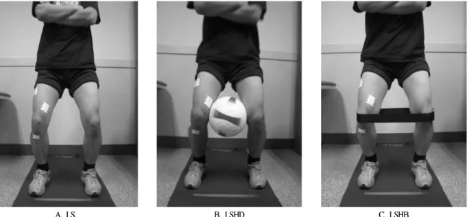
A. LS B. LSHD C. LSHB
Figure 1. Leg Squat Exercise(A), with isometric adduction using a ball(B), and with isometric abduction using a belt(C)
근전도 신호의 표본 추출률 (sampling rate)은 1000 H
Z로 하였고 전극 선의 움직임 신호와 잡다한 생체 신호 잡음을 제거하기 위 한 하한 차단 점 (lower cut-off point)과 전극 부착 부위의 조직 잡 음 (tissue noise)을 제거하기 위한 상한 차단 점(upper cutoff point) 을 이용한 밴드 패스 필터 (band pass filter)를 20-500 H
Z로 사용하 였다 . 그리고 공통 모드 제거(common mode rejection) 능력을 초과 한 측정 환경으로 부터 발생되는 전기적 잡음 (electrical noise)을 제거하기 위해 60 H
Z노치필터 (notch filter)를 사용하였다.
세 가지 운동방법에 따른 근 활성도와 최대 수의적 등척성 수축 (maximal voluntary isometric contraction: MVIC) 시 근 활성 도를 측정하기 위해 근전도 전극 (T246H, BioProtech. Wonju, Korea)을 각각의 근육에 부착하였다. 내측광근의 근전도 두 개의 활성전극 (active electrode)은 슬개골의 상부내측으로 대퇴골의 장 축과 55˚각도를 유지하여 내측으로 4 cm 그리고 상부로 4 cm에 위치하도록 하여 부착하도록 하였으며 외측광근의 근전도 활성 전극은 장축으로부터 15˚를 유지하여 슬개골의 상부 외측에 외 측으로 6-8 cm, 상부로 10 cm 위치하도록 부착하였다. 내․외측광 근의 기준전극 (reference electrode)은 각각 경골 조면(tibial tubercle) 및 비골두 (fibular head)에 부착하였다(Irish et al., 2010).
각 근육에 대한 근 활성도의 표준화 (normalization)를 위해 최대 수의적 등척성 수축 시 근 근 활성도를 도수 근력 검사 (manual muscle testing) 자세에서 세 번 반복 측정하였다(Kendall McCreary, Provance, Rodgers & Romani, 2005). 대상자는 치료용 테이블에 걸 터앉아 최대한 무릎을 신전시킨 후 대상자의 발목 위쪽에 무릎의 굴곡 방향으로 최대 저항을 주고 대상자는 신전 자세를 5초간 유 지하도록 하였다 . 측정 시 대퇴는 회전되지 않도록 대상자에게 지 시하였으며 측정 간에 피로가 발생되지 않도록 30초간의 휴식을 취하도록 하였다 . 세 가지 운동 시 대상자들은 무릎관절이 45˚ 굴 곡된 지점에서 5초간 유지하는 동안 근 활성도를 측정하였다. 세 가지 운동 방법에 따른 각 근육의 근 활성도는 평균평방근 (root mean square: RMS)으로 처리 후 중간 3초간의 각 근육의 근 활성 도를 아래와 같이 백분율 (%MVICRMS)로 환산하였다. 그리고 세 가지 운동방법에 따른 정규화된 근 활성도 값을 가지고 내측광근 과 외측광근의 비 (ratio)를 계산하였다.
%MVICRMS= 운동방법에 따른 평균평방근
× 100 슬관절 신전 최대근수축 시 평균평방근
4. 자료 분석 및 통계 방법
모든 측정값들은 윈도용 SPSS version 14.0 프로그램을 이용 하여 통계 처리하였다 . 세 가지 운동방법에 따른 측정된 내․
외측광근의 근 활성도 그리고 내 ․외측광근 사이의 근 활성도 비 (ratio) 간에 차이가 있는 지 알아보기 위해 반복측정된 일요 인 분산분석 (one-way repeated measured ANOVA)을 사용하였다.
Figure 2. The mean normalized electromyographic (EMG) activity of VMO and VL with standard deviation bars during three exercises. Note. * p <.016, There was a significantly greater VMO activation in comparison with the leg squat with hip abduction ( p =.001), + p <.016, There was a significantly greater VL activation than the leg squat with hip abduction ( p =.001).
VMO: vastus medialis oblique, VL: vastus lateralis, LS: leg squat, LSHD: leg squat with isometric hip adduction, LSHB: leg squat with isometric hip abduction.
유의수준
p<.05에서 통계적 유의성을 검증하였다. 이 검증을 통 해 유의한 차이가 나타난 경우 사후 검증으로 본훼르니 수정 (bonffernii correction)으로 짝-비교 검증을 실시하였다. 유의수준 은 Bonferroni 수정하여 0.016(0.05/3)로 정하였다.
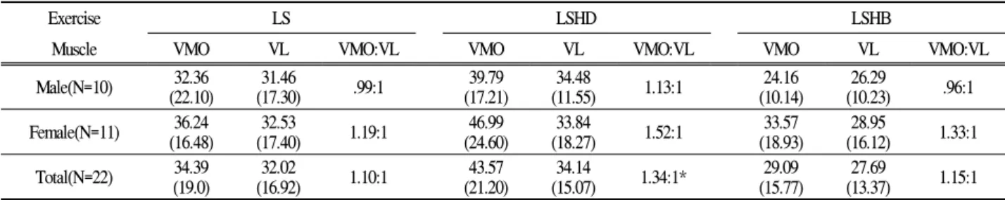
Exercise LS LSHD LSHB
Muscle VMO VL VMO:VL VMO VL VMO:VL VMO VL VMO:VL
Male(N=10) 32.36
(22.10) 31.46
(17.30) .99:1 39.79
(17.21) 34.48
(11.55) 1.13:1 24.16
(10.14) 26.29
(10.23) .96:1 Female(N=11) 36.24
(16.48) 32.53
(17.40) 1.19:1 46.99
(24.60) 33.84
(18.27) 1.52:1 33.57
(18.93) 28.95
(16.12) 1.33:1 Total(N=22) 34.39
(19.0) 32.02
(16.92) 1.10:1 43.57
(21.20) 34.14
(15.07) 1.34:1* 29.09
(15.77) 27.69
(13.37) 1.15:1 Note. Values are mean(SD), No significant difference in VMO:VL ratios between males and females for three exercises( p >.05), *Significantly greater VMO:VL ratio than that of LS( p =.014.). VMO: vastus medialis oblique, VL: vastus lateralis.
Table 1. The electromyographic values(%MVIC) for muscle activity and VMO:VL ratios for the leg squat(LS), the leg squat with hip
adduction(LSHD), and the leg squat exercises with hip abduction(LSHB).
Ⅲ. 결 과
본 연구의 결과 등척성 고관절 내전을 동반한 쪼그려 앉기 운동은 고관절 외전을 동반한 쪼그려 앉기 운동 보다 내・외측 광근의 근 활성도가 통계학적으로 유의하게 증가하였고 (
p<.016)
<Figure 2>, 세 가지 운동방법에 따른 내측광근과 외측광근의 비는 성별과는 유의한 차이가 없었으며 (
p>.05), 등척성 고관절 내전을 동반한 쪼그려 앉기 운동이 쪼그려 앉기 운동 시 보다 통계학적으로 유의하게 증가하였다 (
p<.016)(Table 1).
Ⅳ. 논 의
본 연구는 등척성 고관절 내 ․외전을 동반한 쪼그려 앉기 운동 시 내 ․외측광근의 근 활성도의 변화를 알아보고 무릎관 절의 안정성을 위한 내측광근과 외측광근의 근육 균형 (muscle balance) 정도를 알아보기 위해 근 활성도 비를 비교하였다. 연 구 결과 , 등척성 고관절 내전을 동반한 쪼그려 앉기 운동이 등 척성 고관절 외전을 동반한 쪼그려 앉기 운동보다 내측광근의 근 활성도가 유의하게 높았고 내측광근과 외측광근의 비가 가 장 높았다 .
쪼그려 앉기 운동과 같은 닫힌 힘 연쇄 운동은 대퇴사두근 과 뒤넙다리근 (hamstring)과의 동시수축, 전-후 방향의 경골대퇴 전위 (anterior-posterior tibiofemoral translation)의 감소 그리고 경 골대퇴 관절에서의 압박 힘의 감소 , 그리고 열린 힘 연쇄 운동 과 비교하여 내측광근의 선택적인 수축을 포함한 기능적인 특 징 때문에 슬관절 재활을 위해 널리 수행된다 (Neumann, 2002;
Tang et al., 2001). Gresalmer와 Klein(1998)은 슬개대퇴 통증 증 후군 대상자에게 슬관절 0-45˚ 혹은 0-50˚ 굴곡 자세에서 쪼그 려 앉기 운동을 수행해야 한다고 보고하였다 . 따라서 본 연구 에서는 닫힌 힘 연쇄 운동 중 쪼그려 앉기 운동을 선택하였고 슬관절 각도를 45˚로 정하였다.
등척성 고관절 내전을 동반한 쪼그려 앉기 운동이 가장 내 측광근을 선택적으로 활성화시킨다는 본 연구의 결과와 일치하 는 선행연구들이 있다 . Irish et al.(2010)은 22명의 건강한 20대 를 대상으로 열린 힘 연쇄 운동 (open kinetic chain), 등척성 고관 절 내전을 동반한 45˚ 쪼그려 앉기 운동, 런지운동(lunge)의 세 가지 운동방법에서 내 ․외측광근의 근 활성도를 비교해 본 결 과 고관절 내전을 동반한 쪼그려 앉기 운동 시 내측광근과 외 측광근의 비가 1.18:1로 가장 증가하였으며 런지운동 시 1.14:1 이라고 보고하였고 , 따라서 등척성 고관절 내전과 함께 쪼그려 앉기 운동이 내측광근의 선택적 강화에 효과적이라고 하였다 .
본 연구에서는 등척성 고관절 내전을 동반한 쪼그려 앉기 운동 시 내측광근과 외측광근의 비가 1.34:1로 선행연구에 비해 높았 다 . 선행연구와 본 연구의 내측광근과 외측광근의 비가 차이나 는 이유를 몇 가지로 설명할 수 있다 . 선행 연구에서는 등척성 고관절 내전을 위해 무릎 사이에 베게 (pillow)를 활용하였는데 이는 본 연구에서 저항도구로 공을 사용하여 불안정한 면을 제 공하였기 때문이다 . 많은 연구에서 볼, 즉 불안정한 도구를 활 용하여 닫힌 힘 연쇄 운동 시 근육의 동시수축력이 증가하여 관절의 안정성을 유지한다고 보고하였다 (Andrade, Araújo, Tucci, Martins & Oliveira., 2011; Lehman, MacMillan, MacIntyre, Chivers
& Fluter, 2006; Martins et al., 2008). 또한 운동조절 이론을 활용 한 슬개대퇴 통증 증후군을 대상으로 한 Cowan, Hodges와 Bennell(2001)의 연구에서 슬관절의 안정성을 위협을 받았을 때 내측광근이 외측광근보다 근 수축 개시가 먼저 일어난다고 하 였다 . 본 연구에서는 내·외측광근의 근 수축 개시 시간을 분석 하진 않았지만 슬관절의 안정성에 위협을 주는 불안정한 공을 활용하였을 때 내측광근과 외측광근의 근 활성도 비가 증가하 였음을 증명하였다 .
다른 요인으로는 근육의 길이와 장력에 관계 (length-tension relationship)로 설명할 수 있다. 내측광근은 장내전근과 대내전근 의 건에서 기시하고 전내측 근육사이 막 (antromedial intermuscular septum)과 연결되어 있다(Bose, Kangasuntherum & Osman, 1980).
이러한 해부학적인 사실을 근거로 많은 연구자들은 고관절 내전 근과 슬관절 신전근의 동시 수축은 내측광근을 더욱더 안정된 기시를 제공하여 내측광근의 선택적인 근 활성도를 증가시킨다 고 제안하였다 (Hanten & Schulthies, 1990; Hodges & Richardson, 1993; Karst & Jewett 1993). 그 중 Beck과 Wildermuth(1985)은 등척성 고관절 내전이 내측광근을 신장시킴으로써 길이 -장력의 성질을 변화시켜 내측광근의 수축력을 증가시킨다고 하였다 .
선행 연구에서 슬개골 테이핑 (patellar taping)을 내측광근에 적 용 시 정상인에 비해 슬개대퇴 통증 증후군에서 내측광근의 근 활 성도가 증가되었으며 이러한 근 활성도의 변화되는 기전을 피부 구 심성 (cutaneous affarent) 자극으로 설명하였다(Macgregor, Gerlach, Mellor & Hodges, 2005). 비록 본 연구에서는 테이핑을 적용한 것은 아니지만 실험상 공을 슬개골 중앙에 위치하였고 공의 부 피 때문에 내측측광근에 또한 접촉하였다 . 따라서 공이 내측광 근의 구심성 자극이 입력되어 α -운동신경원의 자극의 증가로 근 활성도가 증가된 것으로 사료된다 .
최근 연구에서는 등척성 고관절 내전을 동반한 쪼그려 앉기 운동이 다른 운동에 비해 선택적으로 내측광근의 근 활성도를 증가시키는지에 대해 논란이 있어 왔다 (Coqueiro et al., 2005;
Earl et al., 2001; Hertel et al., 2004). Zakaria, Harburn과
Kramer(1997)는 대퇴사두근 등척성 운동보다 등척성 고관절 내
전을 동반한 대퇴사두근 등척성운동이 내측광근을 선택적으로 수축하지 못했다고 보고되고 있다 . 본 연구와 달리 선행 연구 는 닫힌 힘 연쇄 운동이 아닌 열린 힘 연쇄 운동을 시행했기 때문이다 . Irishi et al.(2010)은 열린 힘 연쇄에서의 슬관절 신전 운동은 내측광근보다는 외측광근의 활성도를 통계학적으로 유 의하게 증가하여 그 결과 내측광근과 외측광근의 비가 1:1미만 이라고 하였다 . 따라서 열린 힘 연쇄 운동에서의 슬관절 신전 운동은 대퇴슬개 통증 증후군 환자에게 해로운 운동이 될 수 있으며 이러한 운동은 과도한 슬개골의 외측 끌림을 야기시킬 수 있다고 제안하였다 . 닫힌 힘 연쇄 운동에서 실시한 Earl et al.(2001)의 연구에서는 정상인에서 쪼그려 앉기 운동과 등척성 고관절 내전을 동반한 쪼그려 앉기 운동 시 내측광근과 외측광 근의 비가 각각 1.28:1과 1.24:1로 유의한 차이가 없었다. 본 연 구에서는 쪼그려 앉기 운동과 등척성 고관절 내전을 동반한 쪼 그려 앉기 운동 시 내측광근과 외측광근의 비가 각각 1.10:1과 1.34:1로 유의하게 증가하였다. 선행 연구에서는 슬관절에서의 저항도구를 주지 않고 등척성 내전을 했기 때문이라 사료된다 .
본 연구의 제한점으로 대상자를 슬개대퇴 틍증 증후군이 아닌 정상인을 대상으로 했다는 점이다 . 따라서 향후 연구에서는 슬개 대퇴 통증 증후근 환자를 대상으로 공을 이용한 등척성 고관절 내전을 동반한 쪼그려 앉기 운동 효과를 알아볼 필요가 있다 . 다 른 제한점으로는 슬개대퇴 통증 증후군의 특징으로 고관절 외전 근과 외회전근의 약화가 있다고 보고되었다 (Baldon et al., 2009;
Boling et al., 2009). 이러한 운동이 고관절의 주위 근육의 근 활성 도에 미치는 변화에 대한 연구도 선행되어져야 할 것이다 .
Ⅴ. 결 론
본 연구는 공을 이용한 등척성 고관절 내전을 동반한 쪼그 려 앉기 운동과 치료용 벨트를 이용한 등척성 고관절 외전을 동반한 쪼그려 앉기 운동 시 내 ․외측광근의 근 활성도와 내측 광근과 외측광근의 비를 비교하였다 . 본 연구의 결과, 내․외측 광근의 근 활성도는 등척성 고관절 내전을 동반한 쪼그려 앉기 운동 시 등척성 고관절 외전을 동반한 쪼그려 앉기 운동 시에 비해 통계학적으로 유의하게 증가하였다 . 또한 내측광근과 외 측광근의 비는 등척성 고관절 내전을 동반한 쪼그려 앉기 운동 시에 가장 증가하였다 . 그러므로 본 연구에서는 공을 이용한 등척성 고관절 내전을 동반한 쪼그려 앉기 운동이 선택적으로 내측광근의 근 활성도를 증가시킬 수 있다는 것을 보여주었다 . 따라서 슬개대퇴 통증 증후군 환자에게 선택적인 내측광근 강 화운동 방법으로 공을 이용한 등척성 고관절 내전을 동반한 쪼 그려 앉기 운동을 추천하는 바이다 .
참고문헌
Andrade, R., Araújo, R. C., Tucci, H. T., Martins, J., & Oliveira, A. S.(2011). Coactivation of the shoulder and arm muscles during closed kinetic chain exercises on an unstable surface.
Singapore Medical Journal,52(1), 35-41.
Baldon, R. M., Nakagawa, T. H., Muniz, T. B., Amorim, C. F., Maciel, C. D., & Serrão, F. V.(2009). Eccentric hip muscle function in females with and without patellofemoral pain syndrome.
Journal of Athletic Training,44(5), 490-496.
Barton, C. J., Levinger, P., Crossley, K. M., Webster, K. E., &
Menz, H. B.(2011). Relationships between the Foot Posture Index and foot kinematics during gait in individuals with and without patellofemoral pain syndrome.
Journal of Foot and Ankle Research
, 14(4), 10.
Beck, J. L., & Wildermuth, B. P.(1985). The female athlete's knee.
Clinics in Sports Medicine,4(2), 345-366.
Bevilaqua-Grossi, D., Monteiro-Pedro, V., de Vasconcelos, R. A., Arakaki, J. C., & Berzin, F.(2006). The effect of hip abduction on the EMG activity of vastus medialis obliquus, vastus lateralis longus and vastus lateralis obliquus in healthy subjects.
Journal of Neuroengineering and Rehabilitation,3, 13.
Boling, M. C., Padua, D. A., & Alexander, C. R.(2009).
Concentric and eccentric torque of the hip musculature in individuals with and without patellofemoral pain.
Journal of Athletic Training,
44(1), 7-13.
Bose, K., Kangasuntherum, R., & Osman, M.(1980). Vastus medialis oblique and anatomical and physiological study.
Orthopaedics,
3, 880-883.
Coqueiro, K. R., Bevilaqua-Grossi, D., Berzin, F., Soares, A. B., Candolo, C., & Monteiro-Pedro, V.(2005). Analysis on the activation of the VMO and VLL muscles during semisquat exercises with and without hip adduction in individuals with patellofemoral pain syndrome.
Journal of Electromyography and Kinesiology,15, 596-603.
Cowan, S. M., Hodges, P. W., & Bennell, K. L.(2001). Anticipatory activation of vastus lateralis and vastus medialis obliqus occurs simultaneously in voluntary heel and toe raises.
Physical Therapy in Sport
, 71-79.
Cowan, S. M., Bennell, K. L., Crossley, K. M., Hodges, P. W.,
& McConnell, J.(2002). Physical therapy alters recruitment
of the vasti in patellofemoral pain syndrome.
Medicineand Science in Sports and Exercise,
34, 1879-1885.
Cowan, S. M., Hodges, P. W., Bennell, K. L., & Crossley, K.
M.(2002). Altered vasti recruitment when people with patellofemoral pain syndrome complete a postural task.
Archives of Physical Medicine and Rehabilitation,
83, 989-995.
Davlin, C. D., Holcomb, W. R., & Guadagnoli, M. A.(1999). The effect of hip position and electromyographic biofeedback training on the vastus medialis oblique: vastus lateralis ratio.
Journal of Athletic Training,34(4), 342-346.
Dixit, S., DiFiori, J. P., Burton, M., & Mines, B.(2007).
Management of patellofemoral pain syndrome.
American Family Physician,75, 194-202.
Earl, J. E., Schmitz, R. J., & Arnold, B. L.(2001). Activation of the VMO and VL during dynamic mini-squat exercises with and without isometric hip adduction.
Journal of Electromyography and Kinesiology,11, 381-386.
Fox, T. A.(1975). Dysplasia of the quadriceps mechanism:
hypoplasia of the vastus medialis muscle as related to the hypermobile patella syndrome.
Surgical Clinics of North America,55, 199-226.
Garcia, F. R., Azevedo, F. M., Alves, N., Carvalho, A. C., Padovani, C. R., & Negrão, R. F.(2010). Effects of electrical stimulation of vastus medialis obliquus muscle in patients with patellofemoral pain syndrome: an electromyographic analysis.
Revista Brasileira de Fisioterapia,14(6), 477-482.
Grelsamer, R. P., & Klein, J. R.(1998). The biomechanics of the patellofemoral joint.
Journal of Orthopaedic and Sports Physical Therapy,28(5), 286-298.
Hanten, W. P., & Schulthies, S. S.(1990). Exercise effect on electromyographic activity of the vastus medialis oblique and vastus lateralis muscles.
Physical Therapy,70(9), 561-565.
Hertel, J., Earl, J. E., Tsang, K. K., & Miller, S. J.(2004).
Combining isometric knee extension exercises with hip adduction or abduction does not increase quadriceps EMG activity.
British Journal of Sports Medicine,38, 210-213.
Hodges, P. W., & Richardson, C. A.(1993). The influence of isometric hip adduction on quadriceps femoris activity.
Scandinavian Journal of Rehabilitation Medicine,
25, 57-62.
Hubbard, J. K., Sampson, H. W., & Elledge, J. R.(1998). The vastus medialis oblique muscle and its relationship to
patellofemoral joint deterioration in human cadavers.
Journal of Orthopaedic and Sports Physical Therapy,
28, 384-391.
Huberti, H. H., & Hayes, W. C.(1984). Patellofemoral contact pressures. The influence of q-angle and tendofemoral contact.
Journal of Bone and Joint Surgery American,66(5). 715-724
Hudson, Z., & Darthuy E.(2009). Iliotibial band tightness and patellofemoral pain syndrome: a case-control study.
Manual Therapy
, 14(2), 147-151.
Irish, S. E., Millward, A. J., Wride, J., Haas, B. M., & Shum, G.
L.(2010). The effect of closed-kinetic chain exercises and open-kinetic chain exercise on the muscle activity of vastus medialis oblique and vastus lateralis.
Journal of Strength and Conditioning Research,1256-1262.
Karst, G. M., & Jewett, P. D.(1993). Electromyographic analysis of exercises proposed for differential activation of medial and lateral quadriceps femoris muscle components.
Physical Therapy,73, 286-295.
Kendall, F. P., McCreary E. K., Provance, P. G., Rodgers, M.
M., & Romani, W. A.(2005).
Muscles: Testing and Function, with Posture and Pain. Philadelphia, PA:
Lippincott Williams & Wilkins.
Kisner, C., & Colby, L. A.(2010).
Therapeutic Exercise. Philadelphia, PA: The F.A. Davis.
Lam, P. L., & Ng, G. Y.(2001). Activation of the quadriceps muscle during semisquatting with different hip and knee positions in patients with anterior knee pain.
American Journal of Physical Medicine and Rehabilitation,80, 804-808.
Lehman, G. J., MacMillan, B., MacIntyre, I., Chivers, M., &
Fluter, M.(2006). Shoulder muscle EMG activity during push up variations on and off a Swiss ball.
Dynamic Medicine,9, 5-7.
Lieb, F. J., & Perry, J.(1968). Quadriceps function. An anatomical and mechanical study using amputated limbs.
Journal of Bone and Joint Surgery,50, 1535-1548.
Luyckx, T., Didden, K., Vandenneucker, H., Labey, L., Innocenti, B., & Bellemans, J.(2009). Is there a biomechanical explanation for anterior knee pain in patients with patella alta?: influence of patellar height on patellofemoral contact force, contact area and contact pressure.
Journal of Bone and Joint Surgery British,91(3). 344-350.
Macgregor, K., Gerlach, S., Mellor, R., & Hodges, P. W.(2005).
Cutaneous stimulation from patella tape causes a differential increase in vasti muscle activity in people with patellofemoral pain.
Journal of Orthopaedic Research,23(2), 351-358.
Martins, J., Tucci, H. T., Andrade, R., Araújo, R. C., Bevilaqua-Grossi, D., & Oliveira, A. S.(2008). Electromyographic amplitude ratio of serratus anterior and upper trapezius muscles during modified push-ups and bench press exercises.
Journal of Strength and Conditioning Research,
22(2), 477-484.
Neumann, D. A.(2002).
Kinesiology of the Musculoskelektal System.St. Louis, Missouri: Mosby.
Ng, E. C., Chui, M. P., Siu, A. Y., Yam, V. W., & Ng, G.
Y.(2011). Ankle positioning and knee perturbation affect temporal recruitment of the vasti muscles in people with patellofemoral pain.
Physiotherapy,97, 65-70
Pal, S., Draper, C. E., Fredericson, M., Gold, G. E., Delp, S. L., Beaupre, G. S., & Besier, T. F.(2011). Patellar maltracking correlates with vastus medialis activation delay in patellofemoral pain patients.
American Journal of Physical Medicine and Rehabilitation,39(3), 590-598.
Peeler, J., Cooper, J., Porter, M. M., Thliveris, J. A., &
Anderson, J. E.(2005). Structural parameters of the vastus medialis muscle.
Clinical Anatomy,18, 281-289.
Reinold, M. M., Macrina, L. C., Wilk, K. E., Fleisig, G. S., Dun, S., Barrentine, S. W., Ellerbusch, M. T., & Andrews, J.
R.(2007). Electromyographic analysis of the supraspinatus and deltoid muscles during 3 common rehabilitation exercises.
Journal of Athletic Training, 42(4), 464-469.
Seraro, F. V., Cabral, C. M. N., Berzin, F., Candolo, C., &
Monteiro-Pedro, V.(2005). Effect of rotation on the electromyographical activity of the vastus medialis oblique and vastus lateralis longus muscles during isometric leg press.
Physical Therapy in Sport, 6, 15-23.
Tang, S. F., Chen, C. K., Hsu, R., Chou, S. W., Hong, W. H.,
& Lew, H. L.(2001). Vastus medialis obliquus and vastus lateralis activity in open and closed kinetic chain exercises in patients with patellofemoral pain syndrome:
an electromyographic study.
Archives of Physical Medicine and Rehabilitation,82(10), 1441-1445.
Taunton, J. E., Ryan, M. B., Clement, D. B., McKenzie, D. C., Lloyd-Smith, D. R., & Zumbo, B. D.(2002). A retrospective case-control analysis of 2002 running injuries.
British Journal of Sports Medicine,36(2), 95-101.
Tria, A. J., Palumbo, R. C., & Alicea, J. A.(1992). Conservative care for patellofemoral pain.
Orthopedic Clinics of North America,23, 545-554.
Willson, J. D., & Davis, I. S.(2008). Utility of the frontal plane projection angle in females with patellofemoral pain.
Journal of Orthopaedic and Sports Physical Therapy,