뮤코지방증 2형 마우스 모델의 특징과 태반에서 추출한 리소좀 효소 투여의 결과
성균관대학교 의과대학 삼성서울병원 소아청소년과1, 녹십자 연구센터2 관동대학교 의과대학 명지병원 소아청소년과3, 아주대학교 의과대학 의학유전학과4 삼성 생의학연구소 임상연구센터5, 서울대학교 의과대학 분당서울대학교병원 진단검사의학과6
이화여자대학교 과학교육과7, 이화여자대학교 의과대학 소아청소년과8
조성윤1ㆍ김기용2ㆍ김수진3ㆍ손영배4ㆍ맹세현1ㆍ김치화5 고아라5ㆍ송정한6ㆍ여성희7ㆍ김경효8ㆍ진동규1
Characterization of a Mucolipidosis Type II Mouse Model and Therapeutic Implication of Lysosomal Enzyme
Enriched Fraction Derived from Placenta
Sung Yoon Cho
1, Ki-Yong Kim
2, Su Jin Kim
3, Young Bae Sohn
4Se Hyun Maeng
1, Chi Hwa Kim
5, Ah-ra Ko
5, Junghan Song
6Sung-Hee Yeau
7, Kyung-Hyo Kim
8, Dong-Kyu Jin
1Department of Pediatrics1, Samsung Medical Center, Sungkyunkwan University School of Medicine, Seoul
Central Research Center2, Green Cross Corp., Yongin,
Department of Pediatrics3, Kwandong University College of Medicine, Myongji Hospital, Goyang, Department of Medical Genetics4, Ajou University Hospital, Suwon,
Clinical Research Center5, Samsung Biomedical Research Institute, Seoul, Department of Laboratory Medicine6, Seoul National University Bundang Hospital, Seoul
Department of Science Education7, Ewha Womans University, Seoul,
Department of Pediatrics8, Ewha Womans University School of Medicine, Seoul, Korea.
I-cell disease (mucolipidosis type II; MIM 252500) and pseudo-Hurler polydystrophy (mucolipidosis type III; MIM 252600) are disorders caused by abnormal lysosomal transport in cells. The presence of numerous inclusion bodies in the cytoplasm of fibroblasts, a lack of mucopolysacchariduria, increased lysosomal enzyme activity in serum, and decreased GlcNAc-phosphotransferase activity are hallmark. Here, we attempted to investigate phenotypical and biochemical characteristics of the knockoutmouse of GlcNAc-phosphotransferase α/βsubunits; in addition, we also attempted to de- termine whether the lysosome enriched fraction derived from placenta can be beneficial to pheno- type and biochemistry of the knockout mouse.We found that the knockout mouse failed to thrive and had low bone density, as is the case in human. In addition, skin fibroblasts from the animal had the same biochemical characteristics, including increased lysosomal enzyme activity in the cul- ture media, in contrast to the relatively low enzyme activity within the cells. Intravenous injection of the lysosome rich fraction derived from placenta into the tail vein of the animal resulted in a gain of weight, while saline injected animals didn’t.In conclusion, our study demonstrated the phe-
1)
Correspondence: Dong-Kyu Jin, M.D., Ph.D.
Department of Pediatrics, Samsung Medical Center, Sungkyunkwan University School of Medicine, 50 Irwon-dong, Gangnam-gu, Seoul, 135-710, Korea
Tel: +82-2-3410-3525, Fax: +82-2-3410-0043, Email: [email protected]
notypical and biochemical similarities of the knockout mouse to a mucolipidosis type II patient and showed the therapeutic potential of the lysosome enriched fraction. We admit that a larger scale animal study will be needed; however, the disease model and the therapeutic potential of the lysosome enriched fraction will highlight the hope for a novel treatment approach to mucopolipidosis type II, for which no therapeutic modality is available.
Key words: I-cell disease, mucolipidosis type II, GNPTA , knockout mouse, GlcNAc-phosphotransferase
Introduction
I-cell disease (mucolipidosis type II; MIM]
252500) and pseudo-Hurler polydystrophy (mu- colipidosis type III; MIM]252600) are disorders caused by abnormal lysosomal transport in cells of mesenchymal origin. The presence of numerous inclusion bodies in the cytoplasm of fibroblasts, a lack of mucopolysacchariduria, increased lyso- somal enzyme activity in serum, and decreased GlcNAc-phosphotransferase activity are hallmarks of these diseases
1, 2). At the biochemical level, mucolipidosis types II and III are caused by a de- ficiency of UDP-N-acetylglucosamine: lysosomal enzyme N-acetylglucosamine-1-phosphotrans- ferase (GlcNAc-phosphotransferase), which adds an α-N-acetylglucosamine 1-phosphate residue to lysosomal enzymes
1, 3-5).
Trafficking of most lysosomal hydrolases in higher eukaryotes is mediated by a mannose-6- phosphate (M6P)-dependent pathway, in which asparagine-linked oligosaccharides on newly syn- thesized lysosomal hydrolases are specifically and uniquely modified to contain an M6P terminal.
The initial and determining step in biosynthesis of this M6P modification is catalyzed by GlcNAc- phosphotransferase, which catalyzes the transfer of GlcNAc-1-phosphate from UDP-GlcNAc to specific α1,2-linked mannoses on lysosomal hy- drolases
6). The second enzyme in this pathway,
N-acetylglucosamine-1-phosphodiester α-N- acetylglucosaminidase, removes the covering GlcNAc, to generate a terminal M6P. Lysosomal enzymes bearing the M6P modification then bind to one of two M6P receptors in the trans-Golgi apparatus and are transferred to lysosomes. In the absence of lysosomal enzyme targeting to ly- sosomes, the substrates of these enzymes accu- mulate in lysosomes, which results in the appea- rance of inclusion bodies that are responsible for the names inclusion-cell disease and I cell disease.
There are three known complementation types (IIIA, IIIB, and IIIC). The relationship between the responsible gene and mucolipidosis type II and III has been simultaneously reported by both our group
7)andthe other group
8). A mouse model showing the loss of the GlcNAc-1-phosphotrans- ferase gene, which results in decreased phospho- rylation of lysosomal acid hydrolases to undetec- table levels, was reported
9).
In this study, we attempted to investigate the
phenotypical and biochemical characteristics of
the knockout (KO) mouse of GlcNAc-Phospho-
transferaseα/β subunits; in addition, we attempted
to determine whether the lysosome rich fraction
derived from placenta can be beneficial to pheno-
type and biochemistry of the KO mouse.
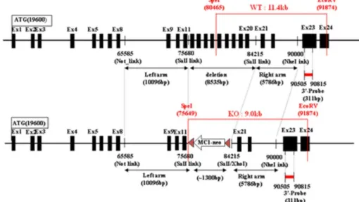
Fig. 1. Design of
GNPTA
knockout vector.Materials and Methods
1. KO mouse modelconstruction of the tar- geting vector (Fig. 1)
Mouse genomic DNA, which was obtained from 129/SvJ mouse J1 embryonic stem cells, was screened by PCR using two-sets of primers for isolation of a 10.1 kb NotI-SalI fragment derived from the GNPTA gene as the 5’ long arm: the forward primer linked NotI (5’gcggccgc_tgaggtac tttcagtactg 3’); the reverse primer linked SalI (5’
gtcgac _agtcactctttagctg 3’) and the 5.8 kb SalI- NheI fragment was derived from the GNPTA gene as the 3’short arm: the forward primer linked SalI (5’gtcgac_agttggatgacatcaga 3’); the reverse pri- mer linked NheI (5’gctagc_ttccttaacaacagttat 3’).
We constructed the targeting vector for deletion of a segment containing a sequence of a region of the GNPTA gene from exon 12 to exon 20 (-8.5 kb), using a 5’10.1 kb long arm fragment and a 3’5.8 kb short arm fragment ligated into the
pOsdupdel vector (6,149 bp). A targeting vector was designed to replace a -8.5 kb genomic frag- ment containing a segment of a region of the GNPTA gene GNPTA knockout mouse from exon 12 to exon GNPTA knockout mouse 20 and a po- sitive selection marker: MC1 promoter and neo- mycin resistance gene. A negative selection marker:
the HSV-1 promoter driven thymidine kinase gene was appended to the construct for selection against non-homologous recombination. The targeting vector was linearized with NotI and electroporated into 129/SvJ mouse J1 embryonic stem cells.
Clones resistant to G418 and gancyclovir were
selected, and homologous recombination was con-
firmed by Southern blotting. The GNPTA gene
was modified in 4 of 336 clones screened. The
four clones containing the targeted mutation were
injected into C57BL_6 blastocysts, which were
subsequently transferred into pseudo pregnant
foster mothers. The resulting male chimeric mice
were bred to C57BL_6 females in order to obtain
heterozygous GNPTA mice. Germ-line transmis-
sion of the mutant allele was verified by Southern
blot analysis of tail DNA from F1 offspring with agouti coat color. Interbreeding of the heterozy- gous mice was performed in order to generate homozygous GNPTA-deficient mice. Offspring generated were wild-type (GNPTA
+/+), homozy- gous GNPTAKO (GNPTA
-/-), and heterozygous (GNPTA
+/-). Mice were maintained under specific pathogen-free conditions at the animal facility of the Samsung Biomedical Research Institute.
2. Preparation of the lysosome enriched fraction
Details of the extraction procedures have been previously described by C. ALQUIER et al
10). We modified the procedure according to our lab. In brief, placental fragments were forced with the bottom of a beaker through a metal sieve (pore size 0.3 mm) and collected in 1 liter of tissue suspension (TS) buffer. The tissue suspension, taken as the homogenate (H) (samples were ho- mogenized for subsequent assays), was centrifuged at 770 g (rav. 12 cm) for 20 min. The supernatant (S1) was eliminated. The resulting pellet was washed by re-suspension in TS buffer, followed by centrifugation at 770 g for 20 min. The pellet, which was re-suspended in 50 mL of TS buffer, represents the 'open follicles' fraction. The open follicles fraction in TS buffer was homogenized in a glass/Teflon Potter-Elvehjem homogenizer with a tight-fitting Teflon pestle, and rotated at 1,500 rev/min with six slow up-and-down strokes.
The resulting material (P1) was centrifuged at 800 g (rav. 11 cm) for 20 min. The supernatant S2 was collected and the pellet P2 was re-sus- pended in 40 mL of TS buffer, followed by centri- fugation under the same conditions. The final P2 pellet was discarded and the two supernatants
S2 (75-80 mL) were pooled and was centrifuged at 4,000 g (rav. 8 cm) for 20 min in order to obtain supernatant S3 and pellet P3. S3 was further centrifuged at 26,000 g (rav 8 cm) for 20 min to give fractions P4 and S4.
3. Lysosomal enzyme activity changes in fibroblast culture media with time
After seeding equal numbers of cells into four culture flasks, the old medium was replaced with fresh medium. Each flask was cultured for 6, 24, 48 and 72hr, and lysosomal enzyme activities in culture medium were determined. Lysosomal en- zyme activities including hexosaminidase, α-N- acetylglucosaminidase, β-galactosidase, β-glu- curonidase, and β-glucosidase in culture media were determined using appropriate fluorogenic substrates and commonly used methods. Enzyme activities were expressed as nmoles of substrate cleaved per mL per hour at 37℃.
Results
1. Characterization of GNPTA KO Mouse
For Southern blot analysis, mouse genomic
DNA obtained from 129/SvJ mouse J1 embryonic
stem cells was screened by PCR using two sets
of primers for isolation of a 311-bp probe region
located just outside the 3’short arm of the KO
vector: the forward primer (5’gaagctagtccagaccg
aatc 3’); the reverse primer (5’agaaaccagcagcttgt
cagg3’). Mouse genomic DNA was isolated from
the tail of the mouse, digested with SpeI and
EcoRV, and hybridized with a 311-bp using this
probe; a 11.4-kb GNPTA wild-type DNA frag-
ment and a 9.0-kb GNPTA mutant DNA fragment
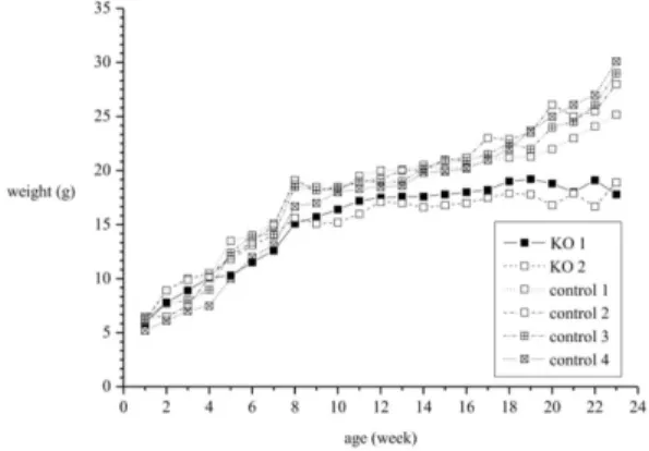
Fig. 2. Unlike wild type mice, knockout mice failed to show progressive weight gain.
Fig. 3. Dual-energy X-ray absorptiometry was performed at 10 weeks for assessment of bone mineral density. (A) DXA of wild type mouse revealed the following results: bone mineral density, 0.0624 g/cm2; bone mineral content, 0.623 g; bone area, 9.98 cm2; lean body mass, 22.5 g; fat mass, 4.6 g; total body mass, 27.2 g; fat percent, 17.1%. (B). DXA of KO mice revealed the following results: bone mineral density, 0.0527 g/cm2; bone mi- neral content, 0.443 g; bone area, 8.40 cm2; lean body mass, 15.1 g; fat mass, 3.2 g; total body mass, 18.2 g; fat percent, 17.3%.
were detected.
2) Weight gain and bone mineral density ofGNTPAKO mice
Although there was no difference in birth weight, after 8 weeks, KO mice failed to show progressive weight gain (Fig. 2). At week 15, average weight of the wild type control mice (n=4) weighed 20 g; however, but that of KO mice (n=2) was 17 g.
This difference of weight was more conspicuous at week 24; weight of wild type mice was between 25-30 gm. However, KO mice still weighed ap-
proximately 18 g. Bone mineral density was as- sessed using DXA (Dual-energy X-ray absorp- tiometry) at 10 weeks. As shown in Fig. 3A and 3B, the mineral content of KO mouse was far lower than that of wild type mouse (bone mineral density; 0.0527 g/cm
2vs. 0.0624 g/cm
2, bone mi- neral content: 0.443 g vs. 0.623 g).
3. Biochemical analysis of skin fibroblasts in GNPTAKO mouse (Fig. 4)
When skin fibroblasts were cultured from KO mouse and several lysosomal enzymes were mea- sured, an increase in the enzyme contents of the culture media was observed, while lysosomal en- zymes inside the cells were relatively lower than those of cultured fibroblasts of wild type animals.
This was illustrated by the increased enzyme ac-
tivities of hexosaminidase (A), N-acetylglucosa-
minidase (B), β-galactosidase (C), and β-glu-
curonidase (D) of KO mouse than those of the
wild type mouse. β-glucosidase(E), which is not
the lysosomal enzyme, did not differ significantly
from that of wild type mouse. With time, the in-
crease of lysosomal enzyme activity was more
Fig. 4. Skin fibroblasts were cultured from the knockout mouse and the several lysosomal enzymes were measured. The enzyme activities of hexosaminidase (A), N-acetylglucosaminidase (B), β-galacto- sidase (C), and β-glucuronidase (D) of knockout mouse increased than those of the wild type mouse. While, β-glucosidase (E), which is not the lysosomal enzyme, did not differ significantly from that of wild type mouse. With time, the increase of the lysosomal enzyme activity was more pronounced, while that of the β-glucosidase was stationary.
Table 1. Enzyme Content Analysis of Placental Ex- tract of P4
Lysosomal enzyme P4 unit
Arylsulfatase A Arylsulfatase B α-Galactosidase β-Galactosidase β-Glucosidase Hexosaminidase
5.25 4.9 0.11
0.1 90.8
6.5
nmol/min/mg protein nmol/min/mg protein nmol/min/mg protein nmol/min/mg protein pmol/min/mg protein nmol/min/mg protein
pronounced, while that of β-glucosidase was sta-
tionary.
4. Composition of lysosomal enzymes in placental extract
Several fractions of the placental extracts were
tested for measurement of lysosomal enzyme con-
tents (Table 1). Of these fractions, fraction No 4
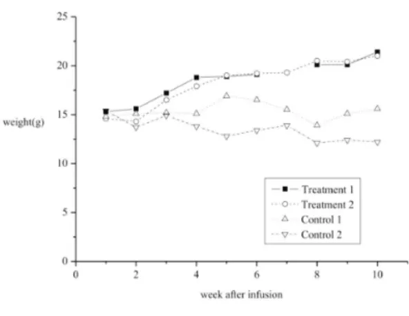
Fig. 5. In vivo efficacy test using fraction of pla- cental extracts (P4 in the method) was performed. A steady gain of weight was observed in knockout mice receiving this fraction of placental extracts, while, the mice receiving saline did not have weight gain at all (n=2 each group).
(Pellet4 suspended in buffer) had the richest con- tent of lysosomal enzymes. Therefore, fraction No 4 was used for the efficacy experiment of the next experiment.
5) In vivo efficacy test using fraction No 4 of placental extracts (Fig. 5)
To demonstrate the efficacy of the placental extract, the No 4 fraction was administered into the tail vein of 6-week old KO mice (n=2) at the dose of 2 mg/kg/dose bi-weekly 4 times, and weight gain of KO mice was measured and com- pared with the weight of KO control mice, which were administered with the same amount of saline (n=2). Remarkably, a steady gain of weight was observed in KO mice receiving fraction No 4 of placental extracts and weight reached almost 20 gm at 10 weeks from the start of placental ad- ministration. However, mice receiving saline did not show any weight gain.
Discussion
In this study, we found that KO mice failed to thrive and had weak bone density, as is the case in human disease of mucolipidosis type II. In ad- dition, skin fibroblasts from the KO mouse had the same biochemical characteristics, including in- creased lysosomal enzyme activity in the culture media, in contrast with the relatively low enzyme activity within the cells. More importantly, for the first time, we demonstrated a gain of weight by intravenous injection of the lysosome rich fraction derived from placenta into the tail vein of the ani- mal, while saline injected animals maintained low body weight.
Phenotypical description of our KO mice is quite similar to that of previously reported animals
9). In the previous report, a comprehensive phenotypic analysis was performed on wild type, heterozy- gous, and homozygous animals. At all ages exa- mined, heterozygous mice were comparable to their wild type counterparts. GNPTA
-/-mice were easily discernible from their control littermates by their small size. Mean body weight and length were sig- nificantly reduced in homozygous animals, along with a reduction in total tissue mass and lean body mass (Fig. 3A, 3B). Therefore, we can say that failure to thrive and poor weight gain are common characteristics of the GNPTA
-/-KO mouse.
Other important characteristics of the disease
are manifested by the biochemical features of
cultured fibroblasts from the animal. Patients with
ML II or -III show significantly elevated levels
of lysosomal enzymes in their sera due to the
inability to synthesize the M6P recognition marker,
which is essential for proper targeting of these
enzymes to lysosomes. This trafficking defect
results in hyper-secretion of enzymes into the blood. Enzyme data from the previous study
9)show that, compared with wild-type mice, GNPTA
-/-mice exhibit 6.7- to 13.9-fold increased levels of lysosomal enzymes, as would be expected if GlcNAc-1-phosphotransferase activity were de- fective in homozygous mice and is consistent with observations in humans. This was also the case in our study. In addition, we can observe the ty- pical inclusion bodies in fibroblasts as well.
The formula for the lysosomal rich fraction has been previously reported
10)and we followed the exact steps of purification as those described in the report. Of particular interest, the lysosomal enzyme activity of the fraction was substantially high enough. Therefore, we proceeded to admi- nister the fraction to the KO mice mouse. After 3 weeks of intravenous injections, we observed a gain of weight, while saline injected animals maintained low body weight.
The beneficial effects of the administered en- zyme enriched fraction suggest that a properly enriched fraction of lysosome derived from pla- centa can be safe as well as effective as a po- tential drug for amelioration of symptoms and failure to thrive in human patients too, although further validation will be needed. However, Ce- redase
Ⓡ(Genzyme) derived from placenta had been used to treat Gaucher disease before Cerezyme
Ⓡ(Genzyme) became available by recombinant CHO technology. Therefore, placenta can be said to be abundant in lysosomal enzymes.
We admit the limitation of our study. Because we had a problem in obtaining human placenta and maintenance of quality control with the crude ex- tract of the placenta, we failed to show a larger scale of the efficacy test. Statistical difference was not evident with data using two mice from
each group. Use of a larger amount of placenta and a refined fraction will provide clearer results, compared with what we have observed.
In conclusion, our study demonstrated the phe- notypical and biochemical similarities of KO mice to human mucolipidosis type II patients and showed the therapeutic potential of the lysosome enriched fraction derived from placenta. We admit that a larger scale animal study will be needed; however, the disease model and the therapeutic potential of the lysosome rich fraction will highlight the hope for a novel treatment approach to mucopolipidosis type II for which no therapeutic modality is avail- able.
Acknowledgements
The study was supported by the Korea Health- care Technology R&D Project; Grant sponsor:
Ministry for Health, Welfare and Family Affairs, Republic of Korea; Grant number: A080588; Grant sponsor: Samsung Biomedical Research Institute;
Grant numbers: C-A9-240-2 Grant sponsor: In- Sung Foundation for Medical Research.
요 약
I 세포 질환(뮤코지방증 2형; MIM 252500)과
pseudo-Hurler polydystrophy (뮤코지방증 3형; MIM
252600)는 세포내 비정상적인 리소좀 관련 운송으로
인해 발병한다. 특징적인 소견으로는 섬유아 세포의 세
포질에 다수의 봉입체, 뮤코다당뇨의 부재, 혈청 내 리
소좀 효소 활성도의 증가, GlcNAc-phosphotransfe-
rase 활성도의 감소를 보인다. 이 연구에서는 GlcNAc-
phosphotransferase 알파/베타 아형에 대한 knockout
마우스의 표현형과 생화학적 특징을 조사하였다. 또한,
태반으로부터 추출한 리소좀 농축 분획을 knockout 마
우스에 투여하였을 때 체중 증가에 대한 효과를 볼 수
있는지에 대해 알아보고자 하였다. knockout 마우스 는 뮤코지방증 2형 환자에서 그렇듯이 정상적인 체중 증가를 보이지 않았고 낮은 골밀도를 보였다. 게다가 knockout 마우스의 피부 섬유 아세포의 배양액에서는 리소좀 효소 활성도가 증가한 반면, 세포 내에서는 리 소좀 효소 활성도가 감소되어 있는 것을 확인할 수 있 었으며 이러한 특징은 뮤코지방증 2형 환자에서 볼 수 있는 특징과 일치한다. knockout 마우스의 꼬리 정맥 내로 태반에서 추출한 리소좀 농축 분획을 투여한 결 과, 체중이 증가하는 것을 확인할 수 있었고, 반면 생 리식염수를 투여한 knockout 마우스의 경우는 체중이 증가하지 않았다. 결론적으로, knockout 마우스의 표 현형과 생화학적 특징이 뮤코지방증 2형 환자와 유사 하다는 것을 확인하였으며, 리소좀 농축 분획의 치료적 가능성을 증명하였다. 더 큰 범위의 동물 실험을 진행 할 필요가 있으나, 이 연구는 질병에 대한 동물 모델을 개발하고 리조솜 농축 분획의 치료적 가능성을 제시하 는 것을 통해 현재까지 치료가 불가능한 뮤코지방증 2 형의 새로운 치료 방법의 가능성을 열었다고 볼 수 있 다.
참 고 문 헌
1) Kornfeld S. Trafficking of lysosomal enzymes in normal and disease states. J Clin Invest 1986;77:
1-6.
2) Maroteaux P, Lamy M. [Hurler's pseudo-polydy- strophy]. Presse Med 1966;74:2889-92.
3) Raas-Rothschild A, Cormier-Daire V, Bao M, Genin E, Salomon R, Brewer K, et al. Molecular basis of
variant pseudo-hurler polydystrophy (mucolipidosis IIIC). J Clin Invest 2000;105:673-81.
4) Sly WS. The missing link in lysosomal enzyme tar- geting. J Clin Invest 2000;105:563-4.
5) Bao M, Elmendorf BJ, Booth JL, Drake RR, Canfield WM. Bovine UDP-N-acetylglucosamine:lysosomal -enzyme N-acetylglucosamine-1-phosphotransferase.
II. Enzymatic characterization and identification of the catalytic subunit. J Biol Chem 1996b;271:
31446-51.
6) Couso R, Lang L, Roberts RM, Kornfeld S. Pho- sphorylation of the oligosaccharide of uteroferrin by UDP-GlcNAc: glycoprotein N-acetylglucosa- mine-1-phosphotransferases from rat liver, Acan- thamoeba castellani, and Dictyostelium discoideum requires alpha 1,2-linked mannose residues. J Biol Chem 1986;261:6326-31.
7) Paik KH, Song SM, Ki CS, Yu HW, Kim JS, Min KH, et al. Identification of mutations in the GNPTA (MGC4170) gene coding for GlcNAc-phosphotrans- ferase alpha/beta subunits in Korean patients with mucolipidosis type II or type IIIA. Hum Mutat 2005;
26:308-14.
8) Tiede S, Storch S, Lubke T, Henrissat B, Bargal R, Raas-Rothschild A, et al. Mucolipidosis II is caused by mutations in GNPTA encoding the alpha/beta GlcNAc-1-phosphotransferase. Nat Med 2005;11:
1109-12.
9) Gelfman CM, Vogel P, Issa TM, Turner CA, Lee WS, Kornfeld S, et al. Mice lacking alpha/beta subunits of GlcNAc-1-phosphotransferase exhibit growth retardation, retinal degeneration, and secre- tory cell lesions. Invest Ophthalmol Vis Sci 2007;
48:5221-8.
10) Alquier C, Guenin P, Munari-Silem Y, Audebet C, Rousset B. Isolation of pig thyroid lysosomes. Bio- chemical and morphological characterization. Bio- chem J 1985;232:529-37.