• 검색 결과가 없습니다.

The Usefulness of Bolus of Radiation Therapy in Patients with Whole Breast Cancer

N/A
N/A
Protected

Academic year: 2021

Share "The Usefulness of Bolus of Radiation Therapy in Patients with Whole Breast Cancer"

Copied!
5
0
0

로드 중.... (전체 텍스트 보기)

전체 글

(1)

◈ Original Article ◈

The Usefulness of Bolus of Radiation Therapy in Patients with Whole Breast Cancer

Jung Whan Min

1

ㆍJin Hyun Son

1

ㆍHoon Hee Park

1

ㆍKyung Rae Dong

2,3

1

Department of Radiological Technology, Shingu University·

2

Department of Radiological Technology, Gwangju Health College University·

3

Department of Nuclear Engineering, Chosun University

1)

Abstract

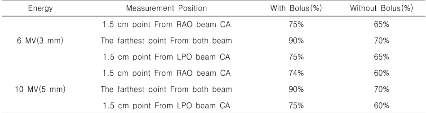
Radiation Therapy has been used in the treatment of breast cancer for over 80 years. Technically, it should include a part or all of such areas as chest wall or breast, axilla, internam mammary nodes and supraclavicular nodes. The purpose of this study is treated breast cancer patient to use 6 MV, 10 MV with bolus so that we observe changing of skin dose and evaluate those usefulness. Using woman's phantom, after CT simulate scanning, Through RTP system to make treatment plan, select three any place. And then, we measure that dose rate. After moving the phantom to linac, we put for TLD to three point same as RTP system which we put on the phantom. We exposed 6 MV, 10 MV with bolus and without so that it is measured dose by TLD device(4000 Harshaw). As a reult expose 6 MV,10 MV, it differences 10%, 15% according to bolus and withoout bolus where lateral point from RAO, LPO beam, other one is 20% where the furthest from both beams. To use bolus in the hospital is material to include closely part at skin among tissue of breast cancer. Acquired skin dose from RTP system is uncertainity.

So it has to test another system likely TLD or other dosimetry system. Also exposed field of breast cancer is included inhomogeneity such as lung, bone and so on. Therefore it has to be accomplished a dose calculating of inhomogeneity part from treatment plan.

Key Words : Radiation therapy, Breast cancer, Bolus, TLD

Ⅰ. Introduction

The total number of breast cancer patients in 1996 was 3,801. In 2006, it increased to 11,275 which is more than 3 times in 10 years. Although

Received May 12, 2011/ 1st Revised May 29, 2011/ 2nd Revised June 14, 2011/ Accepted for Publication July 03, 2011 Corresponding Author: Kyung Rae Dong

Department of Radiological Technology, Gwangju Health College University

(506-701) 683, Shinchang-dong, Gwangsan-gu, Gwangju, Republic of Korea

Tel: 062) 958-7668 Fax: 062) 958-7669 E-mail: [email protected]

there is not an accurate proof of rapid

increasement of breast cancer, there are some

well known causes; obesity by eating too much

westernized high calorie food, decrease of birth

rate and reluctance of breast feeding.

1~3

There are

several breast cancer treatments such as surgical

treatment, chemical treatment, radiation therapy

and etc.

2~6

Bolus is an instrument that is used in

breast cancer radiation treatment. It exposed 6

and 10 MV which increases the surface dose

value. The following experiment will assess the

effectiveness of bolus by measuring the surface

dose value.

(2)

1 4

3 2

Fig. 1. The position of plastic catheter (① Medial : midsternum ② Lateral : midaxillary line

③ Caudal: 1.2 cm below the inframammary line

④ Cephalad : at the base of the clavicle heads)

Fig. 2. 6 MV, with bolus 3 mm, 1.5 cm point from RAO beam CA(Small white arrow), The farthest point from both beam(Big white arrow),

1.5cm point From LPO beam CA(Black arrow)

Fig. 3. 6 MV without Bolus 3 mm Fig. 4. 10 MV with Bolus 5 mm

Ⅱ. Material & Method

1. Material

As experimental subjects, woman phantom(Anthro pomorpic Phantom, Huestis) was used. It executed computerization treatment Computed Tomography (Brilliance Big-Bore, CT-Sim, Philips) in order to acquire image. According to expose high energy (6~10 MV) and existence of bolus and nonexi- stence of bolus, the dose measured through The RTP-system. Linear accelerator was used for exposing phantom(C linac 2100 C, 6 MV, 10 MV X-ray, Varian, USA). Breast board was used to fix the phantom.

2. Computed Tomography scan and treat- ment plan method

Plastic catheter was put into phantom to acquire

CT image. It was to appear outer contours of human body we scanned the phantom using CT.

Measurement position was same as Figure 1.

CT image was sent to RTP system. It made a

treatment plan for using image. For it measured

effect of bolus, We made four occasions. and

then, we measured a dose acquired from treat-

ment plan. There were four occasions. The first

was exposed 6 MV with bolus. The second was

exposed 6 MV without bolus. The third was

exposed 10 MV with bolus. The last was exposed

10 MV without bolus. Measurement position were

1.5 cm point from Right anterior oblique beam

center area, 1.5 cm point from left posterior

oblique beam center area and the furthest point

from both beams(Fig. 2, 3, 4, 5).

(3)

Energy Measurement position With bolus(%) Without bolus(%)

6 MV(3 mm)

1.5 cm point from RAO beam CA 75% 50%

The farthest point from both beams 90% 60%

1.5 cm point from LPO beam CA 75% 60%

10 MV(5 mm)

1.5 cm point from RAO beam CA 75% 50%

The farthest point from both beams 90% 50%

1.5 cm point from LPO beam CA 75% 50%

Table 1. The dose which acquiring from RTP-system Fig. 5. 10 MV, without bolus, 1.5 cm point from RAO beam CA(Small arrow), The farthest point from both beam(Big arrow), 1.5 cm point

from LPO beam CA(Black arrow)

Fig. 6. The experiment which uses the linear accelerator. ① 1.5 cm point from RAO beam CA, ② The farthest point from both beam,

③ 1.5 cm point from LPO beam CA

3. The method of dose evaluation after treatment through Linac

The phantom was moved to treatment room. we put a thermo luminescence dosimeter(TLD) on the phantom. position was same as measurement point which had used RTP system(Fig. 6).

It was exposed four times by linac. The first exposure was 6 MV with bolus. Second one was 6 MV without bolus and third on was 10 MV with bolus. The last was 10 MV without bolus. Through TLD measurement device, we measured dose of TLD which was exposed by Linac. Measurement device was 4000 Harshaw.

Ⅲ. Result

1. The dose evaluation which using RTP system

Analyzing acquired dose distribution from RTP system. The dose of exposure as 6 MV with bolus were 15~30% higher than same energy without bolus. The dose of exposure as 10 MV with bolus were 25~40% higher than same energy without bolus. The result was same as Table 1.

2. The dose evaluation which using TLD

There was no big difference between the result

which measured and the result which measured

from RTP system. Difference of dose that accor-

(4)

Energy Measurement Position With Bolus(%) Without Bolus(%)

6 MV(3 mm)

1.5 cm point From RAO beam CA 75% 65%

The farthest point From both beam 90% 70%

1.5 cm point From LPO beam CA 75% 65%

10 MV(5 mm)

1.5 cm point From RAO beam CA 74% 60%

The farthest point From both beam 90% 70%

1.5 cm point From LPO beam CA 75% 60%

Table 2. The dose acquiring TLD

ding to energy(6, 10 MV), existence of bolus and nonexistence of bolus was 20%. Measurement position is that the furthest point from both beams. Difference of 6 MV exposure were 10%

each, but difference of 10 MV exposure were 14%, 15%. Measurement position were both 1.5 cm point from RAO beam CA and 1.5 cm point from LPO beam CA. The result was same as Table 2.

Ⅳ. Discussion

There was difference of dose between RTP system and TLD. The uncertainty of Surface dose from computerization treatment plan system is high. Therefore it has to test another system likely TLD or other dosimetry system.

7~8

However, the part which is confirmed as TLD is a point as of surface part and thickness of bolus is impossible to measure deeper than this.

Uncertainty of TLD is ± 3%. It will be able to make an error while it verify dose.

9~10

Ⅴ. Conclusion

Use of bolus in treating a breast cancer patient as high energy(6~9 MV) is able to pull a build up depth as far as surface. Therefore, use of bolus can be essential. However, in case of 4 MV exposure does not need a bolus. Because Depth of build up is close to surface. Use of bolus is able to useful as treatment other cancer where close to the surface.

References

1. Lee YH, Park KR, Lee JY, Lee IJ, Vahc YW, Lee KK. Consideration of Surface Dose and Depth of Maximum Dose Using Various Detec- tors for High Energy X-rays. Radiat Oncol J 2003; 21: 322-9.

2. Kweon DC, Lee JS, Goo EH, Kim MJ, Jung JE, Dong KR, et al. An Overall Stem Effect, including Stem Leakage and Stem Scatter, for a TM30013 Farmer-type Chamber. Journal of the Korean Physical Society 2011; 58: 1688-96.

3. Kweon DC, Lee JS, Goo EH, Kim MJ, Jung JE, Chung WK, et al. Comparison of the Measured Stem Leakage Correction Factor for an Ionization Chamber in Air to the Monte Carlo Simulation. Journal of the Korean Physical Society 2011; 58: 1184-94.

4. Lee JS, Kim GH, Jeong KH, Kweon DC, Goo EH, Park CW, et al. Manufacture of Immo- bilization Device (Vac-lok) and the Usefulness of Evaluation for Reproducibility of Patients in the Radiotherapy. J Korea Asso Radiat Prot 2011; 36: 8-15.

5. Kron T, Metcalfe P, Wong T. Thermolumi- nescence dosimetry of therapeutic x-rays with Lif ribons and rods. Phys Med Biol 1993; 38:

833-45.

6. Dong KR, Kweon DC, Chung WK, Goo EH,

Kevin D, Choe JH. Study on the angular

dependence of personal exposure dosimeter (

Focus on thermoluminescent dosimeter and

(5)

photoluminescent dosimeter) Annals of Nuclear Energy 2011; 38: 383-8.

7. Mobit P, Nahum A, Mayles P. A Monte Carlo study of the quality dependence factors of common TLD materials in photon and electron beams. Phys Med Biol 1998; 43: 2015–32.

8. Han EO, Kwon DM, Dong KR, Han SM. A Model for Protective Behavior against the Harmful Effects of Radiation based on Medical Institution Classifications. J Korea Asso Radiat Prot 2010; 35: 157-62.

9. Pradhan AS. Thermoluminescence dosimetry and its applications. Radiat Prot Dosim 1981;

1: 153–67.

10. Jung Y, Dong KR, Kweon DC, Kevin D, Goo

EH, Ahn SY. A Study on the Effects of

Scattering Dose on Eyes and Thyroid for

Panoramagraphy (Focus on TLD and PLD). J

Korea Asso Radiat Prot 2010; 35: 1-5.

수치

Fig.  2.  6  MV,  with  bolus  3  mm,  1.5  cm  point  from  RAO  beam  CA(Small  white  arrow),  The  farthest  point  from  both  beam(Big  white  arrow),
Table  1.  The  dose  which  acquiring  from  RTP-systemFig.  5.  10  MV,  without  bolus,  1.5  cm  point from  RAO  beam  CA(Small  arrow),  The  farthest point  from  both  beam(Big  arrow),  1.5  cm  point
Table  2.  The  dose  acquiring  TLD

참조

관련 문서

• Hormone Therapy With or Without Combination Chemotherapy in Treating Women Who Have Undergone Surgery for Node-Negative Breast Cancer (The TAILORx Trial)..

Specifically, the prevalence and incidence rates of cancer among chronic diseases are continuously increasing, while early diagnosis and treatment of cancer

(Background) The standard triple therapy used as the first-line treatment for Helicobacter pylori infection are a combination of proton pump inhibitor(PPI),

In this study, to investigate how applying a critical pathway to stomach cancer patients affects their recovery and treatment, the clinical effect of the critical

Kameoka, “Sentinel lymph node biopsy for breast cancer patients using fluorescence navigation with indocyanine green,” World Journal of Surgical Oncology, Vol..

verify that USF2 interacts with BRCA1 in HeLa cells, and investigate changing interaction between.. BRCA1 and USF2 after treatment of ionizing radiation (IR), we

To assess the clinical usefulness of performing Q-PCR in practice as a diagnostic technique, we compared blindly the Q-PCR results using blood samples of the

• The kinetic energy of ejected electrons increases linearly with the frequency of the incident radiation but is independent of the intensity of the radiation. • Even at