© 2015 The Korean Ophthalmological Society
This is an Open Access article distributed under the terms of the Creative Commons Attribution Non-Commercial License (http://creativecommons.org/licenses /by-nc/3.0/) which permits unrestricted non-commercial use, distribution, and reproduction in any medium, provided the original work is properly cited.
Original Article
Dacryoscintigraphic Findings in the Children with Tearing
Hyung Chul Kim1, A Ran Cho2, Helen Lew1
1Department of Ophthalmology, Bundang CHA Hospital, CHA University, Seongnam, Korea
2Seoul St. Mary’s Eye Hospital, Suwon, Korea
Purpose: To investigate the diagnostic effectiveness of dacryoscintigraphy in children with tearing; to evaluate tear clearance rate as a diagnostic factor of dacryoscintigraphy in children with tearing; and to analyze the re- sults of treatment according to dacryoscintigraphic findings in children with tearing.
Methods: Between January 2010 and April 2014, 176 eyes of 88 children with tearing (49 boys and 39 girls;
mean age, 23.81 ±14.67 months; range, 12 to 72 months) were studied retrospectively. Of these, 37 of 88 chil- dren with tearing were bilateral cases, and 51 were unilateral cases. None of the patients had a history of cra- niofacial disorder or trauma. The chief complaint of tearing with or without eye discharge and delivery mode, past history of neonatal conjunctivitis, syringing, or probing were collected from parents, grandparents, or pre- vious hospital data. The drainage pattern of the nasolacrimal duct was analyzed, and the clearance rate of 50 μCi 99m technetium pertechnetate was measured by dacryoscintigraphy.
Results: According to the dacryoscintigraphy results, 98 of 125 eyes (78.4%) with tearing showed nasolacrimal obstruction and 29 of 51 eyes (56.9%) without tearing showed patency. There was a significant difference between tearing eyes and normal eyes (p = 0.001). The clearance rate difference after 3 and 30 minutes was 16.41 ± 15.37% in tearing eyes and 23.57 ±14.15% in normal eyes. There was a significant difference between epiphoric eyes and normal eyes (p = 0.05). Based on the dacryoscintigraphic findings, nasolacrimal-duct obstruction was treated with probing or silicone-tube intubation. The majority of patients showed symptom im- provement (75.2%) during the two months of follow-up.
Conclusions: Dacryoscintigraphy is a non-invasive method of qualitatively and quantitatively diagnosing nasolacrimal duct obstruction in children with tearing.
Key Words: Dacryoscintigraphy, Nasolacrimal duct, Obstruction, Tearing
Tearing is a common symptom encountered in ophthal- mology and is usually due to an obstruction of the lacrimal
excretion system [1]. There are a variety of causes of tear- ing within the pediatric population: central nervous sys- tem-related hypersecretion, trichiasis, epiblepharon, kera- toconjunctivitis and other ocular inflammation, functional epiphora due to nasolacrimal duct stenosis or ostium par- tially blocked by hypertrophic or laterally-inclined inferior turbinate, congenital nasolacrimal duct obstruction, or congenital atresia [2-4].
For cases of congenital nasolacrimal duct obstruction, spontaneous resolution is rapid during the first month of
Received: July 14, 2014 Accepted: August 18, 2014
Corresponding Author: Helen Lew, MD. Department of Ophthalmology, Bundang CHA Hospital, CHA University, #59 Yatap-ro, Bundang-gu, Seongnam 463-712, Korea. Tel: 82-31-780-5330, Fax: 82-31-780-5333, E-mail: [email protected]
This study was presented at the 110th meeting of Korean Ophthalmology Society at Goyang, Korea.
life, and 96% resolve in the first year without intervention.
In addition, probing and syringing the nasolacrimal duct at age 12 to 14 months was effective compared with sponta- neous resolution at 15 months. It is preferable to wait until at least age 10 to 12 months before probing unless there is a congenital dacryocele or acute dacryocystitis [2-4].
Diagnostic methods for tearing are canaliculus irrigation test, probing, ultrasound and dacryoscintigraphy. A cana- liculus irrigation test for children older than 12 months is sometimes risky because of possible drainage to the cana- liculus or periocular tissue and can be stressful for children even when using local anesthesia in the clinic. Dacryoscin- tigraphy has some advantages, including a lower dose of radiation, better demonstration of the lacrimal apparatus physiology, and increased safety compared to dacryocys- tography [5].
Patients who complain of epiphora without tear overpro- duction, but show easy passage on syringing are said to have functional lacrimal duct obstruction. This delayed or absent excretion of tears without anatomical obstruction of the lacrimal system has been shown to be caused by steno- sis of the lacrimal system, aberrant punctum location, blocked flow through the punctum, or dysfunction of the lacrimal excretion pump [5-10]. Dacryoscintigraphy is an effective method of diagnosing functional lacrimal duct obstruction. The goal of this study was to investigate the diagnostic effectiveness of dacryoscintigraphy in children with tearing; to evaluate tear clearance rate as a diagnostic factor of dacryoscintigraphy in children with tearing; and to analyze treatment according to dacryoscintigraphic findings in children with tearing.
Materials and Methods
Between January 2010 and April 2014, 176 eyes of 88 children with tearing (49 boys and 39 girls; mean age, 23.81
± 14.67 months; range, 12 to 72 months) were studied ret- rospectively. None of the patients had a history of cranio- facial disorder or trauma. The chief complaint of tearing with or without eye discharge, delivery mode, past history of neonatal conjunctivitis, syringing, or probing were col- lected from parents, grandparents, or previous hospital data. Thirty-seven of 88 children with tearing were bilater- al cases, and 51 were unilateral cases. Nasolacrimal duct drainage was analyzed, and the clearance rate of 50 μCi
99m technetium pertechnetate was measured by da- cryoscintigraphy using Symbia Intevo (Siemens Health- care, Erlangen, Germany) nuclear imaging system (Fig. 1).
Dacryoscintigraphy was performed in all patients. For pa- tient requiring sedation, 10% chloral hydrate (0.8 mL/kg) was used. Patients were supported by adults in order to maintain a standing position in front of the gamma cam- era. After instillation of two drops of 50 μCi 99m techne- tium pertechnetate in the lateral portion of each eyeball, we recorded bilateral eyeball images and determined clear- ance rate at the nasal cavity at 3, 5, 7, 10, 15, 20, and 30 minutes after instillation (Fig. 2).
Based on the dacryoscintigraphic findings, nasolacrimal duct obstruction with tearing was treated using probing or silicone tube intubation. Children with tearing from other causes were observed. Statistical analysis was carried out using the PASW Statistics ver. 18.0 (SPSS Inc., Chicago, IL, USA). Fisher’s exact test, the Mann-Whitney U-test, and Kruskal-Wallis test were used for analyses. All p-val- ues are reported as corrected p-values, and p < 0.05 was considered to indicate statistical significance.
Results
Of the 176 eyes of children with tearing, 51 were normal eyes and 125 were tearing eyes. Sex distribution in normal eyes was 28 boys and 23 girls, and that in tearing eyes was 70 boys and 55 girls. There were no significant differences in sex distribution (p = 0.48), mean age (p = 0.81), or age distribution (p = 0.69) (Table 1).
According to the results of dacryoscintigraphy, 98 of 125 eyes with tearing (78.4%) showed nasolacrimal duct ob- struction and 29 of 51 eyes without tearing (56.9%) showed obstruction. There was a significant difference in duct ob- struction between tearing eyes and normal eyes (p = 0.001).
In addition, 27 of 125 eyes with tearing (21.6%) showed pa- tency, and 22 of 51 eyes without tearing (43.1%) showed patency, a significant difference (p = 0.001). The sensitivi-
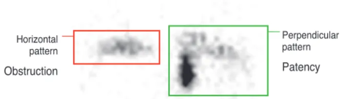
Perpendicular pattern Patency Horizontal
pattern Obstruction
Fig. 1. Patency pattern of dacryoscintigraphic findings.
60 50 40 30 20 10
00 1 2 3 4 5 6 7 8 9 10 11 12 13 14 15 16 17 18 19 20 21 22 23 24 25 Time (min)
Counts/sec
ty of dacryoscintigraphy was 78.4% and the specificity was 43.1% in our study (Table 2).
The clearance rate after 3 minutes was 14.21 ± 13.86% in tearing eyes and 12.68 ± 11.98% in normal eyes. The clear- ance rate after 30 minutes was 30.56 ± 18.31% in tearing eyes and 35.02 ± 18.31% in normal eyes. There were no significant differences in the clearance rate after 3 minutes or 30 minutes. The difference between clearance rate after 3 minutes and 30 minutes was 16.41 ± 15.37% in tearing eyes and 23.57 ± 14.15% in normal eyes, a significant dif- ference between (p = 0.05) (Fig. 3).
Of the 125 tearing eyes, 27 eyes showed patency and 98 eyes showed obstruction. Seventeen tearing eyes with pat- ent dacryoscintigraphic findings were observed, and 13 of 17 had improved tearing two months after dacryoscintigra- phy. In the dacryoscintigraphic findings of four tearing eyes with patency, differences between clearance rate after 3 and 30 minutes were less than 30%. Therefore, four tear- ing eyes with a patent pattern were treated with probing, of which three were improved. Five eyes showing delayed clearance rcompared with the contralateral side received silicone-tube intubation. Of these, four improved. Three eyes with epiblepharon received correction operation, all of which improved (Table 3).
Regarding the obstructed eyes, 29 were observed be- cause there were no symptoms of tearing or there was only slight tearing; therefore, the patients’ parents didn’t want their children to be treated with procedures. In 29 ob- structed eyes treated with observation, six were absent of tearing and discharge more than two months after da- cryoscintigraphy, and 13 were treated with probing, all of whom were improved.
Fig. 2. (A) Clearance rate measured in the nasal cavity area after 3, 5, 7, 10, 15, 20, and 30 minutes. (B) Time activity curve.
60 50 40 30 20 10
00 1 2 3 4 5 6 7 8 9 10 11 12 13 14 15 16 17 18 19 20 21 22 23 24 25 Time (min)
Counts/sec
Table 1. Demographic characteristics of children with tearing Characteristics Normal eyes
(n = 51) Tearing eyes
(n = 125) p-value
Male : female 28 : 23 70 : 55 0.48*
Age (mon) ≤24 35 87 0.69*
>24 16 38 0.69* Total 24.15 ± 15.07 23.70 ± 14.28 0.81† Values are presented as number or mean ± SD.
*Fisher exact test; †Mann-Whitney U-test.
Table 2. Dacryoscintigraphic findings in children with tearing Findings (%) Normal eyes
(n = 51) Tearing eyes
(n = 125) p-value* Obstruction† 29 (56.9) 98 (78.4) 0.001
Patent 22 (43.1) 27 (21.6) 0.001
Total 51 (100) 125 (100)
*Fisher exact test; †Obstruction was defined as the absence of per- pendicular pattern in dacryoscinitigraphy.
Table 3. Clinical outcomes of children with tearing after treatment Success* / cases (%) Tearing eyes (n=125)
Patent
(n=27) Obstruction (n=98) Observation 13 / 17 (76.5) 6 / 29 (20.7) Probing 3 / 4 (75.0) 13 / 13 (100.0) Silicone tube intubation 4 / 5† (80.0) 50 / 52 (96.2) Correction of epiblepharon 3 / 3 (100.0) 8 / 8 (100.0)
Total 21 / 27 (77.8) 73 / 98 (74.5)
*Success: absence of tearing and discharge more than 2 months after treatment; †Delayed clearance rate compared with the con- tralateral side.
A B
3 min 2.37 19.86
5 min 39.43 69.89
7 min 52.14 69.86
10 min 60.68 64.22
15 min 68.57 74.97
20 min 74.75 76.16
30 min 76.87 79.12
Of the 52 eyes treated with silicone-tube intubation, 50 were improved. All of the eight eyes with epiblepharon re- ceived a corrective operation, and all improved. The total success rate of patent eyes was 77.8%, comparted with the total success rate of obstructed eyes at 74.5%. The flow-sheet used to manage children with tearing is shown in Fig. 4.
Discussion
Dacryocystography is the traditional radiological inves- tigation for epiphora. However, like a canaliculus irrigation test, it is an invasive procedure and requires a high dosage of radiation be applied to children [11]. Dacryoscintigraphy
has a more physiologic method of illustrating the lacrimal apparatus and is safer than dacryocystography [1,12-18]. A total of 52 eyes in 43 children with an obstructed pattern and five eyes in four children with a delayed-clearance pattern underwent silicone-tube intubation, and 54 eyes experienced improved symptoms (94.7%).
In tearing eyes with nasolacrimal duct obstruction eval- uated by dacryoscintigraphy, most obstructions were at the level of the proximal nasolacrimal duct or distal nasolacri- mal duct. In tearing eyes with nasolacrimal duct patency evaluated by dacryoscintigraphy, the majority of cases were believed to be conjunctivitis. Empirically, patients with tearing eyes and a patent nasolacrimal duct on da- cryoscintigraphy had conjunctival injection and follicles or papillas on the conjunctiva seen on slit-lamp examination or gross inspection. Tearing symptoms were mostly im- proved through treatment with antibiotics or anti-inflam- matory eye drops. This suggests that the major cause of tearing eyes with patent nasolacrimal duct was conjuncti- vitis. Others potential causes were functional nasolacrimal obstruction or epiblepharon, which were treated with prob- ing, silicone-tube intubation, or correction of epiblepharon.
In congenital nasolacrimal duct obstruction, the success rate of probing was 75.0% in children aged 6 to 15 months [19] and 50% in children aged 7 and 30 months [20]. The success rate of silicone-tube intubation [21-23] was 89% in children between 12 to 48 months [24] and 83.33% in chil- dren between 6 to 30 months [25]. In this study, the success rate of probing was 75.0%, silicone-tube intubation was 80.0% and epiblepharon correction was 100.0% in the tear- ing eyes with nasolacrimal duct patency by dacryoscintigra- phy. The success rates of probing and silicone-tube intuba- tion in this study were greater than or equal to the success rates of previous studies. In the previous studies, the probing or silicone-tube intubation was performed according to tear- ing symptoms. Therefore, tearing eyes with patent nasolac- rimal duct could be also regarded as cases successfully treated by probing or silicone-tube intubation. The age range of these patients was 12 to 72 months in this study, and the outcome of probing or silicone-tube intubation in this study was better than those of previously published reports with younger patients (age range, 6 to 48 months) [19-25]. In nor- mal eyes with nasolacrimal duct obstruction by dacryoscin- tigraphy, the causes were suggested to be a false-positive re- sult of dacryoscintigraphy or dry eyes with functional nasolacrimal duct obstruction.
Fig. 3. Tear clearance rate 3 minutes and 30 minutes after instil- lation according to dacryoscinitigraphy. *p = 0.05.
60 50 40 30 20 10
00 Clearance after 3 min
%
12.68 ± 11.9814.21 ± 13.86
35.02 ± 18.31 30.56 ± 18.31
23.57 ± 14.15 16.41 ± 15.37*
Clearance after 30 min Clearance difference (30 min - 3 min) -10
Normal eyes Epiphora eyes
Fig. 4. Flow-sheet for the management in children of tearing in children older than one year. *30 min - 3 min.
Tearing
Physical examination (-)
30%↓ 30%↑
(+)
Symptom(+) Symptom(-)
Obstruction
Obstruction with
Patency
·Conjunctivitis
·Epiblepharon
Probing History(-) Probing
History(+)
Dacryoscintigraphy
Silicon tube
intubation Medications
*Clearance rate Difference <30%
Probing Epiblepharon
correction Relative difference of clearance rate compared with the contralateral side
Dacryoscintigraphy is a non-invasive modality available to children. According to the study by Heyman et al. [26], the absorbed radiation dose to the lens of the eye in da- cryoscintigraphy (estimated to be 4-14m rads/100 μCi of technetium pertechnetate) is considerably less than that of a skull X-ray (360m rads) or dacryocystography (3,000m rads). This procedure is a qualitative diagnosing method with 78.4% sensitivity and 43.1% specificity. In addition, it can be a quantitative diagnosing method by measuring the technetium-pertechnetate clearance rate difference after 3 and 30 minutes. The clearance rate after 3 minutes was low- er in normal eyes than tearing eyes, but the clearance rate after 30 minutes in normal eyes was similar to the clearance rate in tearing eyes. Differences in the clearance rate be- tween 3 and 30 minutes were significantly less in tearing eyes than normal eyes. We found that tear clearance from the nasal cavity requires at least 3 minutes. This is the first report about dacryoscintigraphic clearance rates in children with tearing.
This study has the limitation of being a retrospective study. The results of this work lay the foundation for further prospective study, which should be performed to confirm the usefulness of dacryoscintigraphy in children with tear- ing. In addition, this study was limited by measuring clear- ance only at the nasal cavity, as 50 μCi 99m technetium pertechnetate drained from the interpalpebral fissure into the nasal cavity. Measuring both the interpalpebral fissure and the nasal cavity can be useful in diagnosing functional nasolacrimal duct obstruction.
In conclusion, dacryoscintigraphy is a non-invasive me- hod of qualitatively and quantitatively diagnosing nasolacri- mal duct obstruction in children with tearing.
Conflict of Interest
No potential conflict of interest relevant to this article was reported.
References
1. Chung YA, Yoo IR, Oum JS, et al. The clinical value of da- cryoscintigraphy in the selection of surgical approach for patients with functional lacrimal duct obstruction. Ann Nucl Med 2005;19:479-83.
2. Schnall BM. Pediatric nasolacrimal duct obstruction. Curr Opin Ophthalmol 2013;24:421-4.
3. Lorena SH, Silva JA, Scarpi MJ. Congenital nasolacrimal duct obstruction in premature children. J Pediatr Ophthal- mol Strabismus 2013;50:239-44.
4. Olitsky SE. Update on congenital nasolacrimal duct ob- struction. Int Ophthalmol Clin 2014;54:1-7.
5. Rossomondo RM, Carlton WH, Trueblood JH, Thomas RP.
A new method of evaluating lacrimal drainage. Arch Oph- thalmol 1972;88:523-5.
6. Montanara A, Catalino P, Gualdi M. Improved radiological technique for evaluating the lacrimal pathways with spe- cial emphasis on functional disorders. Acta Ophthalmol 1979;57:547-63.
7. Wearne MJ, Pitts J, Frank J, Rose GE. Comparison of da- cryocystography and lacrimal scintigraphy in the diagnosis of functional nasolacrimal duct obstruction. Br J Ophthal- mol 1999;83:1032-5.
8. Conway ST. Evaluation and management of “functional”
nasolacrimal blockage: results of a survey of the American Society of Ophthalmic Plastic and Reconstructive surgery.
Ophthal Plast Reconstr Surg 1994;10:185-7.
9. Rosenstock T, Hurwitz JJ. Functional obstruction of the lac- rimal drainage passages. Can J Ophthalmol 1982;17:249-55.
10. Chung WS, Park NG. Functional obstruction of the lacri- mal draings system. J Korean Ophthalmol Soc 1995;36:
1435-8.
11. Shin CH, Woo KI, Chang HR. Evaluation of the functional nasolacrimal duct obstruction with digital subtraction da- cryocystography. J Korean Ophthalmol Soc 2003;44:529-33.
12. Chavis RM, Welham RA, Maisey MN. Quantitative lacri- mal scintillography. Arch Ophthalmol 1978;96:2066-8.
13. Amanat LA, Hilditch TE, Kwok CS. Lacrimal scintigra- phy. II. Its role in the diagnosis of epiphora. Br J Ophthal- mol 1983;67:720-8.
14. Hanna IT, MacEwen CJ, Kennedy N. Lacrimal scintigra- phy in the diagnosis of epiphora. Nucl Med Commun 1992;13:416-20.
15. Ziccardi VB, Charron M, Ochs MW, Braun TW. Nuclear dacryoscintigraphy: its role in oral and maxillofacial sur- gery. Oral Surg Oral Med Oral Pathol Oral Radiol Endod 1995;80:645-9.
16. Sahlin S, Chen E. Gravity, blink rate, and lacrimal drain- age capacity. Am J Ophthalmol 1997;124:758-64.
17. Gencoglu EA, Dursun D, Akova YA, et al. Tear clearance measurement in patients with dry eye syndrome using
quantitative lacrimal scintigraphy. Ann Nucl Med 2005;19:581-7.
18. Park DI, Shin HM, Lee SY, Lew H. Tear production and drainage after botulinum toxin A injection in patients with essential blepharospasm. Acta Ophthalmol 2013;91:e108-12.
19. Miller AM, Chandler DL, Repka MX, et al. Office probing for treatment of nasolacrimal duct obstruction in infants. J AAPOS 2014;18:26-30.
20. Alanon-Fernandez MA, Alanon-Fernandez FJ, Marti- nez-Fernandez A, et al. Comparative study of primary in- tention lacrimal probing with and without nasal endoscopy.
Acta Otorrinolaringol Esp 2014;65:297-301.
21. Angrist RC, Dortzbach RK. Silicone intubation for partial and total nasolacrimal duct obstruction in adults. Ophthal
Plast Reconstr Surg 1985;1:51-4.
22. Katowitz J, Hollsten D, Linberg J. Silicone intubation of the nasolacrimal drainage system. Lacrimal Surg 1988;109-23.
23. Huh D, Son MG, Kim YD. Silicone intubation for func- tional nasolacrimal duct obstruction. J Korean Ophthalmol Soc 2000;41:2303-7.
24. Memon MN, Siddiqui SN, Arshad M, Altaf S. Nasolacri- mal duct obstruction in children: outcome of primary intu- bation. J Pak Med Assoc 2012;62:1329-32.
25. Welsh MG, Katowitz JA. Timing of Silastic tubing removal after intubation for congenital nasolacrimal duct obstruc- tion. Ophthal Plast Reconstr Surg 1989;5:43-8.
26. Heyman S, Katowitz JA, Smoger B. Dacryoscintigraphy in children. Ophthalmic Surg 1985;16:703-9.