PERSISTENT CHEMICAL PERITONITIS RESULTING FROM SPONTANEOUS RUPTURE OF AN OVARIAN MATURE CYSTIC TERATOMA
Hyun Sung Yang, MD
1, Tae Hun Song, MD
1, Hyun Chul Bang, MD
1, Jun-Ho Park, MD
1, Chae Hyeong Lee, MD
1, Ju-Won Roh, MD, PhD
1, Eo-Jin Kim, MD, PhD
2, Yong Seok Lee, MD, PhD
3, Seung-Su Han, MD
4Departments of 1Obstetrics and Gynecology, 2Pathology, 3Radiology, Dongguk University College of Medicine; 4Department of Obstetrics and Gynecology, Chung-Ang University College of Medicine, Seoul, Korea
Spontaneous rupture of mature cystic teratoma occurs rarely, but may lead to a chemical peritonitis. Once rupture of mature cystic teratoma is diagnosed, immediate surgical intervention is necessary. Removal of ruptured ovarian cystic teratoma and copious lavage of abdominal cavity are usually suffi cient to prevent prolonged chemical peritonitis. We report here a rare case of spontaneously ruptured ovarian cystic teratoma diagnosed by computed tomography scan obtained before and after the rupture, and in which chemical peritonitis lasted over 2 months after surgery.
Keywords:
Mature cystic teratoma; Spontaneous rupture; Chemical peritonitis
Received: 2011. 5.30. Revised: 2011. 8. 9. Accepted: 2011. 9. 8.
Corresponding author: Chae Hyeong Lee, MD
Department of Obstetrics and Gynecology, Dongguk University Ilsan Hospital, Dongguk University College of Medicine, 814 Siksa- dong, Goyang 410-773, Korea
Tel: +82-31-961-7368 Fax: +82-31-961-7155 E-mail: [email protected]
Th is is an Open Access article distributed under the terms of the Creative Commons Attribution Non-Commercial License (http://creativecommons.org/licenses/
by-nc/3.0/) which permits unrestricted non-commercial use, distribution, and reproduction in any medium, provided the original work is properly cited.
Copyright © 2011. Korean Society of Obstetrics and Gynecology
Mature cystic teratomas are the most common ovarian tumors, accounting for 20% of adult ovarian tumors and 50% of pediatric ovarian tumors. The most frequent complication of an ovarian mature cystic teratoma is torsion, but rupture occurs rarely with an estimated incidence of 0.3-2.5% [1,2]. Its spontaneous or iat- rogenic intraperitoneal rupture may lead to a chemical peritonitis.
We report a case of intraperitoneal rupture of an ovarian mature cystic teratoma, diagnosed by repeated computed tomography (CT) at short intervals, and resultant chemical peritonitis which persisted for over 2 months after surgery.
Case Report
A 50-year-old woman, gravida 3, para 2, presented to our emer- gency room with several hours history of abdominal pain and nausea. On physical examination, she was afebrile and vital signs were stable. Abdominal examination revealed mild tenderness in lower abdomen. No obvious muscle guarding or rebound tender- ness was noted. Total white blood cell count was 12,980/mm
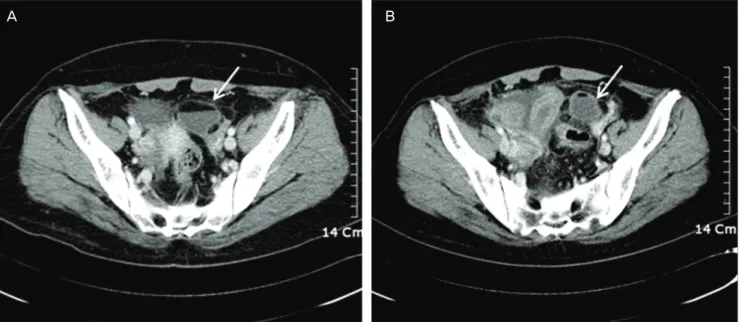
3and C-reactive protein (CRP) was 12.9 mg/dL. Abdominal CT scan showed cystic mass measuring 4.2 cm in maximal diameter with fat-fl uid level in left adnexa, suggestive of a cystic teratoma,
and small amount of ascites in pelvic cavity (Fig. 1A). No other abnormalities suspicious of torsion or rupture of a cystic teratoma were found. Diagnostic laparoscopy had been recommended for further evaluation, but she signed herself out of the hospital against medical advice for personal reasons. Two days later, she readmitted to the emergency room with progressively worsen- ing abdominal pain. Physical examination revealed a distended abdomen with marked tenderness and rebound tenderness in the lower abdomen. The body temperature was elevated to 38.8
oC.
The serum CRP rose to 33.4 mg/dL. Serum CA 19-9 was 75.6 U/
http://dx.doi.org/10.5468/KJOG.2011.54.11.726 pISSN 2233-5188 · eISSN 2233-5196
mL (normal value 0-27 U/mL) and CA-125 was 39.7 U/mL (normal value 0–35 U/mL). A repeated CT scan of the abdomen and pelvis demonstrated a left ovarian cystic teratoma measuring 3 cm in maximal diameter surrounded by fat globules (Fig. 1B). The size of an ovarian cyst and the amount of fat component within the cyst were decreased compared with previous CT scan. Thickening of peritoneum, diffuse wall thickening of small bowels, and increased amount of ascites were also noted. Based on CT fi ndings as well as the clinical symptoms, a diagnosis of ruptured ovarian cystic
teratoma and resultant chemical peritonitis was made.
Single-port laparoscopic surgery using OCTO Port (Dalim SurgNet, Seoul, Korea) was performed. Operative fi ndings revealed about 1,500 mL of yellowish ascites containing adipose component. The peritoneum, uterus, and bowel surfaces were covered by diffuse, thick, white to yellowish plaque-like lesion (Fig. 2). Multiple fl imsy adhesions were present between the omentum and bowel loops.
The left ovarian cyst was adherent to the pelvic sidewall. Left oo- phorectomy was performed and meticulous peritoneal irrigation with 18 L of warm saline was carried out until no more particles of fatty tissue could be detected in the solution. Pathologic examina- tion confi rmed the diagnosis of mature cystic teratoma. The ascitic fl uid cytology was negative for malignant cells. During the periop- erative period, she received intravenous cefotaxime 2 g once daily and metronidazole 500 mg every 8 hours.
After an uneventful immediate postoperative course, the patient complained of abdominal pain, fever, nausea, and diarrhea from the postoperative day 4. With a diagnosis of antibiotic-induced pseudomembranous colitis, oral metronidazole was started. How- ever, stool enzyme immunoassays for Clostridium difficile toxins were negative and pseudomembranes were not visualized by colo- noscopy. Blood cultures for aerobic and anaerobic bacteria did not produce any growth and stool culture demonstrated only normal flora. A CT scan revealed large amount of ascites in abdominal cavity. Ascites were drained by percutaneous placement of a pig- tail catheter and sent for analysis. Examination of the ascitic fl uid
Fig. 1. (A) Computed tomography (CT) scan shows a left ovarian mass (arrow) with fat-fl uid level with small amount of ascites. (B) CT scan after rup- ture shows a left ovarian mass (arrow) surrounded by fat globules, decreased amount of fat component within the cyst, thickening of peritoneum, dif- fuse wall thickening of small bowels, and increased amount of ascites.A B
Fig. 2. Intraoperative laparoscopic fi nding shows left ovarian cyst. The peritoneum, uterus, and bowel surfaces are covered by diffuse, thick, white to yellowish plaque-like lesion.
showed a white blood cell count of 1,600/μL (52% lymphocytes and 25% polymorphonuclear leukocytes) and was negative for acid-fast bacilli and gram stain. A culture of the fl uid for bacteria was sterile. The adenosine deaminase level was 30 IU/L and poly- merase chain reaction for M. tuberculosis was negative. The fever over 38oC continued and the severity of abdominal pain, nausea, vomiting, and diarrhea increased. The CRP level, which had been gradually decreased to 12.5 mg/dL on postoperative day 7, in- creased to 19.7 mg/dL on postoperative day 9.
Persistent chemical peritonitis was suspected and, following ex- tensive discussion and journal search, prednisolone at a dose of 40 mg orally daily was initiated from postoperative day 9. After administration of prednisolone, all of her symptoms gradually im- proved. She was discharged on postoperative day 16. However, 5 weeks later when the dosage of prednisolone had been tapered to 10 mg per day, the symptoms and signs of peritonitis redeveloped.
Therefore, prednisolone 15 mg per day was administered for an- other one month and then tapered off. CRP level was normalized.
At present, one year after the surgery, the patient is under follow- up at the out-patient clinic, and is doing well, with no symptoms or signs of chemical peritonitis.
Discussion
Mature cystic teratoma of the ovary is the most common ovarian neoplasm, accounting for approximately 5-25% of all ovarian tumors [2]. It presents most commonly during the reproductive years and is bilateral in 8-15% of cases. Most of the patients with teratoma are asymptomatic, and the adnexal mass is usually discovered on a routine ultrasonographic examination or with cal- cifi cations in the routine abdominal X-ray image. Complications of mature cystic teratoma of the ovary are torsion (16%), malignant degeneration (2%), rupture (1-2%), and infection (1%) [3].
Spontaneous rupture is an extremely rare complication of mature cystic teratoma because of its usually thick capsule. The exact cause of the rupture is mostly unknown, but torsion with infarction of the tumor, direct trauma or prolonged pressure from pregnancy or delivery, infection of the dermoid contents, malignant change, and internal pressure from rapid growth of the cyst are cited as plausible explanations [4,5]. Mature cystic teratoma may rupture into the peritoneal cavities or, less frequently, into the adjacent hollow viscus, such as the bladder, small bowel, rectum, sigmoid colon, vagina, and even through abdominal wall [3].
Leakage of sebaceous material from the intraperitoneal rupture of
mature cystic teratoma causes an aseptic infl ammatory peritoneal reaction (chemical peritonitis). The incidence of chemical peritoni- tis after rupture or leakage of cystic fl uid in the peritoneum is less than 0.5% [2]. The clinical presentation may be divided into two categories; acute and chronic [4]. Acute peritonitis caused by sud- den rupture of tumor contents may result in acute abdominal crisis and shock, and is usually associated with torsion, trauma, infec- tion, labor, or physical exercise [3,6]. Chronic granulomatous peri- tonitis, which is more common than acute episode, results from a tiny perforation and slow leakage from a breach in the cyst wall [7]. The symptoms and signs might be subtle and marginal in the early period, however, the patient would complain of progressive abdominal distention, low abdominal pain, and gastrointestinal disturbances such as anorexia, nausea, vomiting, and diarrhea [4].
This is in good agreement with our case in which insidious leak- age of contents into the peritoneal cavity, although not visualized on initial CT scan, might have caused mild peritonitis at fi rst visit.
Chronic granulomatous peritonitis is characterized by thick white to yellowish plaque-like lesion on the visceral peritoneum, espe- cially on the surface of the uterus and rectum, dense adhesions, and variable ascites that simulate peritoneal carcinomatosis or tuberculous peritonitis [8].
An accurate diagnosis of a ruptured ovarian teratoma can be accomplished when the discontinuity of the wall is noted at ul- trasonography (US), CT, and magnetic resonance (MR) imaging [9]. The presence of ascites and a distorted or fl attened shape of the tumor suggest tumor rupture. The diagnosis of ruptured ovar- ian teratomas using CT imaging is fairly straightforward because this modality is very sensitive for detection of intraperitoneal fatty implant, most commonly around liver surface [3,9,10]. However, the early diagnosis of intraperitoneal rupture of ovarian teratoma as happened in this case is diffi cult. In the present case, we per- formed abdominal CT at short intervals and could detect appar- ent changes associated with tumor rupture, that is, decrease in tumor size, decreased amount of fat component within the cyst, intraperitoneal fatty implant around the cyst, thickening of peri- toneum, diffuse wall thickening of small bowels, and increased amount of ascites. Acute or chronic peritonitis caused by rupture of an ovarian cystic teratoma can manifest as ascites, diffuse or focal omental infi ltration, and infl ammatory masses involving the omentum and bowel, which mimic peritoneal carcinomatosis and tuberculous peritonitis [4,9].
The treatment of choice, once rupture of an ovarian cystic tera-
toma is diagnosed, is surgical intervention. The cases of spontane-
ously ruptured ovarian cystic teratoma have a favorable prognosis
if obvious intraoperative signs of peritonitis are not seen, because prompt removal of a spontaneously ruptured ovarian cyst with thorough peritoneal lavage is sufficient to prevent prolonged chemical peritonitis. However, there is very limited information in published literature on the management of chronic granuloma- tous peritonitis following rupture of an ovarian cystic teratoma.
Although there are reports of chemical peritonitis caused by intra- operative rupture of an ovarian cystic teratoma that required reop- eration [11-13], most cases could be managed conservatively if all the cyst contents were completely removed macroscopically during laparoscopy [14]. The use of systemic corticosteroids may improve postoperative resolution in chronic granulomatous peritonitis [15]. Koshiba [14] reported a case of chemical peritonitis caused by spontaneously ruptured ovarian teratoma which had been left untreated for >2 weeks, in which they used prednisolone and im- munosuppressive drug, azathioprine, for 1 year after surgery and successfully controlled granulomatous peritonitis.
In conclusion, although spontaneous rupture of an ovarian mature cystic teratoma is a rare condition, the importance of correct di- agnosis cannot be overemphasized. We presented the computed tomography images of a mature cystic teratoma obtained before and after rupture which provided useful objective information in diagnosing such case. In patients with signs of peritonitis after surgery of ovarian cystic teratoma, chemical peritonitis should be included in the differential diagnosis and the use of corticosteroids should be considered.
References
1. Iwata A, Matsubara K, Umemoto Y, Hashimoto K, Fukaya T.
Spontaneous rupture of benign ovarian cystic teratoma in a premenarcheal girl. J Pediatr Adolesc Gynecol 2009;22:e121-3.
2. Peterson WF, Prevost EC, Edmunds FT, Hundley JM Jr, Morris FK. Benign cystic teratomas of the ovary; a clinico-statistical study of 1,007 cases with a review of the literature. Am J Ob- stet Gynecol 1955;70:368-82.
3. Fibus TF. Intraperitoneal rupture of a benign cystic ovarian ter- atoma: fi ndings at CT and MR imaging. AJR Am J Roentgenol
2000;174:261-2.
4. Phupong V, Sueblinvong T, Triratanachat S. Ovarian teratoma with diffused peritoneal reactions mimicking advanced ovar- ian malignancy. Arch Gynecol Obstet 2004;270:189-91.
5. Quer EA, Dockerty MB, Mayo CW. Ruptured dermoid cyst of the ovary simulating abdominal carcinomatosis; report of case.
Proc Staff Meet Mayo Clin 1951;26:489-98.
6. Chang YT, Lin JY. Intraperitoneal rupture of mature cystic ovarian teratoma secondary to sit-ups. J Formos Med Assoc 2009;108:173-5.
7. Bhatla N, Khanna R, Bhargava VL. Intraperitoneal rupture of benign cystic teratoma. Int J Gynaecol Obstet 1993;40:163-4.
8. Suprasert P, Khunamornpong S, Siriaunkgul S, Phongnarisorn C, Siriaree S. Ruptured mature cystic teratomas mimicking ad- vanced stage ovarian cancer: a report of 2 cases study. J Med Assoc Thai 2004;87:1522-5.
9. Park SB, Kim JK, Kim KR, Cho KS. Imaging findings of com- plications and unusual manifestations of ovarian teratomas.
Radiographics 2008;28:969-83.
10. Nitinavakarn B, Prasertjaroensook V, Kularkaew C. Spontane- ous rupture of an ovarian dermoid cyst associated with intra- abdominal chemical peritonitis: characteristic CT fi ndings and literature review. J Med Assoc Thai 2006;89:513-7.
11. Achtari C, Genolet PM, Bouzourene H, De Grandi P. Chemical peritonitis after iatrogenic rupture of a dermoid cyst of the ovary treated by coelioscopy. Apropos of a case and review of the literature. Gynakol Geburtshilfl iche Rundsch 1998;38:146- 50.
12. Clement D, Barranger E, Benchimol Y, Uzan S. Chemical perito- nitis: a rare complication of an iatrogenic ovarian dermoid cyst rupture. Surg Endosc 2003;17:658.
13. Rubod C, Triboulet JP, Vinatier D. Ovarian dermoid cyst com- plicated by chemical peritonitis: case report. Gynecol Obstet Fertil 2007;35:651-3.
14. Koshiba H. Severe chemical peritonitis caused by spontaneous rupture of an ovarian mature cystic teratoma: a case report. J Reprod Med 2007;52:965-7.
15. Stuart GC, Smith JP. Ruptured benign cystic teratomas mimick-
ing gynecologic malignancy. Gynecol Oncol 1983;16:139-43.
성숙난소기형종의 파열로 인해 발생한 만성 화학적 복막염 1예
동국대학교 의과대학 1산부인과학교실, 2병리학교실, 3영상의학교실, 4중앙대학교 의과대학 산부인과학교실 양현성1, 송태훈1, 방현철1, 박준호1, 이채형1, 노주원1, 김어진2, 이용석3, 한승수4
난소의 성숙낭성기형종은 젊은 여성에서 가장 흔한 난소종양이며 염전, 악성변화, 파열, 감염 등의 합병증을 일으킬 수 있다. 성숙낭성기 형종의 자연적인 파열은 매우 드물지만, 이로 인해 화학적 복막염이 생길 수 있다. 화학적 복막염은 항생제에 반응하지 않으므로 치료가 어려우며, 수술 시 충분한 복강세척을 통한 예방이 가장 중요하다. 본 저자들은 컴퓨터전산화단층촬영으로 진단된 성숙낭성기형종의 파 열과 이로 인해 발생한 화학적 복막염 1예를 경험하였기에 문헌 고찰과 함께 보고하는 바이다.
중심단어: 성숙낭성기형종, 난소파열, 화학적 복막염