자궁경부에 전이된 반지세포암종 - 1례 보고-
을지대학교 보건과학대학 임상병리학과
1
․한양대학교 구리병원 병리과2
연세대학교 의과대학 병리학교실
3
김 태 전
1
․김 성 철2
․한 경 희3
Uterine Cervix Metastasized from Signet Ring Cell Adenocarcinoma - 1 Case Report -
Tai-Jeon Kim 1 , Sung-Chul Kim 2 , and Kyung-Hee Han 3
Department of Biomedical Laboratory Sciences, College of Health Sciences, Eulji University, Sungnam 461-713, Korea 1
Department of Pathology, Hanyang University Guri Hospital1, Guri 471-701, Korea 2 Department of Pathology, Collage of Medicine, Yonsei University, Seoul 120-752, Korea 3
This study is a report about a specific patient whose primary stomach adenocarcinoma metastasized to uterine cervix adenocarcinoma. A thirty-nine year old female patient was initially diagnosed as having metastatic adenocarcinoma in the supraclavicular lymph node. Upon further examination, she was diagnosed with stomach adenocarcinoma. 8 months later, a cervix punch biopsy was performed. The stains used for examination were H&E stain, PAS stain, Alcian blue stain, Mucicarmine stain, Papanicolaou’s (Pap.) stain, and as immunohistochemical stains, cytokeratin 7 and 20 were done. In the H&E stain, the tumor cells showed prominent and eccentric nuclei, thin nuclear membrane in abundant mucous cytoplasm, and cylinder shape. In the PAS stain, intracytoplasmic mucin vacuoles were stained with pink, and in Alcian blue and Mucicarmine stains, intracytoplasmic mucin vacuoles were stained with blue and red. As in the above results, she was diagnosed with undifferentiated adenocarcinoma. As found on the cytologic smear preparation of the uterine cervix stained by Papanicolaou's stains, the background was relatively clear, the number of malignant cells was relatively low, and large and eccentric nuclei in abundant cytoplasm were observed. Upon observing the tissue preparation of the uterine cervix biopsy by H&E stain, a clear background, large and eccentric nuclei, and a signet ring cell types were observed, and the number of malignant cells were fewer than in the primary uterine cervix adenocarcinoma. The vacuoles in cytoplasm were observed. The nuclear membrane and chromatin were thick and very rough, and upon observation by cytokeratin 7 and 20 of immunohistochemical stain, the tumor cells indicated a positive rate of 70% and 20%, respectively. According to these results, also she was diagnosed with metastasized uterine cervix adenocarcinoma. In summary of the results of pathologic findings on stomach biopsy and cytologic, histopathologic, and immunohistochemical finding on uterine cervix biopsy, the adenocarcinoma of her uterine cervix could assert the adenocarcinoma of signet ring cell type that was metastasized from the primary undifferentiated adenocarcinoma in stomach.
Key Words : Adenocarcinoma, Metastasized, Papanicolaou’s stain, Cytokeratin 7, Cytokeratin 20
교신저자 : 김성철, (우) 471-701. 경기도 구리시 교문동 249-1 한양대학교구리병원 병리과
Tel : 031-560-2648 E-mail: [email protected]
I. 서 론
위암종은 우리나라 전 인구에서 발생하는 악성종양 중 가장 흔한 암으로서 이의 진단과 치료에 대하여 많은 연 구가 이루어지고 있어 다른 암종과 같이 조기 진단과 적 절한 치료가 중요하며 암세포의 침윤정도와 조직학적 분 류 및 림프절로의 전이유무 등이 환자의 예후에 중요하 다. 특히 위 선암종 중에서 인환세포 암종은 침윤성 성장 을 잘하는 조직형으로서(Antonioli와 Goldman, 1982), Lauren(1965)의 분류에 의하면 미만형에 속하고, Ming (1977)의 분류에서는 침윤형에 속하며, 세계보건기구의 분류에서는 독립적으로 분류된 위 선암종의 한 형태로서 종양세포의 세포질내에 점액과립이 다량 함유되어 핵이 편중된 세포들이 낱개 또는 소군집을 이루면서 섬유화를 동반하기도 한다(Mulligan와 Rember, 1954; Jass 등, 1990). 위의 선암종은 세포가 선을 형성하지 못하고 낱개 세포로 흩어져서 자라며 종양세포의 세포질내에 가득찬 점액이 세포를 팽창시키며 핵을 납작하게 눌러 마치 반 지모양으로 보이는 인환세포로 이루어진 위의 원발성 선 암이다. 자궁외암은 자궁을 둘러싸고 있는 주위의 장기에 서 여러 가지 자궁외 악성종양들이 발생할 수 있다. 이 종양들은 난관내강 또는 자궁내막 내강을 통하여 질내로 직접 탈락될 수가 있다. 그러므로 질후원개나 자궁경관에 서 채취한 세포학적 검사물에는 일반적으로 원발성 자궁 종양에서 탈락한 세포들이 존재하지만 드물게는 자궁외 암에서 탈락한 세포들이 존재하기도 하는데(Treszeza- msky 등, 2003), 이들은 일반적으로 선세포암종들이고 아 주 드물게는 편평상피세포암종들과 육종세포들도 나타날 수 있다.
본 저자들은 위의 원발성 미분화 선암종이 자궁경부로 전이된 특이한 환자 1예를 보고 하고자 한다.
II. 증 례
한양대학교 구리병원에 내원해 쇄골 위쪽에 위치한 림 프절 세침흡입검사에서 전이 선암종으로 진단되었으며, 위 내시경을 통한 조직검사를 실시한 결과, 위 선암종으 로 진단된 39세의 여성 환자를 대상으로 조사를 시행하
였다. 8개월 후 자궁경부의 세포도말 검사와 조직 생검을 시행하여 조직검사를 실시하였다. 세포학적 염색으로는 Papanicolaou(Pap.) 염색, 조직학적 염색으로는 Hematoxylin 과 Eosin(H&E)염색, 조직화학적 염색으로는 Periodic Acid Shiff(PAS) 염색, Alcian blue 염색, Mucicarmine 염 색을 표준화된 염색법으로 시행하였다(제갈승주 등, 1992).
면역조직화학적 염색으로는 EnVision detection system(DAKO, Denmark)법을 이용하였으며, 3% H 2 O 2 -methanol을 사용 하여 내인성 인자의 활성을 억제시키기 위해 10분간 처 리하고, 1차 항체를 Cytokeratin 7(Dako, Cytomation, Denmark)과 20(Dako, Cytomation, Denmark)을 30분 동 안 반응시키고, EnVision(DAKO, Denmark)을 10분 동안 반응시켰다. pH 7.6 Tris 완충액으로 수세한 후, 발색제인 3.3-Diaminobenzidine tetrachloride(DAB)를 사용하여 광 학현미경으로 관찰하며 발색한 후, Mayer's hematoxylin 으로 핵에 대조 염색을 실시한 후 Canada balsam으로 봉 입하여 광학현미경하에서 관찰하였다.
III. 결 과
1. 위 내시경 생검 조직의 H&E 염색과 조직화학적 염색들의 결과
위 내시경 생검 조직 표본에 H&E 염색을 실시한 결과
세포질 내에는 종양의 점액분비가 왕성하여 많은 양의
세포의 점액이 축적되어 점액웅덩이(mucin pool)을 형성
하여 핵이 한쪽으로 편재되어 있고, 세포들이 낱개 또는
소군집을 이루고 있음을 관찰할 수 있었다 (Fig. 1, A). 조
직화학적 염색인 PAS 염색, Alcian blue 염색, Muci-
carmine 염색을 실시한 결과 핵은 세포질의 한쪽으로 밀
려 있었고, 공포내에 점액들이 각각 강한 적색(Fig. 1, B),
청색(Fig. 2, A), 적색(Fig. 2, B)을 나타내고, 악성세포의
형태로 미루어 미분화 선암종임을 알 수 있었다.
A B
Fig 1. In the H&E stain, the tumor cells showed prominent and eccentric nuclei, thin nuclear membrane in abundant mucous
cytoplasm, and cylinder shape (arrow, A. × 400). In the PAS stain, intracytoplasmic mucin vacuoles stained with deep red color(arrow, B. × 400).A B
Fig 2. In Alcian blue and Mucicarmine stains, intracytoplasmic mucin vacuoles staine with blue color (Arrow, A. × 400) and red
color (arrow, B. × 400).2. 자궁경부의 도말세포진과 생검 조직에 세포학적 인 Pap. 염색과 H&E 염색 및 면역조직화학적 염 색의 결과
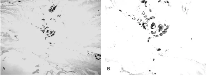
자궁경부에 도말세포진의 Pap. 염색 결과 도말배경이 비교적 깨끗하고, 암세포의 수가 적게 나타났으며 세포질 이 풍부한 편재된 큰 핵을 볼 수 있었다(Fig. 3, A). 그리 고 생검 조직의 H&E 염색 결과 자궁경부에서 원발한 선 암종보다 암세포의 수가 적고, 깨끗한 배경과 크고 편재
된 핵, 그리고 인환세포가 관찰되었다(Fig. 3, B). CK 7,
CK 20에 대한 면역조직화학적 염색 결과 암세포의 세포
질에서 CK 7은 70%, CK 20은 20%의 양성율을 나타냈
다(Fig. 4, A, B).
A B
Fig 3. In finding on the cytologic smear preparation of the uterine cervix stained by Pap. stains, the back ground and the number
of malignant cells were relatively clear and a few eccentric nuclei in abundant cytoplasm were shown (A. × 400). In finding on tissue preparation of the uterine cervix biopsy by H&E stain, the clear back ground, the large and eccentric nuclei, and the signet ring cell types were observed, and the number of malignant cells than the primary uterine cervix adenocarcinoma. The vacuoles in cytoplasm were observed and the nuclear membrane and chromatin were thick and very rougher (arrow, B. × 400).A B
Fig 4. In finding on cytokeratin 7 and 20 of immunohistochemical stain, the tumor cells were indicated each positive rate of 70%
and 20% (A.ⅹ 200, B.ⅹ400 ).