ISSN 1225-6552
Available online at http://kosves.re.kr
< Case Report >
Veterinary Service
*Corresponding author: Jeong-Hee Han, Tel. +82-33-250-8691, Fax. +82-33-256-3722, E-mail. hanjh@kangwon.ac.kr
닥스훈트의 원발성 비장 혈관주위세포종 증례
김성재ㆍ양돈식ㆍ한정희*
강원대학교 수의과대학 동물종합연구소
Primary splenic hemangiopericytoma in a dachshund dog
Sung-Jea Kim, Don-Sik Yang, Jeong-Hee Han*
College of Veterinary Medicine and Institute of Veterinary Science, Kangwon National University, Chuncheon 200-701, Korea
(Received 9 December 2011; revised 26 March 2012; accepted 17 May 2012)
Abstract
A 7-year-old, castrated male dachshund dog with tumor of the spleen portion was referred to the Kangwon National University. The tumor removed surgically and tumor size was 6∼7 cm. Histopatho- logically, this neoplasm was the presence of perivascular whorls of fusiform cells. The cells also ar- ranged in interlacing bundles or storiform patterns. And this neoplasm consisted of spindle cells that often formed distinct whorls around a central capillary. Immunohistochemical analysis revealed multi- focally immunoreactivity for α-smooth muscle actin, whereas not immunoreactive for desmin, S-100, von Willebrand factor. On the base of the histological and immunohistochemical results, this neoplasm was diagnosed as a canine hemangiopericytoma.
Key words : Hemangiopericytoma, Spleen, S-100, α-Smooth muscle actin
서 론
Canine hemangiopericytoma (CHP)는 혈관 주위세포 에서 유래하며, 피하직에 빈발하는 간엽조직계 종양 으로 개의 피부 및 피하직 종양의 4.2%를 차지한다 (Bostock, 1986; Pérez 등, 1996). CHP는 성견부터 노 령견에 이르기까지 빈발하며 평균발생 연령은 10년 령이다(Bostock, 1986; Evans, 1987). 이 종양은 성장이 느리고, 국소침윤이 일반적이며, 전이는 드문 종양이 다(Mazzei 등, 2002; Richardson 등, 1983). 육안적으로 는 지방을 가지고 있는 분엽화된 형태가 특징적이고 (Graves 등, 1998), 조직학적으로는 다양한 형태를 나 타내는데, 방추형의 종양세포가 혈관주위에 나선형을 형성하거나 콜라겐을 산생하고, 다양한 형태의 분열 상을 보인다(Evans, 1987). 또 세포질 돌기를 가지고
있는 방추형의 종양세포가 혈관주위에서 나선형으로 배열되어 ‘지문’ 패턴을 형성한다(Graves 등, 1998;
Mazzei 등, 2002).
CHP와 유사한 조직학적 소견을 보이는 peripheral nerve sheath tumor (PNST), fibrosarcoma, histiocytoma 와 leiomyosarcoma와 감별진단을 하기 위해서 면역조 직화학염색법을 사용하며 대표적인 면역염색 표지인 자는 desmin, S-100, von Willebrand factor (VWF), α-smooth muscle actin (SMA)이다(Hilmas와 Gillette, 1976; Madevell와 Theilen, 1987).
이번 증례는 개의 비장에서 발생한 CHP로 주로 피
부에서 발생하는 기존의 종양과 다른 드문 증례여서
임상적 소견, 병리학적 및 면역조직화학 염색 소견을
보고하자고 한다.
Fig. 1. Abdominal radiographs. (A, B) (ventrodorsal and right later- al view): Enlarged spleen silhouette was observed in the ab- dominal cavity (arrow).
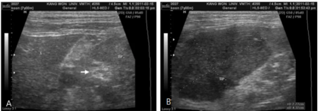
Fig. 2. Abdominal ultrasonogra- phy. (A) The spleen showed inhomogeneous parenchyma. And hyper- echoic nodule was asso- ciated with the spleen (arrow). (B) Enlargement of the spleen was ob- served (2.77×4.32 cm).
Fig. 3. Note the solitary large irregular mass in the spleen, size 6×7 cm.
증 례
종양이 발견된 닥스훈트는 8살 된 수컷으로 활동적 이고, 식욕은 정상이었으며, 외형적으로 특별한 병변 은 없었다. 수술 전 혈액검사 및 혈청검사에서의 대 부분의 수치가 정상을 나타내었으나 이상 소견으로 는 RBC 수치의 증가, lymphocyte 수치의 감소가 있었 다. 단순 방사선 촬영의 배복상(dorsoventral view)에서 비정상적으로 확장된 비장 음영이 관찰되었으며, 우 측 외측상(right lateral view)에서도 비장의 음영이 정 상보다 비정상적으로 확장된 모습을 관찰할 수 있었 다(Fig. 1). 비장 음영의 비정상적인 확장에 대한 확진 을 위하여 초음파 검사를 하였다. 초음파 결과, 비장 은 비균질한 실질이었고, 비장의 중심부에 고에코성 의 결절이 관찰되었다(Fig. 2A). 비장의 크기는 정상 보다 확장된 모습으로 관찰되었다(Fig. 2B).
종양은 비장 정중앙에 있었으며, 크기는 약 6×7 cm 였다. 종양이 있는 부분은 다른 부분에 비해 종대되
고 단단했으며, 정상 비장조직들과 구분되어 있었다 (Fig. 3). 이 종양에 대한 진단을 위해 조직학적, 면역 학적인 검사를 하였다. 조직학적 검사를 위해 수술적 으로 잘라낸 종양 조직을 10% 중성포르말린에 고정 한 후 조직처리 과정을 거쳐 파라핀으로 포맷하였다.
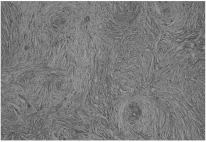
조직은 3 μm 조직 절편으로 제작하여 H&E 염색을 하였다. 조직학적 검사 결과, 방추형 세포들이 모세 혈관 주위를 감싸면서 소용돌이 또는 나선 형태를 이 루었으며 그 사이에 교원질성 기질과 점액소 기질이 드문드문 관찰되었다. 방추형의 종양성 세포들은 비 교적 균일한 모양이었으며, 유사분열상은 거의 관찰 되지 않았다(Fig. 4).
면역조직화학적 반응을 보기 위해 면역염색을 하
였다. 일차 항체로는 desmin (Dako, USA), S-100 (Dako,
USA), vWF (Dako, USA), SMA (Dako, USA)를 사용
하였다(Table 1). 각 절편은 xylene과 농도별 ethanol에
서 탈파라핀 과정과 탈수 과정을 거쳤다. 10 mmol/L
citrate buffer (pH 6.0)에 조직절편을 담그고 랩을 씌
워서 다시 전자레인지에 20분간 끓이는 항원 복원과
Fig. 5. The immunoreactivity for α-smooth muscle actin were pres- ent at neoplastic spindle cells in the hemangiopericytoma.
Immunostain and counterstain with Harris hematoxylin,
×200.
Table 2. Result of immunohistochemical staining
Desmin S-100 protein
von Willebrand factor
α
-smooth muscle actin
- - - +
Fig. 4. Neoplastic spindle cells were arranged whorl and storiform patterns. Collagenous stroma and mucinous matrix were lo- cated between spindle cells arranged. H&E stain, ×100.
Table 1. Primary antibodies used for immunohistochemical staining
Antibody Clone Type Dilution
Desmin D33 Rabbit 1:50
S-100 protein A1/A6/B Rabbit 1:400
von Willebrand factor Polyclonal Rabbit 1:400
α-smooth muscle actin 1A4 Mouse 1:75
정을 거쳤다. 얼음에서 식힌 후, TPBS (1 L PBS, 5 µl Tween 20)에 20분간, phosphate buffer saline (PBS; pH 7.4, 137 mM NaCl, 2.7 mM KCl, 10 mM Na
2HPO
4, 2 mM KH
2PO
4)에서 5분간 두 번 수세를 한 후, 20분간 실온에서 0.3% hydrogen peroxide로 처리하였다. 일차 항체와 이차 항체를 붙이는 과정은 ABC kit (Vector Laboratories, USA)의 지시대로 진행하였다. 발색은 DAB substrate kit (Zymed, USA)를 이용하였고 Harris hematoxylin을 이용하여 대조염색을 하였다. 면역조직 화학적 염색 결과, desmin, S-100, vWF는 음성이었고, SMA의 경우 종양조직의 방추형 세포들의 세포질이 염색되었다(Table 2, Fig. 5).
고 찰
CHP는 개에서 일반적인 피부 종양으로 중년기나
노년기에 잘 발생하며, 주로 사지의 관절 주위 피하 조직에 발생한다(Goldschmidt와 Hendrick, 2002). 이 종양은 성장이 느리고, 국소침윤이 일반적이며, 전이 는 잘 발생하지 않는다(Mazzei 등, 2002; Richardson 등, 1983). 이번 증례는 피부에서 종양과 관련된 피부 병변을 관찰할 수 없었으며 영상진단학적인 진단 결 과 다른 장기에 전이한 흔적이 없이 비장에서만 발생 하였다.
CHP는 다른 간엽성 종양과 감별 진단하는 것이 중 요하다. 조직학적 검사와 면역화학염색으로 다른 혈 관 종양 및 fibrosarcoma, PNST와 leiomyosarcoma를 감별할 수 있다. Fibrosarcoma는 CHP보다 세포들이 비정형으로 핵의 다형태성과 과염색성이 잘 관찰되 며, 유사분열상도 잘 관찰되었는데(Yager와 Wilcock, 1994), 이번 증례에서는 비교적 종양 세포의 핵들이 균일하였으며, 유사분열상도 거의 관찰되지 않았다.
vWF는 다중결합구조를 가진 large glycoprotein으로
주로 비장, 신장, 간에서 발생하는 혈관육종을 진단
하기 위해서 사용된다(Rodeghiero, 2002). 이번 증례의
면역 염색 결과 부분적으로 반응이 나타나는 부분이
있었지만, 일반적으로 혈관육종에 나타나는 것처럼
광범위한 반응은 나타나지는 않았다는 것을 통해 혈
관육종과 감별 진단할 수 있었다. Desmin은 class III 해당하는 intermediate filament로 3가지 종류의 근세포 (skeletal, cardiac and smooth muscle)에 양성의 반응이 나타나게 된다(Goebel와 Warlo, 2000). 이번 증례에서 음성의 결과를 토대로 leiomyosarcoma와 감별할 수 있었다.
조직학상으로 CHP는 혈관 주위를 방추형 종양세 포가 소용돌이 형태나 나선형 형태로 배열되어 ‘지 문’ 패턴을 형성하거나 방추형 종양세포가 다발을 이 루어 서로 교차 및 나선상으로 배열하는 것이 특징이 다(Graves 등, 1998; Mazzei 등, 2002; Evans, 1987).
PNST는 CHP와 달리 개에서 드물게 발생하고 조직학 상 방추형 세포들이 소용돌이 형태를 띠는 것이 CHP 보다 명확하지 않으며 대부분의 소용돌이가 모세혈 관보다 경화된 콜라겐 섬유를 둘러싸고 있다. 그러나 종양성 방추형 세포가 배열하는 형태가 CHP와 상당 히 유사하여 감별하기가 힘들다(Goldschmidt와 Hen- drick, 2002).
이번 증례에서는 PNST와의 정확한 감별진단을 위 해 S-100 protein, SMA를 이용하여 면역염색을 하였 다. S-100은 calcium binding protein으로 세포 내 및 세포 외 기능을 하며 schwannoma와 astrocytoma 등 신경세포 관련 종양에 반응을 보인다(Arumugam와 Logsdon, 2011). Chijiwa 등(2004)은 S-100이 PNTS에 서 73%의 양성반응을 보였으며 CHP에서 60%의 양 성반응을 보였지만 CHP의 경우 국소부위나 조직의 20% 미만에서 양성을 보였다고 하였다. 이번 증례에 서 종양조직이 S-100에 반응을 보이지 않아 CHP일 가능성이 높았지만 확진하기에는 미흡하였다. SMA 은 주로 혈관벽, 장관의 점막 근육층, 다양한 샘의 근 상피 세포 등에서 양성반응이 나타나게 되어 근성연 조직유래 종양을 진단하는데 가장 유용한 표지인자 이다(Rizeq 등, 1994). 또한, 단일클론항체를 사용하 여, 면역 금 전자 현미경으로 관찰하면 여러 위치의 혈관주위세포에서 SMA을 관찰할 수 있다(Pérez 등, 1996; Skalli 등, 1986). Chijiwa 등(2004)은 SMA이 CHP에서 80%의 양성반응을 보였지만 PNTS에서 반 응을 보이지 않았으며 SMA가 CHP와 PNTS를 감별 진단하는데 유용한 표지인자라고 하였다. 이번 증례 에서 종양조직의 면역염색 결과, 종양세포들이 지문 의 패턴을 보이며 세포질이 강하게 염색되었으며 CHP로 확진할 수 있었다.
원발성 CHP에서는 세포의 다형성과 유사분열이 낮게 나타나며 재발한 CHP에서는 세포의 비정형성,
다핵세포, 유사분열의 수가 증가한다(Goldschmidt와 Hendrick, 2002). 유사분열지수는 CHP의 전이와 재발 가능성을 예상하는데 유용한 지표이다(Kuntz 등, 1997).
유사분열지수가 8 이상인 CHP는 25%의 재발률과 2%의 전이율을 보였으며 9 이상인 CHP는 62%의 재 발률과 15%의 전이율을 보인다(Bostock와 Dye, 1980).
이번 증례는 종양 세포의 형태가 비교적 균일하였고, 다핵세포와 유사분열상도 거의 관찰되지 않았다. 또 한, 다른 장기로 전이도 이루어지지 않았다. 임상증 상과 조직학적 검사 결과로 미루어 보아 이번 증례는 원발성 CHP이며 재발할 가능성과 전이 가능성이 낮 은 양성 종양으로 판단된다.
결 론
8살 된 수컷 닥스훈트의 비장에서 발생한 종양을 진단하기 위해 임상검사, 조직학적 검사, 면역조직화 학염색법을 실시하였다. 조직학적 검사한 결과, 혈관 주위세포종이 의심되어 SMA을 이용한 면역조직화학 염색을 통해 확진하였다. 조직학적 검사 결과와 SMA 에 대한 면역 반응 양성 결과를 바탕으로 비장에서 발생한 원발성 혈관 주위세포종으로 최종 진단하였 다.
감사의 글
이 연구는 강원대학교 동물의학종합연구소의 지원 으로 수행되어 이에 감사드립니다.
참 고 문 헌
Arumugam T, Logsdon CD. 2011. S100P: a novel therapeutic tar- get for cancer. Amino Acids 41: 893-899.
Bostock DE. 1986. Neoplasms of the skin and subcutaneous tis- sues in dogs and cats. Br Vet J 142: 1-19.
Bostock DE, Dye MT. 1980. Prognosis after surgical excision of canine fibrous connective tissue sarcomas. Vet Pathol 17: 581-588.
Chijiwa K, Uchida K, Tateyama S. 2004. Immunohistochemical evaluation of canine peripheral nerve sheath tumors and other soft tissue sarcomas. Vet Pathol 41: 307-318.
Evans SM. 1987. Canine hemangiopericytoma: a retrospective analysis of response to surgery and orthovoltage radia-
tion. Vet Radiol 28: 13-16.
Goebel HH, Warlo IA. 2000. Progress in desmin-related my- opathies. J Child Neurol 15: 565-572.
Goldschmidt MH, Hendrick MJ. 2002. Tumor of the skin and soft tissue. pp. 45-118. In: Meuten DJ(ed.). Tumors in do- mestic animals. 4th ed. Blackwell, Iowa.
Graves GM, Bjorling DE, Mahaffey E. 1998. Canine hemangio- pericytoma: 23 cases (1967-1984). J Am Vet Med Assoc 192: 99-102.
Hilmas DE, Gillette EL. 1976. Radiotherapy of spontaneous fi- brous connective-tissue sarcomas in animals. J Natl Cancer Inst 56: 365-368.
Kuntz CA, Dernell WS, Powers BE, Devitt C, Straw RC, Withrow SJ. 1997. Prognostic factors for surgical treat- ment of soft-tissue sarcomas in dogs: 75 cases (1986- 1996). J Am Vet Med Assoc 211: 1147-1151.
Madevell BR, Theilen GH. 1987. Tumors of the skin and sub- cutaneous tissues. Part III. Skin tumors of mesenchymal origin. pp. 282-325. In: Thielen GH, Madewell BR(ed.).
Veterinary cancer medicine. 2nd ed. Lea & Febiger, Philadelphia.
Mazzei M, Millanta F, Citi S, Lorenzi D, Poli A. 2002.
Hemangiopericytoma: histological spectrum, immunohis- tochemical characterization and prognosis. Vet Dermatol
13: 15-21.
Pérez J, Bautista MJ, Rollón E, de Lara FC, Carrasco L, Martin de las Mulas J. 1996. Immunohistochemical character- ization of hemangiopericytomas and other spindle cell tumors in dog. Vet Pathol 33: 391-397.
Richardson RC, Render JA, Rudd RG, Shupe RE, Carlton WW.
1983. Metastatic canine hemangiopericytoma. J Am Vet Med Assoc 182: 705-706.
Rizeq MN, van de Rijn M, Hendrickson MR, Rouse RV. 1994.
A comparative immunohistochemical study of uterine smooth muscle neoplasms with emphasis on the epi- thelioid variant. Hum Pathol 25: 671-677.
Rodeghiero F. 2002. von Willebrand disease: still an intriguing disorder in the era of molecular medicine. Haemophilia 8: 292-300.
Skalli O, Bloom WS, Ropnaz P, Azzarone B, Gabbiani G. 1986.
Cytoskeletal remodeling of rat aortic smooth muscle cells in vitro: relationships to cubtune conditions and analogies to in vivo situations. J Submicnosc Cytol 18:
481-493.
Yager JA, Wilcock BP. 1994. Color atlas and text of surgical pathology of the dog and cat: dermatopathology and skin tumors. vol 1. Wolfe Publishing Ltd, London.