THP-1 대식세포에서 Ox-LDL과 LPS 병용 처리에 의한 3,3'-Diindolylmethane(DIM)의 거품세포 형성 억제 효과
⁃ 연구노트 ⁃
임영선*․구현지*․윤정미 전남대학교 식품영양학과
Inhibition Effect of 3,3'-Diindolylmethane (DIM) on Foam Cell Formation by Co-Treatment of Ox-LDL and LPS in THP-1 Derived Macrophage
Young-Sun Im * , HyunJi Gu * , and Jung-Mi Yun Department of Food and Nutrition, Chonnam National University
ABSTRACT Foam cell formation characterized by lipid accumulation is a hallmark of the early stages of atherosclero- sis. Foam cells form fatty streaks of plaques in the arteries and lead to atherosclerosis. 3,3′-Diindolylmethane (DIM) is a dietary agent derived from cruciferous vegetables such as broccoli and cauliflower. DIM has been shown to exhibit anti-cancer and anti-inflammatory properties. But the inhibitory effects of DIM on foam cell formation are not fully understood. In this study, we investigated the effect of DIM on cholesterol efflux and lipid accumulation in THP-1 foam cells and its underlying molecular mechanism. We exposed a THP-1 derived macrophage to oxidized low-density lipoprotein (Ox-LDL, 0 μg/mL) and lipopolysaccharides (LPS, 500 ng/mL) to initiate foam cell formation and carried out an analysis using MTT assay and western blotting. DIM decreased the expression of cluster of differ- entiation 36, lectin-like oxidized low-density lipoprotein receptor-1, and nuclear factor-κB, while it increased liver X receptor α, peroxisome proliferator-activated receptor-γ, and ATP-binding cassette cholesterol transporter A1 ex- pressions compared to the co-treatment of Ox-LDL and LPS. Taken together, DIM inhibited foam cell formation via the induction of cholesterol efflux and lipid accumulation. Also, DIM inhibited the inflammation induced by foam cells. Thus, DIM may be a potent candidate for the treatment and prevention of inflammation and atherosclerosis.
Key words: 3,3′-diindolylmethane, foam cell, oxidized low density lipoprotein, lipopolysaccharides
Received 5 March 2021; Revised 25 March 2021; Accepted 26 March 2021
Corresponding author: Jung-Mi Yun, Department of Food and Nutrition, Chonnam National University, 77, Yongbong-ro, Buk-gu, Gwangju 61186, Korea, E-mail: [email protected]
Author information: Young-Sun Im (Graduate student), HyunJi Gu (Graduate student), Jung-Mi Yun (Professor)
*
These authors contributed equally to this work.Copyright ⓒ 2021 by The Korean Society of Food Science and Nutrition. All rights Reserved.
This is Open Access article distributed under the terms of the Creative Commons Attribution Non-Commercial License (https://creativecommons.org/licenses/
by-nc/4.0) which permits unrestricted non-commercial use, distribution, and reproduction in any medium, provided the original work is properly cited.
서 론
죽상동맥경화증은 동맥 내막에 거품세포가 축적되는 것을 특징으로 하는 만성 염증성 질환이다(Libby 등, 2002). 거품 세포가 축적되면 혈관이 막혀 혈관이 좁아지고 일부에서 병 변에 혈전이 동반된다(Linton 등, 2019). 거품세포는 대식 세포에서 scavenger receptor를 통해 지질 흡수뿐만 아니 라 콜레스테롤 유출에 의해 형성되며 형성될 때 염증반응이 유발되고 플라크 형성을 가속화한다(Li와 Glass, 2002). 막 단백질인 ATP-binding cassette cholesterol transporter A1(ABCA1)은 거품세포에서 콜레스테롤 유출을 매개하므 로 ABCA1 활성의 상향 조절은 항동맥경화성을 나타낸다고
보고된다(Wang과 Tall, 2003). ABCA1뿐만 아니라 liver X receptor α(LXRα)와 peroxisome proliferator-activated receptor-γ(PPARγ)도 거품세포에서 콜레스테롤을 유출시 킨다(Chawla 등, 2001). Cluster of differentiation 36(CD 36)은 이와 반대로 지질을 축적하는 유전자로 lectin-like oxidized low-density lipoprotein receptor-1(LOX-1)과 함께 scavenger receptor로 불리며 지질 항상성 및 면역 반응의 중요한 조절 역할을 한다(Zingg 등, 2000; Pirillo 등, 2013). 죽상동맥경화증의 제어 전략 중 하나는 콜레스테 롤 유출을 증가시키고 염증과 지질 축적을 막는 것이다(Ball 등, 1995).
3,3′-Diindolylmethane(DIM)은 브로콜리, 콜리플라워 등
에서 DIM은 알츠하이머 마우스에서 항염증 효과를 보였다 (Wu 등, 2021). 또한, DIM은 위암세포의 증식을 억제하고 세포자가사멸(apoptosis)을 촉진한다고 보고되었다(Ye 등, 2021). 그러나 현재까지 DIM의 대식세포에서 거품세포 형 성 억제 효과에 대해서 알려진 바가 거의 없다. 따라서 본 연구에서는 DIM의 항염증 효과와 거품세포 형성 억제 효과 를 알아보기 위해 인간 단핵구 THP-1 세포를 대식세포로 분화시킨 후, lipopolysaccharides(LPS)와 oxidized low- density lipoprotein(Ox-LDL)을 병용 처리해 염증과 거품 세포 형성을 유도하고 지질 축적 관련 유전자 및 콜레스테롤 유출 관련 유전자 단백질 발현에 대한 영향을 확인해보고자 한다.
재료 및 방법
실험재료
DIM, LPS, phorbol 12-myristate 13-acetate(PMA), thiazolyl blue tetrazolium bromide(MTT), Oil red O는 Sigma-Aldrich Co.(St. Louis, MO, USA)에서 구입하였다.
Ox-LDL은 Thermo Fisher Scientific(Waltham, MA, USA)에서 구입하였고, 그 외 나머지 재료들은 Sigma- Aldrich Co. 또는 Biosesang Inc.(Seongnam, Korea)에서 구입하였다.
세포배양
실험에 사용한 인간 단핵구 THP-1 세포는 한국세포주은 행(KCLB; Korean Cell Line Bank, Seoul, Korea)에서 구 입하였다. 세포는 37°C, 5% CO
2
조건의 incubator에서 10%fetal bovine serum(FBS; WelGENE Co., Daegu, Korea), 1% antibiotics(WelGENE Co.)가 함유된 RPMI-1640 me- dium(WelGENE Co.)을 사용하였다.
세포 생존율
세포 생존율은 MTT assay를 사용하여 측정하였다. 24- well plate에 THP-1 세포를 1×10
6
cells/mL 농도로 분주 하여 PMA(1 μM)를 48시간 동안 처리해 배양한 후 DIM(5, 10, 25, 50 μM)을 농도별로 48시간 처리하였다. MTT를 분석하기 24시간 전에 LPS(500 ng/mL)를 24시간 동안 처 리하고 MTT solution 100 mg/mL를 첨가하여 2시간 반응 시킨 후, 상층액을 제거하고 100% dimethyl sulfoxide (Biosesang Inc.) 1 mL로 formazan을 녹여준 후 분광광도 계(EZRead 400 microplate reader, Biochrom, Cam- bridge, UK)로 570 nm에서 흡광도를 측정하였다.Western blot 분석
LPS, Ox-LDL, DIM이 처리된 세포를 phosphate-buf- fered saline(PBS; Biosesang Inc.)으로 씻은 후 harvest
hibitor cocktail(Thermo Fisher Scientific)로 lysis 하여 단백질을 추출하였고, 세포질 단백질은 cytosolic lysis buffer(10 mM HEPES, 10 mM KCl, 0.1 mM EDTA, 0.1 mM EGTA, 1 mM dithiothreitol, 0.5 mM phenylmethyl- sulfonyl fluoride, 10% NP-40)로 추출하였고 핵단백질은 nuclear extraction buffer(20 mM HEPES, 0.4 mM NaCl, 1 mM EDTA, 1 mM EGTA, 1 mM dithiothreitol, 1 mM phenylmethylsulfonyl fluoride, 10% NP-40)로 추출하였 다. 그리고 BCA protein assay kit(Thermo Fisher Scien- tific)을 이용해서 정량하였다. 단백질(10~20 μg)은 6~12%
polyacrylamide gel을 사용하고 nitrocellulose mem- brane(Merck Millipore Ltd., County Cork, Ireland)에 전 이시킨 후 5% non-fat dry milk가 포함된 blocking buffer 에서 2시간 반응하였다. nuclear factor-κB(NF-κB), ABCA1, LOX-1, CD36, PPARγ, LXRα 항체로 반응한 후 Western Blotting Luminol Reagent(Santa Cruz Biotech- nology, Dallas, TX, USA)를 이용해 ChemiDoc XRS+
system(Bio-Rad, CA, USA)으로 결과를 분석하였다. 단백 질 발현 강도 분석을 위해서 Image J(Wayne Rasband Na- tional Institutes of Health, Bethesda, MD, USA)프로그램 을 사용하였다.
Oil red O 염색
THP-1 세포에 Ox-LDL(20 μg/mL), LPS(500 ng/mL), DIM(0.1, 1 μM)을 각 시간에 따라 처리한 후 배지를 제거하 고 10% 포르말린으로 30분 고정하였다. PBS로 씻은 다음 Oil red O solution 1 mL를 처리해 Leica microscope와 Leica Application Suite X software(Leica Microsys- tems, Wetzlar, Germany)를 사용해 염색을 관찰하였고, 염 색된 지질 밀도는 분광광도계(EZRead 400 microplate reader, Biochrom)를 사용해 520 nm에서 흡광도를 측정하 였다.
통계처리
실험 결과는 3회 이상 실시하며 평균±표준편차로 표시하 였고 유의성 검정은 SPSS 통계프로그램(Statistical Pack- age for the Social Science, ver. 26.0, SPSS Inc., Chica- go, IL, USA)을 이용하였다. 통계적 유의성 검정은 Stu- dent’s t-test로 분석하였고
P
<0.05와P
<0.01 수준으로 유 의성을 표시하였다.결과 및 고찰
LPS로 유도된 THP-1 세포 염증 환경에서 DIM의 항염증 효과
LPS는 그람 음성균의 세포 외막에 존재하는 물질로 대식 세포에 염증을 유도하는 것으로 보고된다(Tucureanu 등,
LPS-, DIM LPS 500 mg/mL+DIM
A
LPS- LPS LPS+
only
DIM (μM) 1 5
NF-κB (nuclear) Histone H1
B
LPS- LPS LPS+
only
DIM (μM) 1 5
0 0.5 1 1.5 2 2.5
R e la tiv e dens ity . (N F -κ B/ H is tone H 1 ) . **
#
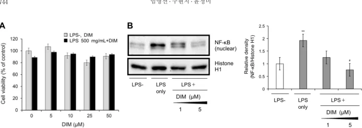
Fig. 1. Anti-inflammation effect of DIM induced LPS in THP-1 derived macrophages. (A) Effect of DIM on cell viability after
48 h was evaluated by the MTT assay. THP-1 monocyte was exposed to 1 μM of PMA for 48 h and incubated for 48 h with DIM (5∼50 μM). And then, THP-1 macrophages were cultured under LPS 500 ng/mL for 24 h prior to harvest. Relative cell viability was determined by the amount of MTT converted into formazan crystals and quantified the percentage of control. The value represents the mean±SD (n=3). (B) Effect of DIM on NF-κB gene expression in LPS induced inflammation in THP-1 derived macrophages. Cell lysates were prepared for western blotting with NF-κB. The blot was stripped and then reprobed with histone H1 as loading control. The band density was quantified with Image J. The value represents the mean±SD (n=3, independent).* P<0.05,
** P<0.01 vs. LPS-; # P<0.05, ## P<0.01 vs. LPS only.
2018). LPS 자극에 의해 면역 반응에 작용하는 전사인자인 NF-κB가 활성화되어 염증이 일어난다(Harada 등, 2003).
우리의 첫 번째 실험은 LPS 처리에 의한 염증 환경에 DIM의 세포독성을 알아보는 것으로, LPS(500 ng/mL) 처리군과 비처리군에 DIM(5, 10, 25, 50 μM)을 처리하고 MTT 분석 방법을 통해 세포 생존율을 측정하였다(Fig. 1A). 그 결과 LPS 처리군과 LPS 비처리군 모두에서 DIM에 대한 세포독 성은 나타나지 않았다(Fig. 1A). 따라서 이후 실험에서 세포 독성이 나타나지 않는 안전한 범위 내에서 후속 실험을 진행 하였다. LPS로 유도된 핵 내 NF-κB 발현이 DIM 처리에 의해서 저해되는지를 알아보기 위해 THP-1 대식세포에 1 μM과 5 μM의 DIM을 48시간 동안 처리한 후 LPS(500 ng/mL)를 처리하였고, 그 결과를 western blot을 통해 확인 하였다. Fig. 1B와 같이 LPS 처리로 유도된 염증 환경에서 NF-κB 발현이 증가하였고, 이러한 발현 증가는 DIM에 의 해서 감소하였다. 이러한 결과는 염증반응에 관여하는 NF- κB의 핵 내 발현 저하를 통해 DIM이 염증 억제 효과가 있었 다는 것을 보여준다. 우리의 연구와 유사하게 Ox-LDL로 유 도된 THP-1 대식세포에서 saikosaponins-a가 NF-κB 단 백질 발현을 억제했고(He 등, 2016), Tesoriere 등(2013) 의 연구에서도 7-ketocholesterol로 유도된 THP-1 세포 에서 indicaxanthin은 NF-κB 단백질 발현 억제를 통해 염 증이 억제된 것을 보여주었다.
DIM 처리에 의한 거품세포 억제
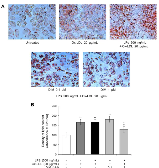
Oil red O 염색은 현미경을 통해 거품세포에 존재하는 세포질 내의 지방 입자를 관찰할 수 있으며 지질 축적이 될 수록 염색이 더 진하게 되는 비형광 염색법이다(Chi 등, 2014). 무처리군(untreated)에서는 Oil red O 염색에 대한 양성반응이 관찰되지 않았고, Ox-LDL(20 μg/mL) 단독 처
리군과 Ox-LDL과 LPS(500 ng/mL) 병용 처리군에서는 진 한 Oil red O 염색 양성반응이 관찰되었다(Fig. 2A). 이의 지질 염색 정도는 분광광도계를 통해 분석한 결과, Ox-LDL 과 LPS 병용 처리군은 지질 밀도가 무처리군에 비해 유의적 으로(
P
<0.05,P
<0.01) 높았다. 또한 이러한 지질 밀도 증가 는 DIM 1 μM 처리에 의해 유의적으로(P
<0.05) 제어됨을 확 인할 수 있었다(Fig. 2B). 본 연구와 유사하게 Chi 등(2014) 의 연구에서 phytochemical인 berberine은 Ox-LDL로 유 도된 THP-1 거품세포에 Oil red O 염색을 하여 분석함으로 써 거품세포 형성 억제를 확인하였다. 또한 Fu 등(2016)의 연구에서도 Ox-LDL로 유도된 THP-1 거품세포에 sodium paeonolsilate를 처리시키고 Oil red O 염색을 분석했을 때 지질 축적이 분명히 감소하였다. 따라서 Oil red O 염색 결 과 DIM은 거품세포 형성을 억제한다고 보인다.죽상동맥경화
in vitro유사 환경에서 DIM의 콜레스테롤, 지질 관련 유전자 발현
핵 수용체인 LXRα는 관련 유전자를 조절하여 대식세포 에서 콜레스테롤 항상성을 유지한다(Wang과 Tontonoz, 2018). 즉, LXR 활성화는 ABCA1을 통해 HDL 형성을 증가 시키고 콜레스테롤 유출 및 장내 콜레스테롤 배출을 촉진한 다(Wang과 Tontonoz, 2018). PPARγ의 활성화는 염증반 응과 관련된 매개물의 분비 감소를 유발하고 특정 유전자의 발현을 직접 조절함으로써 면역반응을 조절한다(Kwon 등, 2010). 또한 PPARγ 조절의 직접적인 표적으로 알려진 유일 한 대식세포 유전자는 CD36이고(Tontonoz 등, 1998), CD 36과 LOX-1은 Ox-LDL 흡수에 관여하는 특정 수용체로 알려져 있다(Bruni 등, 2005). 이와 반대로 막 단백질인 ABCA1은 거품세포에서 콜레스테롤 유출을 매개하므로 ABCA1 활성의 상향 조절은 항동맥경화성을 나타낸다고 보
Untreated Ox-LDL 20 μg/mL LPs 500 ng/mL +Ox-LDL 20 μg/mL
DIM 0.1 μM DIM 1 μM LPS 500 ng/mL+Ox-LDL 20 μg/mL
0 50 100 150 200 250
Dens ity of lipid c ont ent . (abs or banc e at 520 nm ) .
**
#
*
**
**
B
LPS (500 ng/mL) Ox-LDL (20 μg/mL) DIM (μM)
- - -
- + -
+ + -
+ + 0.1
+ + 1
Fig. 2. Effect of DIM on foam cell formation in THP-1 derived macrophages cultured LPS and Ox-LDL. DIM ameliorate lipid
accumulation in THP-1 macrophages by increasing cholesterol efflux in vitro. (A) Foam cell formation was determined by Oil Red O staining. (B) Oil Red O areas were calculated the percentage of the stained cell area to the total cell area. The data are presented as the mean±SD (n=3, independent).* P<0.05, ** P<0.01 vs. untreated; # P<0.05, ## P<0.01 vs. LPS+Ox-LDL. Magnification, ×200.
고된다(Wang과 Tall, 2003).
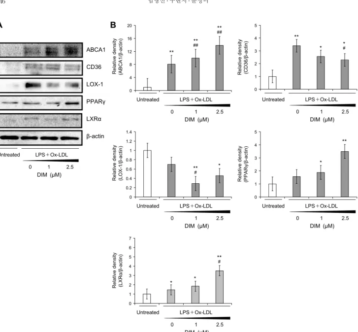
본 연구 결과 Fig. 3에서 보이는 바와 같이 Ox-LDL과 LPS 병용 처리군에서 CD36, LOX-1 유전자 발현은 증가했 고, 이러한 발현 증가는 DIM 처리에 의해 감소하였다. 또한 이와는 반대로 LPS, Ox-LDL 병용 처리 시 ABCA1, PPAR γ, LXRα의 발현이 감소하였고, DIM을 처리했을 때 농도 의 존적으로 발현이 증가하였다.
Zhang 등(2015)과 Seo 등(2015)의 연구 결과에서는 우 리의 결과에서 확인한 바와 유사하게 Ox-LDL이 CD36과 LOX-1 발현을 증가시켰다. Chawla 등(2001)에 따르면 ABCA1, PPARγ, LXRα는 Ox-LDL의 노출에 의해 유도되 고 이러한 상황에서 발현이 증가하는 것으로 보고되고 있다.
Li 등(2017)의 연구에서 Ox-LDL로 유도된 THP-1 대식세
포에 phytochemical인 puerarin 처리 시 본 연구 결과와 같이 ABCA1, PPARγ, LXRα가 증가하는 결과를 보였다.
본 연구에서는 DIM 처리에 의해서 낮아진 ABCA1, PPARγ, LXRα 유전자 상향 조절을 통해 콜레스테롤 유출을 증가시 켰다. 따라서 DIM이 적어도 지질 축적(CD36, LOX-1) 및 콜레스테롤 유출 관련 유전자(ABCA1, PPARγ, LXRα)를 조 절함으로써 적어도 일부 혈중 지질 개선에 효과를 보이는 것으로 생각된다.
요 약
본 연구는 십자화과 채소에 존재하는 DIM의 거품세포 억제 효과와 항염증 효과를 연구하고자 하였다. 이를 위해서 인간
A
Untreated LPS+Ox-LDL 0 1 2.5 DIM (μM)
ABCA1
CD36
LOX-1
PPARγ
LXRα
β-actin
Untreated LPS+Ox-LDL 0 1 2.5 DIM (μM)
0 4 8 12 16 20
R e la tive d e n sity . (A BC A1/ β -a ct in ) .
**
## **
** ##
B
Untreated LPS+Ox-LDL 0 1 2.5 DIM (μM)
0 1 2 3 4 5
R e la tive d e n si ty . (CD3 6 /β -a ct in ) . **
* #
*
Untreated LPS+Ox-LDL 0 1 2.5 DIM (μM)
0 0.2 0.4 0.6 0.8 1 1.2 1.4
R e la tiv e d e n si ty . (L O X -1 /β -a ct in ) .
** #
*
Untreated LPS+Ox-LDL 0 1 2.5 DIM (μM)
0 1 2 3 4 5
R e la tive d e n si ty . (PPAR γ/ β -a ct in ) . **
*
Untreated LPS+Ox-LDL 0 1 2.5 DIM (μM)
0 1 2 3 4 5 6 7
R e la tiv e de ns ity . (L X R α /β -a ct in ) .
** #
* *
Fig. 3. Effects of DIM on the protein expressions of lipid accumulation and cholesterol efflux in THP-1 derived macrophages by
western blot. THP-1 cells were differentiated using 1 μM PMA for 48 h. THP-1 derived macrophages were cultured in the absence or presence of DIM (1, 2.5 μM) then treated with LPS (500 ng/mL) and Ox-LDL (20 μg/mL) for 24 h. (A) Expression of lipid accumulation and cholesterol efflux proteins in THP-1 derived macrophages treated with DIM. Cell lysates were prepared for western blotting with each antibody as described in methods. The blots are stripped and then reprobed with β-actin as loading control.(B) The band density was quantified with Image J. The data are presented as the mean±SD (n=3, independent).
* P<0.05, ** P<0.01
vs. untreated;# P<0.05, ## P<0.01 vs. LPS+Ox-LDL.
단핵구 THP-1 세포를 대식세포로 분화시켰고 이에 Ox- LDL, LPS를 병용 처리하여 죽상동맥경화 세포 환경을 만들 었다. DIM의 연구에 사용된 모든 농도에서 세포독성이 없었 다. Ox-LDL과 LPS 병용 처리군의 lipid uptake를 강하게 보여줌으로써 거품세포가 형성되었음을 Oil red O 염색을 통해서 확인할 수 있었다. 즉, 거품세포의 양성반응이 진하 게 나타났으며 지질 밀도가 유의적으로 높았다. 이러한 거품 세포의 지질축적은 DIM 처리 시 유의적으로 억제됨을 확인 할 수 있었다. 또한 이의 결과와 관련된 분자 수준에서의 유전자 발현을 보면 Ox-LDL과 LPS 병용 처리 시에 콜레스
테롤 유출 관련 유전자는 감소하고 지질 축적 관련 유전자 발현은 증가함을 확인할 수 있었다. 이에 DIM을 처리했을 때 콜레스테롤 유출 관련 유전자인 ABCA1, PPARγ, LXRα 의 발현은 DIM의 농도가 높아질수록 발현이 높아졌으며, 지질 축적 관련 유전자인 CD36, LOX-1 유전자에서는 DIM 의 농도가 높아질수록 발현이 낮아졌다. 결론적으로 본 연구 결과를 통해서 DIM은 거품세포 형성을 억제하는 잠재적 가 능성을 지닌 식이인자로 여겨지며, DIM을 함유한 식품 섭취 를 통해 염증 및 죽상동맥경화증을 억제 또는 예방할 가능성 이 있으리라 생각된다.
이 논문은 2021년도 정부(과학기술정보통신부)의 재원으로 한국연구재단의 지원을 받아 수행된 연구임(No. 2019R1 F1A1060739).
REFERENCES
Ball RY, Stowers EC, Burton JH, Cary NRB, Skepper JN, Mitchinson MJ. Evidence that the death of macrophage foam cells contributes to the lipid core of atheroma. Atherosclerosis.
1995. 114:45-54.
Bruni F, Pasqui AL, Pastorelli M, Bova G, Cercignani M, Palaz- zuoli A, et al. Different effect of statins on platelet oxidized- LDL receptor (CD36 and LOX-1) expression in hypercholes- terolemic subjects. Clin Appl Thromb Hemost. 2005. 11:417- 428.
Chawla A, Boisvert WA, Lee CH, Laffitte BA, Barak Y, Joseph SB, et al. A PPARγ-LXR-ABCA1 pathway in macrophages is involved in cholesterol efflux and atherogenesis. Mol Cell.
2001. 7:161-171.
Chi L, Peng L, Pan N, Hu X, Zhang Y. The anti-atherogenic effects of berberine on foam cell formation are mediated through the upregulation of sirtuin 1. Int J Mol Med. 2014.
34:1087-1093.
Fu W, Fu W, Ding S, Chen Z, Jiang J, Gong Z, et al. Sodium paeonolsilate inhibits ox-LDL induced macrophage foam cell formation and inflammation in atherosclerosis. Int J Clin Exp Med. 2016. 9:1051-1061.
Harada K, Ohira S, Isse K, Ozaki S, Zen Y, Sato Y, et al. Lipo- polysaccharide activates nuclear factor-kappaB through toll- like receptors and related molecules in cultured biliary epi- thelial cells. Lab Invest. 2003. 83:1657-1667.
He D, Wang H, Xu L, Wang X, Peng K, Wang L, et al. Saikosa- ponin-a attenuates oxidized LDL uptake and prompts choles- terol efflux in THP-1 cells. J Cardiovasc Pharmacol. 2016.
67:510-518.
Kwon EY, Park C, Kwon JC, Kim SH, Park SH, Choi SM, et al. Effects of peroxisome proliferator-activated receptor-γ on the production of tumor necrosis factor-α in stimulated human monocoytes. Infect Chemother. 2010. 42:291-295.
Li AC, Glass CK. The macrophage foam cell as a target for therapeutic intervention. Nat Med. 2002. 8:1235-1242.
Li CH, Gong D, Chen LY, Zhang M, Xia XD, Cheng HP, et al. Puerarin promotes ABCA1-mediated cholesterol efflux and
Eur J Pharmacol. 2017. 811:74-86.
Libby P, Ridker PM, Maseri A. Inflammation and atherosclero- sis. Circulation. 2002. 105:1135-1143.
Linton MF, Yancey PG, Davies SS, Jerome WG, Linton EF, Song WL, et al. The role of lipids and lipoproteins in athero- sclerosis. Endotext, South Dartmouth, MA, USA. 2019. p 4.
Pirillo A, Norata GD, Catapano AL. LOX-1, OxLDL, and athe- rosclerosis. Mediators Inflamm. 2013. Article ID 152786.
http://dx.doi.org/10.1155/2013/152786
Seo JW, Yang EJ, Yoo KH, Choi IH. Macrophage differentiation from monocytes is influenced by the lipid oxidation degree of low density lipoprotein. Mediators Inflammation. 2015.
Article ID 235797. http://dx.doi.org/10.1155/2015/235797 Tesoriere L, Attanzio A, Allegra M, Gentile C, Livrea MA. Phy-
tochemical indicaxanthin suppresses 7-ketocholesterol-induced THP-1 cell apoptosis by preventing cytosolic Ca
2+
increase and oxidative stress. Br J Nutr. 2013. 110:230-240.Tontonoz P, Nagy L, Alvarez JGA, Thomazy VA, Evans RM.
PPARγ promotes monocyte/macrophage differentiation and uptake of oxidized LDL. Cell. 1998. 93:241-252.
Tucureanu MM, Rebleanu D, Constantinescu CA, Deleanu M, Voicu G, Butoi E, et al. Lipopolysaccharide-induced inflam- mation in monocytes/macrophages is blocked by liposomal delivery of G
i
-protein inhibitor. Int J Nanomedicine. 2017.13:63-76.
Wang B, Tontonoz P. Liver X receptors in lipid signalling and membrane homeostasis. Nat Rev Endocrinol. 2018. 14:452- 463.
Wang N, Tall AR. Regulation and mechanisms of ATP-binding cassette transporter A1-mediated cellular cholesterol efflux.
Arterioscler Thromb Vasc Biol. 2003. 23:1178-1184.
Wu X, Liu J, Chen C, Huang Z, Zang Y, Chen J, et al. 3,3′-Diin- dolylmethane alleviates acute atopic dermatitis by regulating T cell differentiation in a mouse model. Mol Immunol. 2021.
130:104-112.
Ye Y, Ye F, Li X, Yang Q, Zhou J, Xu W, et al. 3,3′-Diindolyl- methane exerts antiproliferation and apoptosis induction by TRAF2-p38 axis in gastric cancer. Anticancer Drugs. 2021.
32:189-202.
Zhang H, Zhai Z, Zhou H, Li Y, Li X, Lin Y, et al. Puerarin inhibits oxLDL-induced macrophage activation and foam cell formation in human THP1 macrophage. Biomed Res Int. 2015.
Article ID 403616. https://doi.org/10.1155/2015/403616 Zingg JM, Ricciarelli R, Azzi A. Scavenger receptors and modi-
fied lipoproteins: fatal attractions?. IUBMB Life. 2000. 49:
397-403.