1. Introduction
It is important to define planning target volume (PTV) clearly for improvement in efficiency of radiation treatment in case of tumor located within moving organs.Shape of the tumor on computed tomography(CT) images is various in each patient because of different breathing patterns. Consequently, internal target volume (ITV) is needed to be defined in case that tumor is located within moving organs. Four-dimensional computed tomography (4DCT) is able to get each phase of the breathing signal and has presented superiority in designing ITV.
1,2)A report released by the American
Association of Physicists in Medicine Task Group 76 outlines the procedures involved with the acquisition and reconstruction of the CT images with RPM and 4DCT imaging for each phase of the breathing signal, allowing for the range of tumor motion to be determined.
3)More exquisite localization of normal tissue and tumor tissue has been demonstrated through treatment planning with use of 4DCT images and real-time position management (RPM) (Varian Medical Systems, Palo Alto, CA) compared to use of the helical CT images.
4)Although 4DCT imaging is able to define range of the tumor motion by assessing the location of tumor at each breathing phase, the wider application of this procedure to each patient is difficult.
The reason is thatthe more increasing scan time is, the more exposure dose and breathing-artifacts occur on images.
5)However, efficient treatment planning is possible as it reveals the movement of the internal organs
본 논문은 2016년 11월 01일 접수하여 2016년 12월 10일 채택되었음.
책임저자 : 김성은, 분당서울대학교병원 방사선종양학과 경기도 성남시 분당구 구미로 166, 463-707 Tel : 031) 787-2913
호흡동조전산화단층촬영과
콘빔전산화단층촬영의 팬텀 영상 체적비교
분당서울대학교병원 방사선종양학과
목 적 :
Computerized imaging reference systems 동적팬텀을이용한 cone-beamcomputed tomography(CBCT) 영상과 four- dimensionalcomputed tomography(4DCT) 영상의 체적을 비교분석 하고자 한다.대상 및 방법 :
동적팬텀 내에 직경 1, 2, 3 cm 노드를 각각 삽입하고, CT simulator와 TruebeamSTx X-ray Imaging system을 이용하여 4DCT 영상과 CBCT 영상을 얻었다. 4DCT 영상은 maximum intensity projection(MIP), minimum intensity projection(MinIP), 그리고 average intensity projection(AVG)영상으로 재구성 하고 노드의 체적은 Eclipse system의 CT ranger tool로 CT number를 설정하여 측정하였다.결 과 :
CBCT를 기준으로 노드1, 2, 3 cm의 체적을 비교하였을 때 4DCT의 MIP는 0.54~2.33, 5.16~8.06, 9.03~20.11 ml, MinIP는 0.00~1.48, 0.00~8.47, 1.42~24.85 ml, AVG는 0.00~1.17, 0.00~2.19, 0.04~3.35 ml의 차이를 보였다.
결 론 :
노드의 체적을 비교한 결과 CBCT 영상은 4DCT의 AVG 영상과 유사한 것으로 확인되었다.핵심용어 :
Dynamic phantom을 이용한 4DCT와 CBCT 영상의 volume 비교김성은・원희수・홍주완・장남준・정우현・최병돈
of the patients. The 4DCT is used to design treatment planning for patients who undergo special treatment such as volumetric modulated arc therapy (VMAT), stereotactic body radiation therapy (SBRT) and image-guided radiation therapy (IGRT).
6)In treatment room, treatment planning and confirmation scanning procedures such as electronic portal imaging device (EPID) and cone-beam computed tomography (CBCT)are preformed to verifythe treatment position before and after radiation therapy.
7)Nonetheless, Lagerwaard et al.
8)argued that 3D image acquisition with CBCT would involve lengthy acquisition times and require stabilized breathing patterns from patient,
9-11)as irregular breathing patterns, such as a short or long respiratory cycle, would create breathing-artifacts and alter the tumor volumes on imaging.
Wang et al.
12)showed that there was only a 0.01%
difference in tumor volumes between the images acquired with 4DCT and CBCT for the stationary state. In the moving state, this discrepancy increased by up to 7.3% on 4DCT compared to with the CBCT. Li et al.
13)compared the anterior-posterior (AP), superior-inferior (SI) and right-left (RL) margin figures of two-dimensional
(2D) kV and CBCT images using a head and neck phantom. CBCT imaging showed enlargement in the AP, SI and RL directions by 0.12, 0.13 and 0.30 cm, respectively, compared to 2D kV.
In this study, we compared volumes of each images acquired from the reconstruction image-filtering algorithm of 4DCT and CBCT with the dynamic phantom system.
2. MATERIALS AND METHODS
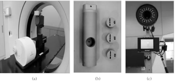
We placed a programmable motion phantom and marker block on the top of the computerized imaging reference systems (CIRS) dynamic thorax phantom model 008A (CIRS, Norfolk, VA) by CT simulator (Brilliance Big Bore 16-slice, Philips, Netherlands) with RPM system. For acquisition of breathing signal, patients were instructed to maintain a respiratory rate of 12 breaths per minute (bpm). We applied acquired breath signal to dynamic phantom system and scanned 4DCT with inserting 3 nodes (1, 2 and 3 cm). The scanning conditions were as follow: 120 kVp and 250 mAs. The 4DCT images that
Fig 1. The set-up of the dynamic phantom used to model the respiratory trajectories and the real-time position management (RPM) systems used to acquire the 4DCT images via the computed tomography (CT) simulator: (a) Dynamic phantom, (b) Insert and image nodal sizes of 1, 2 and 3 cm, (c) RPM System: Infrared camera
(a) (b) (c)
were acquired included stationary and moving images based on movement of the node from 0 to 90%, at 10%
intervals. We reconstructed the images with the following parameters: maximum intensity projection (MIP), minimum intensity projection (MinIP) and average intensity projection (AVG) images. The image-filtering algorithm of the CT simulator and the extracted Digital Imaging and Communications in Medicine files (DICOM) were used to accomplish this task (Fig. 1).
The TruebeamSTx X-Ray Imaging (XI) system (Varian Medical system, Palo Alto, CA) was used for CBCT scanning. The scanning parameters were as follow: 125 kVp, 270 mAs. We acquired five images each for the stationary and moving states, based on the tracking of position and respiratory motion, respectively, for the following nodal sizes: 1, 2 and 3 cm (Fig. 2).
We analyzed the images by using the values of MIP, MinIP and AVG, which were derived from the image-
Fig 2.
The set-up of the dynamic phantom used to acquire the cone-beam computed tomography (CBCT) images with the TruebeamSTx X-Ray imaging (XI) system.
Fig 3.
The measurement of the nodal
volume using the computed
tomography (CT) ranger tool of the
Eclipse system.
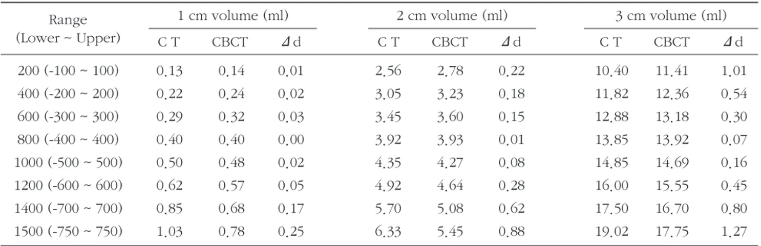
Table 1. A table showing the measurement and comparison of the volumes from the stationary images acquired with computed tomography (CT) and cone-beam computed tomography (CBCT), based on the changes evident over the specified range and the nodal size.
Δdis difference value between CT and CBCT
1 cm volume (ml) 2 cm volume (ml) 3 cm volume (ml)
C T CBCT Δd C T CBCT Δd C T CBCT Δd
200 (-100 ~ 100) 0.13 0.14 0.01 2.56 2.78 0.22 10.40 11.41 1.01
400 (-200 ~ 200) 0.22 0.24 0.02 3.05 3.23 0.18 11.82 12.36 0.54
600 (-300 ~ 300) 0.29 0.32 0.03 3.45 3.60 0.15 12.88 13.18 0.30
800 (-400 ~ 400) 0.40 0.40 0.00 3.92 3.93 0.01 13.85 13.92 0.07
1000 (-500 ~ 500) 0.50 0.48 0.02 4.35 4.27 0.08 14.85 14.69 0.16
1200 (-600 ~ 600) 0.62 0.57 0.05 4.92 4.64 0.28 16.00 15.55 0.45
1400 (-700 ~ 700) 0.85 0.68 0.17 5.70 5.08 0.62 17.50 16.70 0.80
1500 (-750 ~ 750) 1.03 0.78 0.25 6.33 5.45 0.88 19.02 17.75 1.27
Range (Lower ~ Upper)
Table 2. A table showing the measurement and comparison of the volumes from the moving images acquired with computed tomography (CT) and cone-beam computed tomography (CBCT), based on the changes evident over the specified range and the nodal sizes.
MIP = Maximum intensity projection, MinIP=Minimum intensity projection, AVG =Average Intensity Projection and CBCT =Cone-Beam Computed Tomography.
CBCT Values are mean ± stand deviation.
1 cm (ml)
MIP 0.54 0.95 1.27 1.60 1.96 2.42 3.15 3.81
MinIP 0.00 0.00 0.00 0.00 0.00 0.00 0.00 0.00
AVG 0.00 0.00 0.00 0.18 0.50 1.50 2.18 2.18
CBCT 0.00±0.00 0.00±0.00 0.00±0.00 0.00±0.00 0.14±0.01 0.34±0.01 1.01±0.05 1.48±0.06
2 cm (ml)
MIP 6.76 7.92 8.87 9.86 10.83 11.98 13.63 15.21
MinIP 0.00 0.00 0.00 0.00 0.00 0.00 0.00 0.02
AVG 0.00 0.07 0.97 2.40 4.52 7.10 9.88 11.80
CBCT 0.00±0.00 0.00±0.00 0.81±0.08 2.19±0.11 4.03±0.15 6.17±0.16 8.47±0.15 9.61±0.17
3 cm (ml)
MIP 21.96 24.27 26.08 27.91 29.72 31.84 34.83 37.57
MinIP 0.87 1.22 1.48 1.72 1.97 2.42 2.93 3.33
AVG 2.25 4.08 5.78 12.29 17.41 22.89 28.12 31.53
CBCT 2.29±0.15 4.16±0.20 7.16±0.30 11.35±0.42 16.17±0.49 21.07±0.48 25.80±0.46 28.18±0.42
Range 200 400 600 800 1000 1200 1400 1500
(Lower~Upper) (-100~100) (-200~200) (-300~300) (-400~400) (-500~500) (-600~600) (-700~700) (-750~750)
filtering algorithm of the CT simulator. The volumes were acquired from the CBCT images using the Eclipse CT ranger tool (Version 11.0, Varian, Palo Alto, USA) system.
We analyzed with the CT ranger tool and used comparative measuring the volumes by doing auto contouring after establishing the region of interest (ROI) on node and changing the number of ranges into in order of 200, 400, 600, 800, 1000, 1200, 1400 and 1500 (Fig. 3).
3. RESULTS
Table 1 shows the volumes of CT and CBCT in the state of stationary. Minimum difference of the volumes is 0.00, 0.01 and 0.07 ml in 1, 2 and 3 cm. Maximum difference of the volumes is 0.25, 0.88 and 1.27 ml in 1, 2 and 3 cm, respectively. The actual volume for 1, 2 and 3 cm is 0.52, 4.18 and 14.13 ml, respectively. All of these values are similar at range 800 and 1000. In range 800,
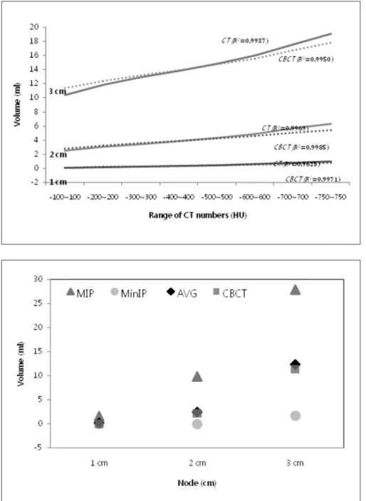
Fig 4.
A graph showing the line of best fit for the computed tomography (CT) and cone-beam computed tomography (CBCT) images for values ranging from 200 to 1500 for the nodes in the stationary state.
Fig 5.
A graph showing the values for the
maximum intensity projection (MIP),
minimum intensity projection
(MinIP), average intensity projection
(AVG) and those of the cone-beam
computed tomography (CBCT), for
the nodes, over a range of 200 to
1500 in the moving state.
difference in volume between CT and CBCT for 1, 2 and 3 cm was 0.00, 0.01 and 0.07 ml, respectively. The changes in the volumes for the specified ranges indicated that the volumes tended to increase as the range widened (Fig. 4).
We compared the volumes acquired from the 4DCT and CBCT images in the moving state, using the reconstruction image-filtering algorithm. In range 800, the volumes for a nodal size of 1 cm were 1.60, 0.00 and 0.18 ml for MIP, MinIP and AVG, respectively, while CBCT was 0.00±0.00 ml. The volumes for a nodal size of 2 cm for MIP, MinIP and AVG were 9.98, 0.00 and 2.40 ml, respectively whereas CBCT was 2.19±0.11 ml. The volumes for a nodal size of 3 cm for MIP, MinIP and AVG were 27.91, 1.72 and 12.29 ml, respectively while CBCT was 11.35±0.42 ml (Table 2). Difference in volumes for MIP, MinIP against CBCT was lager as node size was bigger. It is the result that demonstrated change of the volumes of MIP, MinIP, AVG and CBCT respectively with nodal size 1, 2 and 3 cm in the moving condition according to the change of range (Fig. 5).
4. DISCUSSION and CONCLUSION
We demonstrated that nodal volumes were various significantly when reconstruction images applied to image-filtering algorithm following comparative analysis for the values of MIP, MinIP and AVG volumes of 4DCT and CBCT.
After comparing the volumes in both stationary and moving states, we found that the volumes increased as the range widened. We compared both states for the values of MIP, MinIP, AVG and CBCT with the change in the range of values and we were able to confirm that the AVG volumes were similar to those on CBCT.
Bony anatomy such as spine be able to be used for a reference point of image matching during CBCT scan followed by treatment and they provide well-defined boundaries and their volumes are unaffected by respiratory patterns. Soft tissues such as liver, diaphragm
and heart are susceptible to breathing artifacts and other image-related artifacts such as enlargement or distortion, during imaging. Additionally, they are also affected by volume changes during respiratory cycles and provide unclear margins during CBCT scan.3,
14,15)In patient with liver cancer, the volumes on image are different due to respiratory motion. Matneyet al.
16)indicated that AVG images were similar to 30% phase images, when comparing between 4DCT and CBCT images for 10 lung cancer patients.
There was a limitation about CIRS phantom with problems such as variations in breathing pattern, period and the amplitude of the respiratory cycle in this study.
However, in this study with CIRS phantom, CBCT images were similar to AVG images of 4DCT.
5. REFERENCES
1. Underberg RW, Lagerwaard FJ, Cuijpers JP, Slotman BJ, vanSornsendeKoste JR, Senan S: Four-dimensional CT scans for treatment planning in stereotactic radiotherapy for stage I lung cancer. Int J RadiatOncolBiol Phys. 2004;60:1283-1290
2. Persson GF, Nygaard DE, MunckAfRosenschold P, et al.: Artifacts in conventional computed tomography (CT) and free breathing four-dimensional CT induce uncertainty in gross tumor volume determination. Int J RadiatOncolBiol Phys. 2011;80:1573-1580
3. Keall PJ, Mageras GS, Balter JM, et al.: The management of respiratory motion in radiation oncology. Report of AAPM Task Group 76. Med Phys 2006;33:3874-3900
4. Li R, Lewis JH, Cervino LI, Jiang SB: 4D CT sorting based on patient internal anatomy. Phys Med Biol.
2009;54:4821-4833
5. Vedam SS, Keall PJ, Kini VR, Mostafavi H, Shukla HP,
Mohan R: Acquiring a four-dimensional computed tomography data set using an external respiratory signal. Phys Med Biol. 2003;48:45-62
6. Wagman R, Yorke E, Ford E,et al.: Respiratory gating for liver tumors: use in dose escalation. Int J RadiatOncolBiol Phys. 2003;55:659-668
7. Jaffray DA, Siewerdsen JH, Wong JW,Martinez AA:
Flat-panel cone-beam computed tomography for image-guided radiation therapy. Int J RadiatOncolBiol Phys. 2002;53:1337-1349
8. Lagerwaard FJ, Van Sornsen de Koste JR, Nijssen- Visser MR, et al.: Multiple “Slow”CT Scans for Incorporating Lung Tumor Mobility in Radiotherapy Planning. Int J RadiatOncolBiol Phys. 2001;51:932-937
9. Godfrey D, Yin F, Wang Z, Yoo S, Oldham M, Willett C: Rapid low-dose 3D image-guided treatment verification of sites prone to respiratory motion using breath-hold on-board digital tomosynthesis (DTS). Med Phys 2006;33:2268
10. Wang Z, Yin F, Marks L, Wu Q, Yoo S, Willett C:
Intra- and Inter-breath-hold Position Variations for OBI Guided Amplitude Gating Treatment with Breath Hold. Med Phys 2006;33:2040
11. Yin F, Marks L, Wang Z, et al.: A technique for cone- beam CT guided stereotactic body radiation therapy.
Med Phys 2006;33:2063
12. Wang Z, Wu QJ, Marks LB, Larrier N, Yin FF: Cone- beam CT localization of internal target volumes for stereotactic body radiotherapy of lung lesions. Int J RadiatOncolBiol Phys. 2007;69:1618-1624
13. Li H, Zhu XR, Zhang L, et al.: Comparison of 2D radiographic images and 3D cone beam computed tomography for positioning head-and-neck
radiotherapy patients. Int J RadiatOncolBiol Phys.
2008;71:916-925
14. Heinzerling JH, Anderson JF, Papiez L, et al.: Four- Dimensional computed tomography scan analysis of tumor and organ motion at varying level of abdominal compression during stereotaxic treatment of lung and liver. Int J RadiatOncolBiol Phys.
2008;70:1571-1578
15. Wunderink W, Romero AM, Osorio EM, Boer HC, Levendag PC, Heijmen BJ: Target coverage in image- guided stereotactic body radiotherapy of liver tumors.
Int J RadiatOncolBiol Phys. 2007;68:282-290
16. Matney J, Vedam S,Dong L, et al.: Is average CT a good estimate of mid-ventilation position of surrounding normal structures for proton therapy treatment planning of lung tumors?. Phys Med Biol.
2009;36:2710
Purpose :
The aim of this study was to compare the differences between the volumes acquired with four-dimensional computed tomography (4DCT)images with a reconstruction image-filtering algorithm and cone-beam computed tomography (CBCT) images with dynamic phantom.Materials and Methods :
The 4DCT images were obtained from the computerized imaging reference systems (CIRS) phantom using a computed tomography (CT) simulator. We analyzed the volumes for maximum intensity projection (MIP), minimum intensity projection (MinIP) and average intensity projection (AVG) of the images obtained with the 4DCT scanner against those acquired from CBCT images with CT ranger tools.Results :
Difference in volume for node of 1, 2 and 3 cm between CBCT and 4DCT was 0.54~2.33, 5.16~8.06, 9.03~20.11 ml in MIP, respectively,0.00~1.48, 0.00~8.47, 1.42~24.85 ml in MinIP, respectively and 0.00~1.17, 0.00~2.19, 0.04~3.35 ml in AVG, respectively.Conclusion :
After a comparative analysis of the volumes for each nodal size, it was apparent that the CBCT images were similar to the AVG images acquired using 4DCT.Seong-Eun Kim, Hui-SuWon, Joo-WanHong, Nam-JunChang, Woo-HyunJung, Byeong-DonChoi
Comparison of Volumes between Four-Dimensional Computed Tomography and Cone-Beam Computed
Tomography Images using Dynamic Phantom
Department of Radiation Oncology, Seoul national university bundang hospital, Gyeonggi-do, Korea
Abstract