Clin Endosc 2012;45:177-180
Copyright © 2012 Korean Society of Gastrointestinal Endoscopy 177 CASE REPORT
A Case of Ampullary Perforation Treated with a Temporally Covered Metal Stent
Woo Young Park, Kwang Bum Cho, Eun Soo Kim and Kyung Sik Park
Department of Internal Medicine, Keimyung University School of Medicine, Daegu, Korea
Endoscopic retrograde cholangiopancreatography (ERCP)-related perforation is classified into three or four types based on anatomical location and the mechanism of injury. Although ampullary injury, among them, may be managed nonsurgically, surgical management is required in cases of perforation with retroperitoneal fluid collection and severe condition. Here, a patient with ERCP-related severe am- pullary perforation with retroperitoneal fluid collection that was treated nonsurgically with a covered stent is presented.
Key Words: Perforation; Endoscopic retrograde cholangiopancreatography; Stents Open Access
Received: October 10, 2011 Revised: January 12, 2012 Accepted: January 17, 2012
Correspondence: Kwang Bum Cho
Department of Internal Medicine, Keimyung University School of Medicine, 56 Dalseong-ro, Jung-gu, Daegu 700-712, Korea
Tel: +82-53-250-7088, Fax: +82-53-250-7088, E-mail: [email protected]
cc This is an Open Access article distributed under the terms of the Creative Commons Attribution Non-Commercial License (http://creativecommons.org/
licenses/by-nc/3.0) which permits unrestricted non-commercial use, distribution, and reproduction in any medium, provided the original work is properly cited.
Print ISSN 2234-2400 / On-line ISSN 2234-2443 http://dx.doi.org/10.5946/ce.2012.45.2.177
INTRODUCTION
The endoscopic retrograde cholangiopancreatography (ERCP)-related perforation are rare but sometimes carries 16% to 18% of death rate.1 The incidence of perforation has been reported from 0.3% to 2.2%.2-5 ERCP-related perfora- tion is classified into three or four types based on anatomical location and the mechanism of injury.2,3 The management of ERCP-related perforation has been differentiated by the types of perforation. Guidewire perforation are benign and in general do not require surgery.2,3 Periampullary perforations with the diagnosis of retroperitoneal air may be managed nonsurgically such as by aggressive endoscopic drainage and medical treatment with broad spectrum antibiotics.3,5 But pa- tients with retroperitoneal fluid collection have worse prog- nosis and require surgical intervention.2 Here, we present our experience in a case of ERCP-related severe ampullary perfo- ration with retroperitoneal fluid collection that was treated nonsurgically with a covered metal stent.
CASE REPORT
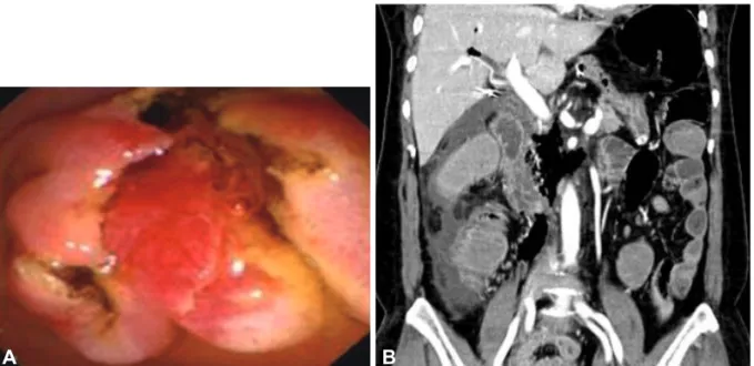
A 61-year-old Korean woman was referred to our hospital for right upper quadrant pain. She had undergone cholecys- tectomy for acute calculous cholecystitis 4 years ago. Initial abdominal computed tomography (CT) showed biliary tree dilatation but, although periampullary swelling was suspect- ed, no definite occluding lesion was evident. ERCP was per- formed to obtain a biopsy specimen from the ampulla, which showed nonspecific dilatation of the common bile duct (CBD) without a definite mass around the ampulla. A specimen was taken from the ampulla after endoscopic sphincterotomy (Fig.
1A). The day after the ERCP, she developed a severe right flank pain and fever of 39℃, shortness of breath (26/min), and rapid pulse rate (102/min). A physical examination re- vealed decreased bowel sound and whole abdominal disten- sion. CBC showed intense leukocytosis (22,000/mm3) and neutrophila (94.6%). Arterial gas analysis showed hypoxemia (pO2=60.8 mm Hg on room air). Abdominal CT revealed retroperitoneal air and fluid collection suggesting post-ERCP ampullary perforation (Fig. 1B). Although her symptoms were severe, her condition looked stable and there was no sign of sepsis, and conservative treatment was considered.
Subsequent ERCP revealed ampullary edema only, but the cholangiography showed continuous leakage of the radiocon- trast media from the ampullary level. Although the perforat- ing point was not visualized, a 5-cm long, 10-mm in diameter, full-covered metal stent (Niti-s; Taewoong Medical, Seoul,
178 Clin Endosc 2012;45:177-180 Ampullary Perforation and Metal Stent
Korea) was inserted to seal the perforation defect and to pro- vide a lumen for biliary drainage (Fig. 2).The fever and the abdominal pain after the ERCP subsided at day 4 after the stenting. She was fasted for 6 days and intravenous antibiotics and fluids were administered. Her clinical course was un- eventful. Oral feeding was resumed at day 7 after the stenting.
No further surgical intervention was necessary. Retroperito- neal air and fluid collection gradually resolved by follow-up abdominal CT and thus the stent was extracted at day 10 (Fig.
3A). The patient was discharged on day 23 after ERCP and consequent abdominal CT revealed complete resolution of the retroperitoneal fluid collection (Fig. 3B).
DISCUSSION
Traditionally, traumatic periduodenal perforation has been managed surgically. The recent paradigm of management, however, has shifted to a more selective approach that re- quires consideration of perforation type and surgical indica- tions.2,3,6 The majority of patients without free wall duodenal perforation or peritoneal signs are being treated nonsurgical- ly.2,4,7 The retroperitoneal air is a common finding indicating nonsurgical management and the amount of retroperitoneal air is not correlated with clinical course. For those treated conservatively, biliary drainage is the mainstay of treatment to reduce morbidity and mortality3 and is usually consisted of endoscopic nasobiliary drainage (ENBD) and endoscopic ret- Fig. 1. The Immediate post-endoscopic retrograde cholangiopancreatography images. (A) Duodenoscopic finding showing fully cut ampulla of vater after endoscopic sphicterotomy (EST). (B) Computed tomography showing collected air and fluid at right retroperitoneal space caused by EST.
A B
Fig. 2. Images of the inserted stent. (A) Duodenoscopic finding showing the inserted covered-stent at ampulla of vater. (B) Abdominal com- puted tomography showing the metal stent in the common bile duct.
A B
Park WY et al.
179 rograde biliary drainage (ERBD). The finding of retroperito-
neal fluid collection, however, suggests continued bile leak from the site of perforation. It is suggested that patients with retroperitoneal fluid collection have worse prognosis and re- quire surgical intervention.2,6
However, the traditional drainage methods such as ENBD or ERBD may be limited for preventing bile and pancreatic fluid leakage, especially when severe CBD dilatation or a large perforation hole is present. Surgery should be undertaken, therefore, if pain and abdominal signs are prominent, if sup- puration is suspected, or if symptoms do not improve after a brief period of nonoperative management.8
Biliary self-expanding metal stents have the advantage of being inserted with small sizes and provide large diameters for biliary drainage; however, their use in benign conditions has been limited, mainly because of difficulty in extracting them. On the other hand, a covered stent has been occasion- ally used to treat an esophagorespiratory fistula or an esopha- geal rupture.9 In cases of ampullary perforation, a covered stent can provide complete sealing of the perforation defect and the stent lumen may allow physiologic drainage of bile and thus prevent additional fluid leakage. A small sized report suggested that fully covered metal stents were removed with- out any complication after being placed in the CBD for a mean time of over 4 months and that it could be used in the management of benign biliary conditions.10
Recently, a case of persistent duodenal fistula caused by sphincterotomy-related duodenal perforation was reported.
The patient underwent an ERCP and sphincterotomy, after which a retroperitoneal duodenal perforation occurred. She
underwent a laparotomy and drainage of the retroperitoneal space. After that, a high volume duodenal fistula developed.
However, the fistula healed completely after the transient use of stents.11 Based on this concept, we applied a covered stent to the perforation site immediately after the perforation de- veloped, and achieved complete resolution of the retroperito- neal fluid collection and rapid clinical improvement. Howev- er, despite this treatment success, the merits of covered stents for ampullary perforation with retroperitoneal fluid collection have not been fully established because nonsurgical treatment failures have a high complication rate, with a potentially fatal outcome. Therefore, management using a covered stent and serial follow-up by abdominal CT may be useful treatment option for ERCP-related ampullary perforation with retro- peritoneal fluid collection in selected patients.
Conflicts of Interest
The authors have no financial conflicts of interest.
REFERENCES
1. Cotton PB, Lehman G, Vennes J, et al. Endoscopic sphincterotomy complications and their management: an attempt at consensus. Gastro- intest Endosc 1991;37:383-393.
2. Stapfer M, Selby RR, Stain SC, et al. Management of duodenal perfora- tion after endoscopic retrograde cholangiopancreatography and sphin- cterotomy. Ann Surg 2000;232:191-198.
3. Howard TJ, Tan T, Lehman GA, et al. Classification and management of perforations complicating endoscopic sphincterotomy. Surgery 1999;
126:658-663.
4. Enns R, Eloubeidi MA, Mergener K, et al. ERCP-related perforations:
risk factors and management. Endoscopy 2002;34:293-298.
5. Fatima J, Baron TH, Topazian MD, et al. Pancreaticobiliary and duode- nal perforations after periampullary endoscopic procedures: diagnosis Fig. 3. The Follow-up images. (A) Balloon occluded cholangiogrphic finding showing no more leakage of the radiocontrast media at ampul- lary level. (B) Follow-up abdominal computed tomography showing complete resolution of the retroperitoneal air and fluid collection.
A B
180 Clin Endosc 2012;45:177-180 Ampullary Perforation and Metal Stent
and management. Arch Surg 2007;142:448-454.
6. Avgerinos DV, Llaguna OH, Lo AY, Voli J, Leitman IM. Management of endoscopic retrograde cholangiopancreatography: related duodenal perforations. Surg Endosc 2009;23:833-838.
7. Morgan KA, Fontenot BB, Ruddy JM, Mickey S, Adams DB. Endo- scopic retrograde cholangiopancreatography gut perforations: when to wait! When to operate! Am Surg 2009;75:477-483.
8. Chung RS, Sivak MV, Ferguson DR. Surgical decisions in the manage- ment of duodenal perforation complicating endoscopic sphincteroto- my. Am J Surg 1993;165:700-703.
9. Hu HT, Song HY, Kim JH. Immediate placement of a temporary cov- ered stent for the management of iatrogenic malignant esophageal per- foration. Cardiovasc Intervent Radiol 2011;34:886-888.
10. García-Cano J, Taberna-Arana L, Jimeno-AyllÓn C, et al. Use of fully covered self-expanding metal stents for the management of benign biliary conditions. Rev Esp Enferm Dig 2010;102:526-532.
11. Vezakis A, Fragulidis G, Nastos C, Yiallourou A, Polydorou A, Voros D.
Closure of a persistent sphincterotomy-related duodenal perforation by placement of a covered self-expandable metallic biliary stent. World J Gastroenterol 2011;17:4539-4541.