Case Report
Jejunogastric intussusception: a rare complication of gastric cancer surgery
Seung Hyoung Lee, In Gyu Kwon, Seung Wan Ryu, Soo Sang Sohn
Department of Surgery, Keimyung University School of Medicine, Daegu, Republic of Korea
Received September 4, 2014; Accepted October 23, 2014; Epub November 15, 2014; Published November 30, 2014
Abstract: Jejunogastric intussusception (JGI) is a rare condition and less than 200 cases have been published since its first description in 1914. In addition, JGI is potentially lethal complication of gastrectomy or gastrojejunostomy.
We report the case of a 73-year-old man with a history of a Billroth II procedure who presented to the emergency department after 6 hours of epigastric pain and hematemesis. Endoscopy and computed tomography showed in- tussuscepted jejunum through a gastrojejunostomy that required emergency operation. At laparotomy a retrograde type II, JGI was confirmed and managed by resection of involved intestine. Postoperative recovery was uneventful.
This case presents the rare complication of acute jejunogastric intussusception more than 25 years after a Billroth II procedure.
Keywords: Jejunogastric intussusception (JGI), intussusception, gastrojejunostomy
Introduction
Intussusception represents 90% of patients in children and adult intussusception occur only 5% of all intussusceptions [1]. In contrast to intussusceptions in children, a demonstrable etiology is found in 70% to 90% of cases in the adult population [1-3]. Among them, jejunogas- tric intussusception (JGI) is a rare complication of all types of gastric resection and occurs in less than 0.1% of gastric resection [4]. Ad- ditionally, JGI is potentially lethal complication of gastrectomy or gastrojejunostomy. The mor- tality rate of this condition is as high as 50% if surgery is delayed for more than 48 hours [5].
We present the case of a patient who present- ed with retrograde JGI occurring 25 years after partial gastrectomy with Billroth II gastroenter- ostomy for gastric cancer and reviews the asso- ciated studies. This is the first report of JGI occurred 25 years after partial gastrectomy with Billroth II gastroenterostomy in a patient with gastric cancer.
Case report
A 73-year-old man was referred to the emer- gency department of Keimyung University
Dongsan Medical center (Daegu, Republic of Korea) from a local hospital, complaining of epi- gastric pain, nausea and hematemesis. He had undergone a partial gastrectomy and Billroth II anastomosis for gastric cancer approximately 25 years ago. Upon arrival at the emergency department, the patient appeared mildly dehy- drated but, he was hemodynamically stable. On physical examination, he was afebrile and his abdomen was soft, mild distended and diffuse- ly tender (particularly in the epigastric area).
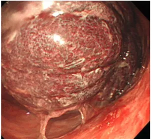
Furthermore, the laboratory findings on arrival were normal. Especially, his white blood cell count was 9080 cell/µl and his hemoglobin was 12.4 g/dL. Emergency endoscopy was per- formed by endoscopist, internal medicine doc- tor and that revealed gastric ulcer (acute stage 2, Forrest type Ib) and a bulky, rounded conges- tive mass that occupied practically the posteri- or wall of the remnant stomach (Figure 1).
Initially, internal medicine doctor interpreted a bulky mass in the stomach as a bezoar.
On the third day in the hospital, the patient complained more epigastric pain and his white blood cell count was elevated 14420 cell/µl and a computed tomography (CT) scan of the abdomen was performed: it showed a distend-
Figure 1. Endoscopic picture: A bulky, rounded con- gestive mass is seen in the stomach and the adja- cent gastric mucosa is petechial changed.
ed stomach containing a long segment of the jejunum with a thickened, strangulated bowel wall and a large quantity of ascitic fluid (Figure 2). These findings were consistent with JGI and an exploratory laparotomy was performed after the CT scan.
The surgical findings showed a markedly dilat- ed afferent loop distal to the previous gastroje- junostomy and approximately 60 cm of efferent loop retrograde invagination in the remnant stomach through the previous gastrojejunosto- my (Figure 3). The intussusception could not be reduced because of severe inflammation and edema of the invaginated jejuna loop. In addi- tion, the segment of involved bowel was found to be necrotic, and therefore resection of involved jejunum was performed. Intestinal continuity was restored with recreation of the gastrojejunostomy just proximal to previous anastomotic site. The pathologic finding of the resected bowel revealed intussusception of the jejunum with congestion and edema (Figure 4).
No leading point was found. The patient recov- ered well after surgery without complications and he was discharged from hospital with a good health status.
Written informed consent was obtained from the patient. The study was approved by the eth- ics committee of Dong San Medical Center, Keimyung University School of Medicine, Dae- gu, Republic of Korea.
Discussion
Jejunogastric intussusception (JGI) was first reported in 1914 by Bozzi in a patient with gas- trojejunostomy [6]. And then, it was also report- ed in patients after Billroth I reconstruction, Billroth II reconstruction and total gastrectomy with Roux-en-Y anastomosis [4, 7-9]. But these literatures have been reported less than 200 cases. Recently, retrograde intussusceptions have been also reported after Roux-en-Y gas- tric bypass, pancreaticojejunostomy and rarely in association with previously placed gastros- tomy tubes [10-12]. These JGI after gastrecto- my is a rare disorder and occurs in only 0.07- 2.1% of individuals who underwent gastrecto- my [13, 14].
There is a wide variation in the duration be- tween the gastric operation and the occurrence of JGI: 6 days to 20 years in patients with gas- tric surgery respectively [15]. In this case, the interval between partial gastrectomy with Bill- roth II reconstruction and JGI was 25 years.
This is the longest period of the JGI occurrence after gastric operation due to gastric cancer that have ever reported.
Up to now, the pathogenesis of JGI is unclear.
There are two major theories such as functional and mechanical. The functional theory that is the most widely accepted is the disordered motility with functional hyperperistalsis trig- gered by spasm or hyperacidity [16]. The mechanical factors have been incriminated such as adhesions, a long mesentery, gastric derangements and a sudden increase of the intraabdominal pressure [17].
Classically, JGI can be classified into four types.
Type I is an antegrade intussusception of the afferent limb (5.5%). Type II is a retrograde intussusception of the efferent limb and is most commonly observed (75.5%). Type III is a combination of types I and II with intussuscep- tion of the afferent and efferent limbs (6.5%).
Type IV consists of an intussusception through a Braun side-to-side jejunojejunal anastomosis (8%) [18]. Our case was classified as type II.
Clinically, JGI can present acutely or chronically ill. Acute presentation is characterized by the sudden epigastric pain, emesis with or without hematemesis, and a palpable and tender
gastric surgery. Among them, hematemesis can occurred because of compromised jejunal vascular supply [19]. In the chronic form, the symptoms may be similar to the acute form but transient, milder and subside spontaneously. In addition, in the chronic form, the symptoms may be vague and physicians can make a wrong diagnosis or delayed diagnosis without any attention of JGI [20].
Because JGI is rare, this condition is not often considered as a diagnosis when patients are first visiting. The diagnosis of JGI can be deter- mined with many imaging studies, such as Figure 2. Computed tomography (CT) scan of the abdomen. A. An abdominal CT scan (coronal view) shows markedly dilated stomach (arrowhead) with mass-like, thick-walled bowel loops (arrow). B. An abdominal CT scan (axial view) shows that mesenteric vessels and fat also entrapped in the dilated lumen (arrow).
Figure 3. Operative photograph shows that efferent limb intussuscepted into the previous gastrojejunos- tomy (arrow).
Figure 4. A gross surgical specimen shows the ne- crotic changed intussusception (arrow).
abdominal mass. These symptoms are the
ies and CT scan of the abdomen and pelvis.
Endoscopy is the diagnostic procedure for the patient with hematemesis. But, the intussus- ception could be mistaken as an immobile clot or a bezoar on endoscopy like in our case [18, 21]. US findings of intussusception classically reveals an mass with echogenic center sur- rounded by concentric echogenic rings with a peripheral rim of hypoechogenicity, described as “pseudokidney” or “doughnut” sign [22]. US is the method of first choice because it can be performed at bedside without ionizing radia- tion. CT allows the differentiation of the distinct types of the JGI and the views given by CT are often more easily accepted by the surgeons.
The typical CT finding of intussusception is a soft tissue mass with a “sausage” or “target”
appearance [22].
The definitive treatment of JGI is the surgical intervention as soon as possible. Surgical option include reduction with correction, resec- tion of involved bowel, revision of the an- astomosis.
Mortality rates increase suddenly with surgical delay and reported mortality ranges from 10%
for treatment within the first 48 hours to over 50% with a 96 hours delay [23]. So, early diag- nosis is very important because of mortality and morbidity. To reduce delays in diagnosis and to minimizing morbidity and mortality, we should keep in mind that JGI is a possible com- plication in a patient with gastrectomy, present- ing abdominal pain, hematemesis, even though many years have passed after gastric surgery.
Conclusion
JGI is a rare complication after all types of gas- tric resection but, this condition is lethal if diag- nosis and surgery is delayed. Therefore, we should be aware of this disease and it is impor- tant to consider JGI in all patients with previous gastric surgery presenting with abdominal pain, vomiting or hematemesis.
Disclosure of conflict of interest None.
Address correspondence to: Dr. Seung Wan Ryu, Department of Surgery, Keimyung University Dong San Medical Center, 56 Dalseong-Ro, Jung-Gu, Daegu 700-712, Republic of Korea. Tel: +82-53-
250-7322; Fax: +82-53-250-7322; E-mail: questi- [email protected]
References
[1] Azar T and Berger DL. Adult intussusception.
Ann Surg 1997; 226: 134-138.
[2] Nagorney DM, Sarr MG and McIlrath DC. Sur- gical management of intussusception in the adult. Ann Surg 1981; 193: 230-236.
[3] Haas EM, Etter EL, Ellis S and Taylor TV. Adult intussusception. Am J Surg 2003; 186: 75-76.
[4] Wheatley MJ. Jejunogastric intussusception di- agnosis and management. J Clin Gastroenterol 1989; 11: 452-454.
[5] Hovelius L. Jejunogastric intussusception after gastric resection. A report of two cases. Acta chirurgica Scandinavica 1971; 137: 491-494.
[6] Bozzi E. Bull Acad Med 1914; 122: 3-4.
[7] Shiffman MA and Rappaport I. Intussusception following gastric resection. Report of five cas- es. Report of five cases and literature review.
Am Surg 1966; 32: 715-724.
[8] López-Mut JV, Cubells M, Campos S, Miranda V and Rivera P. Jejunogastric intussusception: a rare complication of gastric surgery. Abdom Imaging 1998; 23: 558-559.
[9] Salem MH, Coffman SE and Postlethwait RW.
Retrograde intussesception at the gastrojeju- nal stoma. Ann Surgy 1959; 150: 864-871.
[10] Goverman J, Greenwald M, Gellman L and Ga- daleta D. Antiperistaltic (retrograde) intussus- ception after Roux-en-Y gastric bypass. Am Surg 2004; 70: 67-70.
[11] Whipple OC, Stringer EF, Senkowski CK and Hartley M. Retrograde intussusception of the efferent limb after a pancreaticojejunostomy.
Am Surg 2003; 69: 353-355.
[12] Gasparri MG, Pipinos II, Kralovich KA and Mar- golin DA. Retrograde jejunogastric intussus- ception. South Medl J 2000; 93: 499-500.
[13] Marx WJ. Reduction of jejunogastric intussus- ception during upper gastrointestinal examina- tion. AJR Am J Roentgenol 1978; 131: 334- 336.
[14] Narita HFK, Yoshitomi H, Yamamori N, Iguchi T, Hori K, Hato M. Post operative intussusception - report of a case, and a comparison be- tween adult and pediatric intussusceptions af- ter laparotomy. Nihon Rinsyo Gekagekai- gakukaishi 1951; 52: 2125-2395.
[15] Conklin EF and Markowitz AM. Intussuscep- tion, a complication of gastric surgery. Surgery 1965; 57: 480-488.
[16] Robertson DS and Weder CH. Acute jejunogas- tric intussusception. Can J Surg 1968; 11:
210-214.
[17] Bundrick TJ and Turner MA. Retrograde jejuno- gastric intussusception. Rev Interam Radiol 1981; 6: 21-24.
[18] Brynitz S and Rubinstein E. Hematemesis cau- sed by jejunogastric intussusception. En- doscopy 1986; 18: 162-164.
[19] Foster DG. Retrograde jejunogastric intussus- ception; a rare cause of hematemesis; review of the literature and report of two cases. AMA Arch Surg 1956; 73: 1009-1017.
[20] Olsen AK and Bø O. Intussusception as a com- plication of partial gastrectomy. A case report.
Acta chirurgica Scandinavica 1978; 144: 405- 408.
[21] O'Dell KB, Gordon RS and Victory C. Acute je- junogastric intussusception: a rare cause of abdominal pain. Ann Emerg Med 1992; 21:
565-567.
[22] Hammond N, Miller FH and Dynes M. In- tussusception into the enteroanastomosis af- ter Billroth II gastrectomy and Roux-en-Y jeju- nostomy: sonographic and CT findings. AJR Am J Roentgenol 2001; 177: 624-626.
[23] Walstad PM, Ritter JA and Arroz V. Delayed je- junogastric intussusception after gastric sur- gery: an ever-present threat. Am Surg 1972;
38: 172-175.