DOI:10.5125/jkaoms.2010.36.5.434
434
Ⅰ.
서 론Melanotic neuroectodermal tumor of infancy (MNTI)는 주로
1세 이하의 유아에서 발생하는 착색성 양성 신생물이다
1,2.
1918년 Krompecher
3이 처음으로 congenital melanocarcino- ma로 보고한 이래로 이 양성 신생물의 기원에 대해 연구되 어 왔으며, pigmented ameloblastoma, retinal angle tumor, melanotic progonoma, melanoblastoma, melanoameloblas- toma, retinoblastic teratoma 등으로 불리어졌으나, 1966년 Borello와 Gorlin
4이 소변검사에서 vanillylmandelic acid (VMA)가 높게 나타나는 것을 발견하여, 처음으로 신경능 기원의 MNTI로 명명하였다. 그 이후로 여러 가지 조직검
사, 면역화학검사, 전자현미경검사에 의해 이 이론을 증명 할 수 있었다
5-8. MNTI의 90% 이상이 두경부 영역에서 발생 하며, 주로 상악전치부에서 2-4 cm의 종창성 병소로 나타 난다. 그 외에 두부, 하악골, 뇌에서도 발생하며, 드물게 어 깨, 피부, 사지 등에서도 나타난다고 보고되고 있다. 남자 대 여자의 성비는 6:7로 거의 비슷하게 나타나며, 90% 이상 에서 출생 후 6개월 이내에 발생한다. 평균 발현나이는 4.3 개월이지만 23세, 24세, 67세에 발생한 경우도 보고되었다.
MNTI는 양성 종양으로서 주로 보존적 절제술로 양호한 결 과를 얻을 수 있지만, 10-15% 정도로 재발하였다. 또한 1.5- 2.1%의 악성변이를 나타나며, 이 중 5%에서 전이가 나타 났다
9-11. 1988년 Steinberg 등
12은 다발성의 MNTI에 대해 보 고하였는데 문헌고찰에 의하면 전체 200예 중에서 2예에 서 다발성의 MNTI를 발견하였다. 외과적 절제술로 양호한 결과를 얻을 수 있지만, 재발과 다발성이 있다는 것은 임상 적으로 중요한 의미가 있는 것으로 수술 시 이러한 요소를 고려하는 것이 좋은 결과를 얻을 수 있으리라 생각되어 우 리가 경험한 다발성의 MNTI에 대해 소개하고자 한다.
조 영 철
685-714 울산광역시 동구 전하동 290-3 울산대학교 의과대학 구강악안면외과학교실 Yeong-Cheol Cho
Department of Oral and Maxillofacial Surgery, Ulsan university hospital 290-3 Jeonha-dong Dong-gu, Ulsan 682-714, Korea
Tel: +82-52-250-7230 Fax: +82-52-250-7236 E-mail: lovenip@mail.ulsan.ac.kr
Abstract (J Korean Assoc Oral Maxillofac Surg 2010;36:434-7)
다발성 유아기 흑색 신경외배엽성 종양의 치험례
최병환
1∙박수원
1∙장수미
1∙박봉찬
1∙손한나
1∙손장호
1∙성일용
1∙김종렬
2∙조영철
1*
1
울산대학교 의과대학 구강악안면외과학교실,
2온병원 구강악안면외과
Multicentric melanotic neuroectodermal tumor of infancy: a case report
Byoung-Hwan Choi
1, Su-Won Park
1, Soo-Mi Jang
1, Bong-Chan Park
1, Han-Na Son
1, Jang-Ho Son
1, Iel-Yong Sung
1, Jong-Ryoul Kim
2, Yeong-Cheol Cho
1*
1
Department of Oral and Maxillofacial Surgery, College of Medicine, University of Ulsan, Ulsan, Korea
2
Department of Oral and Maxillofacial Surgery, Onhospital, Busan, Korea
A melanotic neuroectodermal tumor of infancy (MNTI) is a uncommon osteolytic pigmented neoplasm that primarily affects the jaws of newborn infants. Most patients (> 90%) present with the tumor in the first year of life. Approximately 65% form in the maxilla, 11% in the mandible, 5% in the brain and elsewhere. MNTI is normally benign, but up to 15% may recur and a few have metastasized. Approximately 200 cases of MNTI have been reported but only 2 of them presented as multifocal. A case of MNTI in a 7 month old boy was encountered. The chief complaint was maxillary anteri- or ridge swelling. The incisional biopsy findings were MNTI. Two months after the first operation, mild swelling of another site was observed. The infant was examined periodically since undergoing two procedures with no recurrence. This case demonstrates the possibility of a multicentric MNTI.
We report a multicentric MNTI with a review of the relevant literature.
Key words: Melanotic neuroectodermal tumor of infancy (MNTI), Multicentric
[paper submitted 2010. 7. 14 / revised 2010. 10. 20 / accepted 2010. 10. 22 ]
다발성 유아기 흑색 신경외배엽성 종양의 치험례
435
Ⅱ.
증례보고�환자: 강 O O, 남아 7개월
�주소: 상악전치부의 종창
�병력: 내원 1개월 전 50 cm 높이에서 추락한 후에 서서 히 무통성의 종창이 성장하였고, 평소 이갈이가 심하 였다.
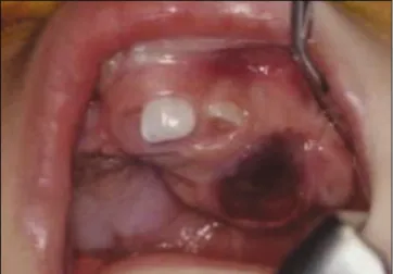
�임상소견: 상악전치부에 2×2 cm 크기의 종창부위로 유중절치가 함입되어 있고, 치조융선의 순구개측으로 종창이 확장되어 있었다. 임상소견으로는 맹출성 낭 종, 치근단 낭종으로 의심되는 상황이었다.(Fig. 1)
�혈액 및 소변검사: 특별히 이상소견은 없었으나, hepatitis (+)로 이는 모체의 수직감염으로 인한 것으로 생각하며 VMA가 9.52 mg/day 1.2-6.5)로 높게 나왔다.
�방사선검사: 구내 표준방사선사진에서 경계가 불분명 한 낭종성 소견과 함께, 유중절치의 변위를 볼 수 있었
다. Computed tomography (CT) 상에서 좌측 유중절치 부위에 순구개측으로 확장된 경계가 분명한 낭종을 관 찰할 수 있었다.(Fig. 2)
�치료경과: 첫 번째 내원하였을 때 시행한 조직검사에 서 MNTI로 진단되어 2주 후 전신마취하에 수술을 하 였다. 수술 시 유치, 병소상방의 치조점막, 주위의 건전 한 상악골까지 포함하는 en bloc excision을 시행하였 다.(Fig. 3) 광범위하게 병소를 제거하고 소파술을 시행 하였으므로 차후 재발은 거의 없고, 영구치 맹출에 장 애가 있으리라고 판단되었다. 수술부위는 특별한 이상 소견 없이 치유되었다. 퇴원하고 약 3주 후의 재내원 시 우측 치조부위에 좌측과 유사한 양상의 종창이 발 견되었다.(Fig. 4) 일단 관찰하기로 하고 주기적 검사를 하였지만 점차 종창이 심해져서 MNTI가 재발한 것으 로 보고 CT검사를 시행하였다. CT 상에서 좌측과 동일 한 양상의 낭종을 발견할 수 있었고, 첫 번째 수술을 시
Fig. 1. Clinical photograph showing swelling on left anterior alveolus.
Fig. 2. Axial computed tomography (CT) of patient showing radiolucent lesion expanding into labial and palatal direction.
Fig. 3. Intraoperative photograph of the resected tumor. Fig. 4. Clinical photograph showing on right anterior alveo-
lus at 2 months after first operation.
J Korean Assoc Oral Maxillofac Surg 2010;36:434-7
436
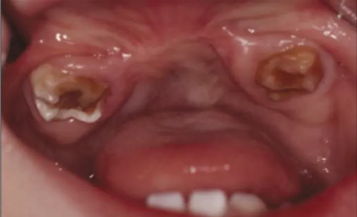
행한 때로부터 10주 후 전신마취하에 우측병소를 동 일한 방법으로 수술, 제거하였다. 병소는 잘 치유되었 다. 두 번째 수술을 시행하고 2달 후 VMA 수치가 1.20 mg/day로 나왔다. 이 후 수년 동안의 주기적 검사에서 특별한 이상은 관찰되지 않았고, 유전치의 상실과 이 로 인한 치조골 성장의 장애가 나타났다.(Fig. 5) 5세 때 전치부의 심미성과 보철수복을 위해 iliac particulate cancellous bone and marrow (PCBM)을 이용한 골이식 을 시행하였다. 이후 발음과 심미성 회복을 위해 가철 성 의치를 사용 중이며. 차후 교정, 보철 등 악기능재건 을 위한 여러 가지 치료가 필요하리라 생각한다.
�병리소견: 종양세포는 small neuroblastic cell과 larger melanin containing epithelial cell로 구성되어 있으며, 이 세포들은 dense fibrous stroma로 지지되고 smaller cell 들은 larger cell로 포위되어 alveolar formation을 형성하 고 있었다.(Fig. 6) 두 번째 수술 후 조직소견도 첫 번째 수술 후 종물보다 smaller neuroblastic cell이 좀 더 많이 나타났지만 거의 유사한 양상이었다.(Fig. 7)
Ⅲ.
고 찰MNTI의 치료는 일반적으로 외과적 절제술과 소파술로 알려져 있다. 그러나 비록 양성 종양이지만, 재발률이 15- 20% 정도이고 1.5-2.1%의 악성 전이를 보이며 급속히 성장 하고 나이가 어린 유아에서 발생한다는 것을 고려하여 수 술 시 5 mm의 건전한 조직을 포함하는 광범위한 절제술을 선호하고 있다. 본 증례에서는 첫 수술을 시행하고 약 2개 월 후 병소가 재발하였는데, 발생부위가 첫 번째와 다른 부 위이기 때문에 재발보다는 다발성의 병소가 발현한 것이 라고 판단되었다.
다발성 MNTI는 200예 중에서 2예에서 보고되었는데 사 실 MNTI에서 재발과 다발성을 구분하는 것은 다소 어려움 이 있다. Jones와 Williams
13은 4예의 다른 부위에서의 MNTI의 재발에 대해서 보고하였다. 재발된 MNTI는 비록 병소부위가 다른 부위라도 재발할 경우 병소가 더 급속히 성장하고, 공격적이고, 주위 골로 침범이 많이 되는 것으로 알려져 있다. Nagase 등
14은 MNTI의 재발에 대해 연구하였 는데 모두 악골에서 재발하였고, 술후 1년 내에 발생한 것 이 90%였다고 보고하였다. 다발성의 MNTI 기전에 대해서 Steinberg 등
12은 원발성 병소와 연결된 색소성 세포의 얇은 돌기가 골소주 내에 존재한다고 하였고, 이러한 이론에 따 라 신경능세포가 상악골의 성장에 따라 이동할 때 종양세 포가 seeding되는 효과에 의한 것이라고 설명하였다. 본 증 례에서는 원발성 병소와 위치가 다르고, 재발된 병소라고 보기에는 어려움이 있어 다발성으로 존재하는 병소가 수 술 후 자극에 의해 발현한 것으로 판단하였다.
MNTI의 진단은 과거력, 임상소견, 방사선소견과 조직검 사로 확진할 수 있다. 감별을 요하는 질병은 치성낭종, 치 성종양(ameloblastoma, Adenomatoid odontogenic tumor (AOT), odontogenic myxoma, odontogenic fibroma)등이 있으 며, 어린 나이에 발생하는 것을 볼 때 ewing sarcoma, rhab-
Fig. 5. Clinical photograph after second operation. There was no recurrence.
Fig. 6. Histopathologic findings of the primary operation showing tumor cells composed of small neuroblastic cells and larger melanin containing epithelial cells.(H&E stain- ing, original magnification ×100)
Fig. 7. Histopathologic findings of the secondary operation
showing similar features with primary operation.(H&E stain-
ing, original magnification ×100)
다발성 유아기 흑색 신경외배엽성 종양의 치험례