pISSN 2383-7837
© 2021 The Korean Society of Pathologists/The Korean Society for Cytopathology
Fine-needle aspiration (FNA) is an excellent, minimally invasive diagnostic technique for evaluating a mass or lesion. In particular, FNA is commonly used for evaluating enlarged lymph nodes (LNs). In many cases, the aspirate can provide conclusive evidence for a diagnosis; sometimes, the cytology reveals only necrosis.
Necrosis of LNs is found in various diseases. Malignant neo- plasms (including lymphomas and metastatic carcinomas) must first be excluded. In addition, numerous benign conditions, such as tuberculosis (TB) and Kikuchi disease, also cause LN necrosis. When necrosis is identified in an LN FNA cytology sample, pathologists should consider various entities and at- tempt to find clues that lead to a final diagnosis. However, LN FNA cytology sometimes reveals necrosis alone, and few studies have investigated this situation.
The aim of this study was to evaluate the causes of necrosis in FNA of enlarged LNs.
MATERIALS AND METHODS
We searched the electronic medical record system of the Keimy- ung University Dongsan Hospital from 2003–2017 to find pa- tients who underwent FNA on cervical LNs. We selected cases with a description of necrotic features on the pathology report. In addition, we also collected the clinical parameters of these cases, including patient age, sex, biopsy findings, other ancillary tests for Mycobacterium tuberculosis (MTB; including acid-fast bacilli staining), and molecular studies carried out using conventional polymerase chain reaction (PCR) or real-time PCR. We classi- fied these cases into five categories: granulomatous inflamma- tion, Kikuchi disease, suppurative inflammation, malignant neoplasm, and necrosis only. When granuloma was included in the microscopic description (e.g., granulomatous inflammation with caseous necrosis, granulomatous inflammation with a necrotic background, or necrosis with a vague granuloma), it was classi-
Causes of necrotic features in fine-needle aspirates from cervical lymph nodes
Young Jin Seo
1, Hyeongchan Shin
1,2, Hye Won Lee
1,2, Hye Ra Jung
1,21Department of Pathology, Keimyung University Dongsan Hospital, Daegu;
2Department of Pathology, Keimyung University School of Medicine, Daegu, Korea
Background: Lymph node fine-needle aspiration (LN FNA) cytology indicates necrosis in various diseases. Dominant necrotic features make the diagnosis of underlying conditions very difficult. Methods: We retrospectively reviewed 460 patients who underwent cervical LN aspiration cytology that revealed necrotic findings at Keimyung University Dongsan Hospital in Daegu, Korea, from 2003–2017. Each specimen was evaluated and analyzed in association with the clinical findings, biopsy findings, and/or other ancillary tests, including acid-fast bacilli staining and molecular testing for Mycobacterium tuberculosis. Results: When necrotic features were noted upon cervi- cal LN FNA cytology, the most common pathologic LN FNA category was necrosis alone (31.5%). The second most common category was granulomatous inflammation (31.3%), followed by Kikuchi disease (20.0%) and malignant neoplasm (8.7%). In cases where the cervical LN FNA revealed necrosis alone, the most common final diagnosis was tuberculosis. In young patients, Kikuchi disease should be considered as one cervical LN FNA category, while metastatic carcinoma should be suspected in older patients. Conclusions: Even when necrosis alone is observed in LN FNA cytology, it is important to determine the cause through further evaluation.
Key Words: Fine-needle aspiration; Lymph node; Necrosis; Tuberculosis; Kikuchi disease Received: July 20, 2020 Revised: September 16, 2020 Accepted: September 28, 2020
Corresponding Author: Hye Ra Jung, MD, PhD, Department of Pathology, Keimyung University Dongsan Hospital, Department of Pathology, Keimyung University School of Medicine, 1095 Dalgubeol-daero, Dalseo-gu, Daegu 42601, Korea
Tel: +82-53-258-7395, Fax: +82-53-258-7382, E-mail: [email protected]
fied as granulomatous inflammation. When the LN FNA result contained content that indicated histiocytic necrotizing inflam- mation, we placed the patient into the Kikuchi disease category.
Cases reported as suspicious for Kikuchi disease with various microscopic descriptions (e.g., necrotizing lymphadenitis, poly- morphous lymphoid cells, and macrophages on a necrotic back- ground) were classified as Kikuchi disease. Malignant neoplasms (e.g., metastatic carcinoma, metastatic sarcoma, metastatic mel- anoma, and malignant lymphoma) with a necrotic background were determined to be malignancies. Cases with suppurative inflammation on a necrotic background, acute inflammatory cells on a necrotic background, aspirated pus, or neutrophilic infiltration with necrotic material were classified as suppurative inflammation.
RESULTS
Clinical characteristics of the patientsWe retrieved the electronic medical reports of 460 patients who underwent LN aspiration cytology at our hospital that revealed
necrotic features. The mean age of the patients was 42.5 years (range, 2 to 86 years; median, 40 years). The male-to-female ratio (M:F ratio) was 0.62. Except for cases of pure necrosis, the most common FNA category was granulomatous inflammation (31.3%), followed by Kikuchi lymphadenitis (20.0%), malig- nancy (8.7%), and suppurative lymphadenitis (6.5%) (Table 1).
Granulomatous inflammation
Of the 460 patients, 144 were classified with granulomatous inflammation from among the LN FNA categories. The M:F ratio was 0.38. This type of inflammation was mainly distrib- uted in people from 20–39 years of age (36.8%). Of the 144 patients with granulomatous inflammation, 117 underwent an- cillary tests for MTB; 82 (70.0%) of these were diagnosed with TB. In addition, MTB testing was performed in 364 of the 460 total cases who underwent cervical LN FNA. Among these pa- tients, 148 cases were MTB-positive, which represented 32% of all cervical LN FNA cases (Table 2).
Table 1. The age and sex distribution of cervical LN FNA categories according to the FNA diagnosis
FNA category Mean age (yr) Age range (yr) Men (person) Women (person) Total (%)
Granulomatous inflammation (31.3%) 46.0 0–19 4 4 8
20–39 14 39 53
40–59 13 32 45
<60 9 29 38
Total 40 104 144
Kikuchi disease (20.0%) 26.4 0–19 15 16 31
20–39 14 37 51
40–59 1 8 9
<60 0 1 1
Total 30 62 92
Suppurative inflammation (6.5%) 40.8 0–19 0 5 5
20–39 5 4 9
40–59 4 7 11
<60 3 2 5
Total 12 18 30
Malignant neoplasm (8.7%) 61.9 0–19 0 0 0
20–39 1 0 1
40–59 8 6 14
<60 24 1 25
Total 33 7 40
Necrosis (31.5%) 44.0 0–19 5 6 11
20–39 22 33 55
40–59 13 36 49
<60 15 15 30
Total 55 90 145
Other (2.0%) 46.0 Total 5 4 9
Total 42.5 175 285 460
LN, lymph node; FNA, fine-needle aspiration.
Kikuchi disease
Ninety-two patients who underwent LN FNA were deter- mined to have Kikuchi disease. Of these 92 people, 89.1% were
young (< 40 years). The M:F ratio was approximately 1:2. Thir- teen of these patients classified as having Kikuchi disease by LN FNA underwent ancillary tests for MTB. Among them, 11
Table 2. The results of ancillary tests for Mycobacterium tuberculosis in the granulomatous inflammation, Kikuchi disease, suppurative lymphadenitis, malignant neoplasm, and necrosis categories of cervical LN FNA findings
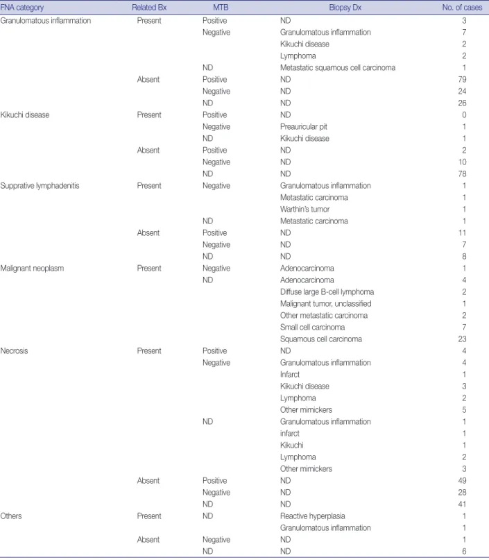
FNA category Related Bx MTB Biopsy Dx No. of cases
Granulomatous inflammation Present Positive ND 3
Negative Granulomatous inflammation 7
Kikuchi disease 2
Lymphoma 2
ND Metastatic squamous cell carcinoma 1
Absent Positive ND 79
Negative ND 24
ND ND 26
Kikuchi disease Present Positive ND 0
Negative Preauricular pit 1
ND Kikuchi disease 1
Absent Positive ND 2
Negative ND 10
ND ND 78
Supprative lymphadenitis Present Negative Granulomatous inflammation 1
Metastatic carcinoma 1
Warthin’s tumor 1
ND Metastatic carcinoma 1
Absent Positive ND 11
Negative ND 7
ND ND 8
Malignant neoplasm Present Negative Adenocarcinoma 1
ND Adenocarcinoma 4
Diffuse large B-cell lymphoma 2
Malignant tumor, unclassified 1
Other metastatic carcinoma 2
Small cell carcinoma 7
Squamous cell carcinoma 23
Necrosis Present Positive ND 4
Negative Granulomatous inflammation 4
Infarct 1
Kikuchi disease 3
Lymphoma 2
Other mimickers 5
ND Granulomatous inflammation 1
infarct 1
Kikuchi 1
Lymphoma 2
Other mimickers 3
Absent Positive ND 49
Negative ND 28
ND ND 41
Others Present ND Reactive hyperplasia 1
Granulomatous inflammation 1
Absent Negative ND 1
ND ND 6
LN, lymph node; FNA, fine-needle aspiration; Related Bx, presence or absence of biopsy on related site of fine needle biopsy; MTB, result of ancillary tests in- cluding acid-fast bacillus stain, polymerase chain reaction or reverse transcription polymerase chain reaction for Mycobacterium tuberculosis; Biopsy Dx, di- agnosis by related site biopsy; ND, not done.
Fig. 1. Smear and biopsy findings of the neck lesion diagnosed as necrosis on aspiration but changed diagnosis by biopsy (A, B). (A) Fine needle aspiration (FNA) shows colloid material. (B) Lymph node (LN) excision specimen was diagnosed as metastatic papillary carcinoma. (C, D) Case 2. (C) FNA shows pinkish amorphous material. (D) LN excision specimen was diagnosed as Warthin tumor. (E, F) Case 3. (E) FNA shows cystic fluid material. (F) LN excision specimen was diagnosed as salivary duct cyst. (G, H) Case 4. (G) FNA shows myxoid stroma. (H) LN excision specimen was diagnosed as schwannoma. (I, J) Case 5. (I) FNA shows red blood cells and fibrin material. (J) LN excision speci- men was diagnosed as reactive hyperplasia.
A
C
E
G
I
B
D
F
H
J
patients were negative, but the remaining two patients had posi- tive results. Therefore, the final diagnosis of 11 patients was TB.
Suppurative lymphadenitis
Of the 460 total patients, 30 (6.7%) were classified with sup- purative lymphadenitis. Twenty-five patients (83.3%) were <60 years of age. The M:F ratio was 0.67. Among these 30 patients, 21 underwent ancillary tests for MTB. Eleven patients received a final diagnosis of TB. Four of the 10 patients who were nega- tive for MTB underwent LN biopsy and were finally diagnosed with granulomatous inflammation, Warthin tumor, and meta- static carcinoma (Table 2).
Malignant neoplasm
In 40 patients, malignant neoplasms were classified into dis- tinct LN FNA categories. The average patient age was 61.9 years.
The M:F ratio was 4:7. All 40 patients underwent LN biopsies in the same region. Metastatic carcinoma accounted for 92.5%
of cases. Among these cases, the incidence of squamous cell carci- noma was 62.2%. Two cases of malignant lymphoma were dif- fuse large B-cell lymphomas. In young patients, many cases were diagnosed as TB, while some older patients had metastatic carci- noma.
Necrosis alone
Of the 460 cases, 145 were classified as having necrosis alone;
94 cases underwent ancillary testing for MTB, and 53 patients (56.4%) were diagnosed with TB. In addition, 27 patients re- ceived an LN biopsy. These remaining cases were finally diag- nosed with granuloma, Kikuchi disease, necrosis, lymphomas, and other disorders (Table 2). Seven other mimickers included reactive hyperplasia (n = 2), metastatic papillary carcinoma (n = 1), Warthin tumor (n = 1), salivary duct cyst (n = 1), schwanno- ma (n = 1), and spindle cell tumor (n = 1) (Fig. 1).
DISCUSSION
FNA is frequently performed as a minimally invasive proce- dure in patients with mass lesions of superficial organs. It is also a very useful test for clinicians to use when deciding on a treat- ment. However, the FNA cytology slides may offer no diagnostic clues. In some cases, the cellularity is too low and reveals only necrotic material. In these situations, the pathologist faces diffi- culties in arriving at a diagnosis. Necrosis is common in benign inflammatory lesions as well as in metastatic malignancies. Ex- tensive necrosis is known to accompany not only metastatic ma-
lignancy, but also acute inflammation or granulomatous inflam- mation. In addition, focal necrosis is seen in LNs associated with systemic lupus erythematosus, infectious mononucleosis, and brucellosis [1].
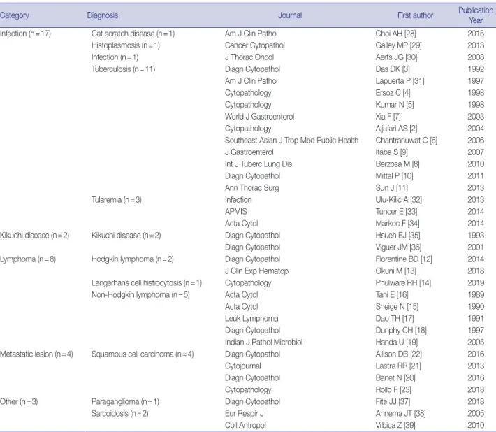
We reviewed the literature on LN FNAs that reveal necrotic features. The most common reports were on TB [2-11], fol- lowed by studies on lymphoma [12-19] and metastatic lesions [20-23] (Table 3). However, most of these studies were case re- ports. There have been very few systematic reports of cases where necrosis is observed or certain diseases must be considered to be more important based on the patient age.
In this study, we analyzed 460 patients who underwent cervi- cal LN FNA cytology that revealed necrotic features. The most common causative disease was TB. Of the 460 patients, 247 underwent ancillary testing for MTB, and 148 (59.9%) were fi- nally diagnosed with TB. Typically, the most characteristic cy- tologic findings of TB are nodular collections of epithelioid his- tiocytes with Langhans giant cells and caseous necrosis. However, either end of the cytologic spectrum may be seen, especially ne- crosis without granulomas [24]. Kumar et al. [5] reported that the presence of acute lymphadenitis does not completely exclude TB. They clinically suspected TB in 263 cases with a cytologic picture that demonstrated acute suppurative inflammation. LN FNA should be repeated in all FNA cases that show acute sup- purative inflammation without granulomas. It has been reported that a repeat LN FNA helps to detect more than 55% of addi- tional TB cases [25]. In this study, among the patients classified with suppurative lymphadenitis according to FNA cytology, 36.7% were finally diagnosed with TB following ancillary tests.
Therefore, it is important to rule out TB when necrotic features are noted on LN FNA cytology.
Necrotic features may also be characteristic of the Kikuchi
disease category in cervical LN FNA. The FNA findings in Ki-
kuchi disease typically reveal a polymorphous lymphoid popu-
lation, crescentic histiocytes, extensive apoptotic bodies, and
necrosis. The combination of crescentic histiocytes and karyor-
rhectic debris suggests the diagnosis of Kikuchi disease. However,
some cases show only karyorrhexis and necrosis, which is not
specific for Kikuchi disease. For example, infectious mononucle-
osis may rarely demonstrate extensive necrosis. Infectious mono-
nucleosis is a self-limited viral disease that frequently involves
the cervical LNs of young patients who present with fever and
pharyngitis. In infectious mononucleosis, a polymorphous infil-
trate with atypical large lymphoid cells is usually noted. The pa-
tient’s clinical history is helpful in arriving at a differential diag-
nosis in these cases [26].
Another important cause of necrotic features in cervical LN FNA cytology is malignant neoplasm. Among our 460 cases, 49 were finally confirmed as malignancies (including metastatic car- cinoma) by tissue biopsy. The mean age of these patients was 62 years. Forty cases were metastatic carcinoma. Among these cases, the most common malignancy was squamous cell carcinoma (62.5%). This trend was consistent with the findings of previous reports. In squamous cell carcinoma, metastatic LNs of the head and neck are often the first signs of malignancy in the inconspic- uous organs of this region [1]. Additionally, cystic changes are often observed in metastatic LNs in squamous cell carcinoma [27]. Necrotic material may also be identified in the cyst spaces;
therefore, pathologists should be mindful during their micro- scopic examinations. In our study, most cases were diagnosed as
malignant neoplasms by FNA alone; the remaining nine cases were diagnosed as suppurative lymphadenitis (n = 1), granuloma (n = 3), and necrosis (n = 5) of the LNs. Therefore, when necrotic features are seen in the cervical LN FNA cytology in older pa- tients, pathologists should consider the possibility of malignancy and carefully correlate their findings with the clinical history of the patient to ensure the proper evaluation is recommended to clinicians, even though there may be a paucity of cells on the smeared slides.
In this study, we identified misdiagnosed cases of cervical LN necrosis on FNA. Following an excision biopsy, these cases were diagnosed as reactive hyperplasia, salivary duct cysts, Warthin tumors, schwannomas, and spindle cell tumors. As demonstrated by these cases, fibrinoid material that is present due to excessive
Table 3. Previously published reports with the keywords “FNA,” “necrosis,” and “lymph node”
Category Diagnosis Journal First author Publication
Year
Infection (n=17) Cat scratch disease (n=1) Am J Clin Pathol Choi AH [28] 2015
Histoplasmosis (n=1) Cancer Cytopathol Gailey MP [29] 2013
Infection (n=1) J Thorac Oncol Aerts JG [30] 2008
Tuberculosis (n=11) Diagn Cytopathol Das DK [3] 1992
Am J Clin Pathol Lapuerta P [31] 1997
Cytopathology Ersoz C [4] 1998
Cytopathology Kumar N [5] 1998
World J Gastroenterol Xia F [7] 2003
Cytopathology Aljafari AS [2] 2004
Southeast Asian J Trop Med Public Health Chantranuwat C [6] 2006
J Gastroenterol Itaba S [9] 2007
Int J Tuberc Lung Dis Berzosa M [8] 2010
Diagn Cytopathol Mittal P [10] 2011
Ann Thorac Surg Sun J [11] 2013
Tularemia (n=3) Infection Ulu-Kilic A [32] 2013
APMIS Tuncer E [33] 2014
Acta Cytol Markoc F [34] 2014
Kikuchi disease (n=2) Kikuchi disease (n=2) Diagn Cytopathol Hsueh EJ [35] 1993
Diagn Cytopathol Viguer JM [36] 2001
Lymphoma (n=8) Hodgkin lymphoma (n=2) Diagn Cytopathol Florentine BD [12] 2014
J Clin Exp Hematop Okuni M [13] 2018
Langerhans cell histiocytosis (n=1) Cytopathology Phulware RH [14] 2019
Non-Hodgkin lymphoma (n=5) Acta Cytol Tani E [16] 1989
Acta Cytol Sneige N [15] 1990
Leuk Lymphoma Dao TH [17] 1991
Diagn Cytopathol Dunphy CH [18] 1997
Indian J Pathol Microbiol Handa U [19] 2005
Metastatic lesion (n=4) Squamous cell carcinoma (n=4) Diagn Cytopathol Allison DB [22] 2016
Cytojournal Lastra RR [21] 2013
Diagn Cytopathol Banet N [20] 2016
Cytopathology Rollo F [23] 2018
Other (n=3) Paraganglioma (n=1) Diagn Cytopathol Fite JJ [37] 2018
Sarcoidosis (n=2) Eur Respir J Annema JT [38] 2005
Coll Antropol Vrbica Z [39] 2010
hemorrhaging, amorphous material of tumor components, or hypocellular myxoid stroma could be confused with a necrotic background.
An accurate diagnosis on cervical LN FNA cytology is impor- tant to determine the most appropriate treatment and to prevent unnecessary surgery. This study was conducted in the Republic of Korea, and we found that the most common cause of necrosis in cervical LN FNA cytology was TB. When there are necrotizing features in the FNA of the cervical lymph nodes in young patients, Kikuchi disease should be considered first. Metastatic carcinoma should always be suspected in older patients, even if they do not have any previous history of malignancy. In addition, when nec- rotizing features are noted in the FNA of the cervical lymph nodes of patients in any age group, MTB testing should be performed in parallel to exclude TB, which has a high prevalence in the Republic of Korea.
Ethics Statement
This study was approved by the Institutional Review Board of the Keimyung University Dongsan Hospital with a waiver of informed consent (DSMC 2020-03-069-002) and performed in accordance with the principles of the Declaration of Helsinki.
ORCID
Young Jin Seo https://orcid.org/0000-0002-9048-0903 Hyeongchan Shin https://orcid.org/0000-0002-3716-7678 Hye Won Lee https://orcid.org/0000-0001-8540-524X Hye Ra Jung https://orcid.org/0000-0002-1477-6606 Author Contributions
Conceptualization: HRJ. Data curation: YJS, HS. Formal analysis: YJS, HRJ. Methodology: YJS, HRJ. Supervision: HRJ. Visualization: YJS, HWL.
Writing—original draft: YJS, HRJ. Writing—review & editing: YJS, HS, HWL, HRJ. Approval of final manuscript: all authors.
Conflicts of Interest
The authors declare that they have no potential conflicts of interest.
Funding Statement No funding to declare.
References
1. van Schaik JE, Muller Kobold AC, van der Laan B, van der Vegt B, van Hemel BM, Plaat BEC. Squamous cell carcinoma antigen con- centration in fine needle aspiration samples: a new method to de- tect cervical lymph node metastases of head and neck squamous cell carcinoma. Head Neck 2019; 41: 2561-5.
2. Aljafari AS, Khalil EA, Elsiddig KE, et al. Diagnosis of tuberculous lymphadenitis by FNAC, microbiological methods and PCR: a comparative study. Cytopathology 2004; 15: 44-8.
3. Das DK, Bhambhani S, Pant JN, et al. Superficial and deep-seated tuberculous lesions: fine-needle aspiration cytology diagnosis of 574 cases. Diagn Cytopathol 1992; 8: 211-5.
4. Ersoz C, Polat A, Serin MS, Soylu L, Demircan O. Fine needle aspira- tion (FNA) cytology in tuberculous lymphadenitis. Cytopathology 1998; 9: 201-7.
5. Kumar N, Tiwari MC, Verma K. AFB staining in cytodiagnosis of tuberculosis without classical features: a comparison of Ziehl- Neelsen and fluorescent methods. Cytopathology 1998; 9: 208-14.
6. Chantranuwat C, Assanasen T, Shuangshoti S, Sampatanukul P.
Polymerase chain reaction for detection of Mycobacterium tuber- culosis in papanicolaou-stained fine needle aspirated smears for di- agnosis of cervical tuberculous lymphadenitis. Southeast Asian J Trop Med Public Health 2006; 37: 940-7.
7. Xia F, Poon RT, Wang SG, Bie P, Huang XQ, Dong JH. Tuberculosis of pancreas and peripancreatic lymph nodes in immunocompetent patients: experience from China. World J Gastroenterol 2003; 9:
1361-4.
8. Berzosa M, Tsukayama DT, Davies SF, et al. Endoscopic ultra- sound-guided fine-needle aspiration for the diagnosis of extra-pul- monary tuberculosis. Int J Tuberc Lung Dis 2010; 14: 578-84.
9. Itaba S, Yoshinaga S, Nakamura K, et al. Endoscopic ultrasound- guided fine-needle aspiration for the diagnosis of peripancreatic tuberculous lymphadenitis. J Gastroenterol 2007; 42: 83-6.
10. Mittal P, Handa U, Mohan H, Gupta V. Comparative evaluation of fine needle aspiration cytology, culture, and PCR in diagnosis of tuberculous lymphadenitis. Diagn Cytopathol 2011; 39: 822-6.
11. Sun J, Teng J, Yang H, et al. Endobronchial ultrasound-guided transbronchial needle aspiration in diagnosing intrathoracic tuber- culosis. Ann Thorac Surg 2013; 96: 2021-7.
12. Florentine BD, Cohen AN. Nodular sclerosing classical Hodgkin lymphoma masquerading as acute suppurative-necrotizing lymph- adenitis. Diagn Cytopathol 2014; 42: 238-41.
13. Okuni M, Yakushijin K, Sakai Y, et al. A case of classical Hodgkin lymphoma with total lymph node infarction. J Clin Exp Hematop 2018; 58: 24-6.
14. Phulware RH, Guleria P, Iyer VK, et al. Cytological diagnosis of Langerhans cell histiocytosis: a series of 47 cases. Cytopathology 2019; 30: 413-8.
15. Sneige N, Dekmezian RH, Katz RL, et al. Morphologic and immu- nocytochemical evaluation of 220 fine needle aspirates of malignant lymphoma and lymphoid hyperplasia. Acta Cytol 1990; 34: 311-22.
16. Tani E, Liliemark J, Svedmyr E, Mellstedt H, Biberfeld P, Skoog L.
Cytomorphology and immunocytochemistry of fine needle aspi- rates from blastic non-Hodgkin’s lymphomas. Acta Cytol 1989; 33:
363-71.
17. Dao TH, Fleury-Feith J, Haioun C, et al. Percutaneous fine needle aspiration cytology and biopsy in the diagnosis and classification of lymphoma: clinical evaluation. Leuk Lymphoma 1991; 5: 237-42.
18. Dunphy CH, Ramos R. Combining fine-needle aspiration and flow cytometric immunophenotyping in evaluation of nodal and extra- nodal sites for possible lymphoma: a retrospective review. Diagn Cytopathol 1997; 16: 200-6.
19. Handa U, Mohan H, Punia RS, Nada R. FNAC in a case of NHL presenting initially as nodal infarction. Indian J Pathol Microbiol 2005; 48: 510-2.
20. Banet N, Rooper LM, Maleki Z. Metastatic HPV-related head and neck squamous cell carcinoma to the lung and mediastinal lymph nodes in aspirated cytology material: a diagnostic pitfall. Diagn Cy- topathol 2016; 44: 206-14.
21. Lastra RR, Pramick MR, Nakashima MO, et al. Adequacy of fine-
needle aspiration specimens for human papillomavirus infection molecular testing in head and neck squamous cell carcinoma. Cy- tojournal 2013; 10: 21.
22. Allison DB, Bishop JA, Ali SZ. Cytopathologic characteristics of SMARCB1 (INI-1) deficient sinonasal carcinoma: a potential diag- nostic pitfall. Diagn Cytopathol 2016; 44: 700-3.
23. Rollo F, Dona MG, Pellini R, et al. Cytology and direct human pap- illomavirus testing on fine needle aspirates from cervical lymph node metastases of patients with oropharyngeal squamous cell car- cinoma or occult primary. Cytopathology 2018; 29: 449-54.
24. Mac DeMay R. The art and science of cytopathology. Chicago: Amer- ican Society for Clinical Pathology, 2012.
25. Kumar N, Jain S, Murthy NS. Utility of repeat fine needle aspira- tion in acute suppurative lesions: follow-up of 263 cases. Acta Cytol 2004; 48: 337-40.
26. Tong TR, Chan OW, Lee KC. Diagnosing Kikuchi disease on fine needle aspiration biopsy: a retrospective study of 44 cases diagnosed by cytology and 8 by histopathology. Acta Cytol 2001; 45: 953-7.
27. Mokhtari S. Mechanisms of cyst formation in metastatic lymph nodes of head and neck squamous cell carcinoma. Diagn Pathol 2012; 7: 6.
28. Choi AH, Bolaris M, Nguyen DK, Panosyan EH, Lasky JL 3rd, Duane GB. Clinicocytopathologic correlation in an atypical pre- sentation of lymphadenopathy with review of literature. Am J Clin Pathol 2015; 143: 749-54.
29. Gailey MP, Klutts JS, Jensen CS. Fine-needle aspiration of histoplas- mosis in the era of endoscopic ultrasound and endobronchial ultra- sound: cytomorphologic features and correlation with clinical labo- ratory testing. Cancer Cytopathol 2013; 121: 508-17.
30. Aerts JG, Kloover J, Los J, van der Heijden O, Janssens A, Tournoy
KG. EUS-FNA of enlarged necrotic lymph nodes may cause infec- tious mediastinitis. J Thorac Oncol 2008; 3: 1191-3.
31. Lapuerta P, Martin SE, Ellison E. Fine-needle aspiration of periph- eral lymph nodes in patients with tuberculosis and HIV. Am J Clin Pathol 1997; 107: 317-20.
32. Ulu-Kilic A, Gulen G, Sezen F, Kilic S, Sencan I. Tularemia in cen- tral Anatolia. Infection 2013; 41: 391-9.
33. Tuncer E, Onal B, Simsek G, et al. Tularemia: potential role of cyto- pathology in differential diagnosis of cervical lymphadenitis: multi- center experience in 53 cases and literature review. APMIS 2014; 122:
236-42.
34. Markoc F, Koseoglu RD, Koc S, Gurbuzler L. Tularemia in differen- tial diagnosis of cervical lymphadenopathy: cytologic features of tularemia lymphadenitis. Acta Cytol 2014; 58: 23-8.
35. Hsueh EJ, Ko WS, Hwang WS, Yam LT. Fine-needle aspiration of histiocytic necrotizing lymphadenitis (Kikuchi’s disease). Diagn Cytopathol 1993; 9: 448-52.
36. Viguer JM, Jimenez-Heffernan JA, Perez P, Lopez-Ferrer P, Gonza- lez-Peramato P, Vicandi B. Fine-needle aspiration cytology of Kiku- chi’s lymphadenitis: a report of ten cases. Diagn Cytopathol 2001; 25:
220-4.
37. Fite JJ, Maleki Z. Paraganglioma: cytomorphologic features, radio- logic and clinical findings in 12 cases. Diagn Cytopathol 2018; 46:
473-81.
38. Annema JT, Veselic M, Rabe KF. Endoscopic ultrasound-guided fine-needle aspiration for the diagnosis of sarcoidosis. Eur Respir J 2005; 25: 405-9.
39. Vrbica Z, Boras Z, Rakusic N, et al. Necrotising sarcoid granuloma- tosis of the spinal cord: case report. Coll Antropol 2010; 34: 713-7.