• 검색 결과가 없습니다.

Primary Malignant Fibrous Histiocytoma of the Chest Wall Misdiagnosed as Traumatic Hemothorax

N/A
N/A
Protected

Academic year: 2021

Share "Primary Malignant Fibrous Histiocytoma of the Chest Wall Misdiagnosed as Traumatic Hemothorax"

Copied!
3
0
0

로드 중.... (전체 텍스트 보기)

전체 글

(1)

Primary Malignant Fibrous Histiocytoma of the Chest Wall Misdiagnosed as Traumatic Hemothorax

Ki-Eun Hwang, M.D.

1

, June-Seong Hwang, M.D.

1

, Kyung-Min Kim, M.D.

1

, Jae-Wan Jung, M.D.

1

, Su- Jin Oh, M.D.

1

, Eun-Kyoung Kim, M.D.

1

, Dae-Woon Ryu, M.D.

2

, Keum-Ha Choi, M.D.

3

, Eun-Taik Jeong, M.D.

1

and Hak-Ryul Kim, M.D.

1

Departments of

1

Internal Medicine,

2

Cardiothoracic Surgery, and

3

Pathology, Institute of Wonkwang Medical Science, Wonkwang University School of Medicine, Iksan, Korea

Malignant fibrous histiocytoma (MFH), a type of sarcoma, is a malignant neoplasm with uncertain origins that arise from both the soft tissues and the bone. The occurrence of MFH on the chest wall is extremely rare. We hereby report a case of a 72-year-old woman who was incidentally detected with MFH after a traffic accident.

Keywords: Histiocytoma, Malignant Fibrous; Thoracic Wall; Hemothorax

Case Report

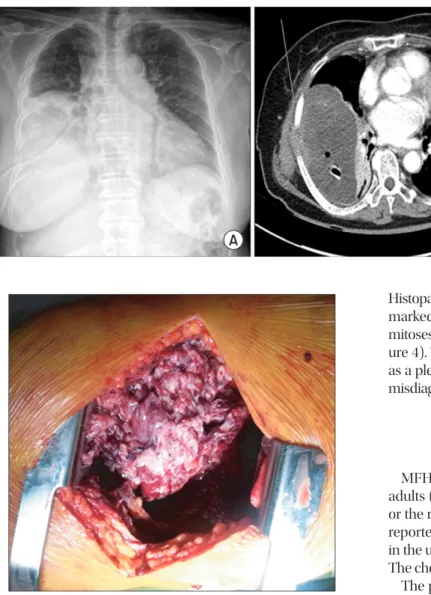
A 72-year-old woman complained of right-sided chest pain and orthopnea following a traffic accident that occurred 1 month previously. Chest radiography and computed tomog- raphy revealed large amounts of right pleural effusion and a

Copyright © 2014

The Korean Academy of Tuberculosis and Respiratory Diseases.

All rights reserved.

Introduction

Malignant fibrous histiocytoma (MFH) is the most common malignant soft tissue tumor. MFH occurs most commonly in the extremities (70−75%), followed the trunk and retroperi- toneum. It is a frequently occurring soft tissue tumor with ag- gressive biological behavior. Primary MFH of the chest wall is especially rare.

We report a case of a 72-year-old woman in whom MFH was detected incidentally after she had a traffic accident. We describe this unusual case and review the literature.

CASE REPORT

http://dx.doi.org/10.4046/trd.2014.76.6.289

ISSN: 1738-3536(Print)/2005-6184(Online) • Tuberc Respir Dis 2014;76:289-291

289

Address for correspondence: Hak-Ryul Kim, M.D.

Department of Internal Medicine, Wonkwang University School of Medicine, 460 Iksan-daero, Iksan 570-974, Korea

Phone: 82-63-859-2583, Fax: 82-63-855-2025 E-mail: [email protected]

Received: Feb. 12, 2014 Revised: Mar. 4, 2014 Accepted: Mar. 12, 2014

cc

It is identical to the Creative Commons Attribution Non-Commercial License (http://creativecommons.org/licenses/by-nc/3.0/).

Figure 1. Chest radiographs showing large amounts of pleural effu-

sion and a fracture in the seventh right rib.

(2)

KE Hwang et al.

290 Tuberc Respir Dis 2014;76:289-291 www.e-trd.org

pathological seventh rib fracture (Figure 1). A hemorrhagic fluid was aspirated at thoracentesis. She was then diagnosed with traumatic hemothorax and a closed thoracostomy was performed. Laboratory examination of the pleural effusion revealed that it was exudative, and had an adenosine deami- nase level of 15.7 IU/L, white blood cell count of 980/mm

3

(lymphocyte count 80%), and red blood cell count of 6,560/

mm

3

. Except for the level of tumor marker cancer antigen-125, the levels of tumor markers such as carcinoembryonic anti- gen and cytokeratin fragment 19 remained within the normal range. After drainage of 1.5 L, the symptoms improved.

Chest radiography performed 7 days after closed thoracos- tomy revealed a large amount of the remaining pleural effusion (Figure 2). We then performed video-assisted thoracoscopic surgery for the persistent retained hemothorax. However, lung decortication and rib resection had to be performed because of the presence of a very large mass at the incision site (Figure 3).

Histopathological examination of the biopsy specimen revealed marked cytological and nuclear pleomorphism with frequent mitoses. The tumor cells stained positive for vimentin only (Fig- ure 4). The mass on the chest wall and pleura was confirmed as a pleomorphic MFH. Thus, pleomorphic MFH was initially misdiagnosed as traumatic hemothorax.

Discussion

MFH is among the most common soft-tissue sarcomas in adults (20−30%), and is principally located in the extremities or the retroperitoneum

1

. In a retrospective series of 167 cases reported by Kearney et al.

2

, 51% arose in the lower limbs, 24%

in the upper limbs, 16% in the trunk, and 9% retroperitoneum.

The chest wall is a rare site of origin

3

.

The peak incidence is in the fifth decade of life, with a pre- dominance of elderly male patients. There is no typical clinical symptom, but the patients with MFH of the chest wall usually experience chest pain, cough, and hemoptysis. However, there were no pre-trauma tumor-related symptoms in our case. In our case, MFH was diagnosed by pleural effusion. Because the majority of the data available in the literature are case reports due to the low frequency of the diseases, the frequency of presence of pleural effusion with MFH is unknown.

The mass was not seen because of large amount of pleu- ral effusion in our case, however, the important diagnostic tool is chest computerized tomography (CT) and magnetic resonance (MR) imaging. The mass usually shows intense enhancement on CT with a clear margin separating it from the surrounding tissue

4

. Furthermore, the mass often shows decreased central attenuation due to necrosis, hemorrhage and mucoid material

5

. On MR imaging, the mass shows an in- termediate to low signal intensity on T1-weighted images and high signal intensity on T2-weighted images, which frequently shows inhomogeneous central intensity

6

. The final diagnosis was confirmed by results of the histopathology and immuno- histochemistry. Immunohistochemical staining for various Figure 2. (A) Chest radiograph showing the remaining pleural effusion in the right thoracic cavity 7 days after perform- ing the closed thoracostomy. (B) Chest computed tomography scans showing remaining pleural effusions after closed thoracostomy.

Figure 3. A very large mass protruded into the thorax, which invad-

ed and damaged the encountered rib.

(3)

Primary MFH misdiagnosed as traumatic hemothorax

http://dx.doi.org/10.4046/trd.2014.76.6.289 291

www.e-trd.org

molecules such as caletinin, CD68, pan-cytokeratin, vimentin were performed to rule out other malignancies.

Wide resection is the first choice treatment for all soft-tissue sarcomas. The mean survival of patients without operation is 11.7 months, those of patients whom underwent operation is 23.2 months

7-10

. The additional chemotherapy and radio- therapy may play a role in treating osseous MFH. The tumor grade, size, and the presence of distant metastases at the initial presentation remain the most important prognostic factor for MFH

11,12

. Favorable factors of MFH was Union for Interna- tional Cancer Control/American Joint Committee on Cancer (UICC/AJCC) stage I and II, superficial location, myxoid type and under 50 years, supporting the rational for radical en- block resection of tumor tissues.

In conclusion, primary MFH of the chest wall is extremely rare tumor and surgery is treatment of choice of MFH. We report a case of a 72-year-old woman in whom MFH was de- tected incidentally after she had a traffic accident.

Conflicts of Interest

No potential conflict of interest relevant to this article was reported.

Acknowledgements

This study was supported by a grant from Wonkwang Uni- versity in 2012.

References

1. Weiss SW, Enzinger FM. Malignant fibrous histiocytoma: an analysis of 200 cases. Cancer 1978;41:2250-66.

2. Kearney MM, Soule EH, Ivins JC. Malifnant fibrous histiocy-

toma: a retrospective study of 167 cases. Cancer 1980;45:167- 78.

3. Hashimoto H. Malignant fibrous histiocytoma: a clinico- pathologic study of 130 cases (author’s transl). Fukuoka Igaku Zasshi 1979;70:585-613.

4. Enjoji M, Hashimoto H, Tsuneyoshi M, Iwasaki H. Malignant fibrous histiocytoma.: a clinicopathologic study of 130 cases.

Acta Pathol Jpn 1980;30:727-41.

5. Tateishi U, Kusumoto M, Hasegawa T, Yokoyama R, Mori- yama N. Primary malignant fibrous histiocytoma of the chest wall: CT and MR appearance. J Comput Assist Tomogr 2002;26:558-63.

6. Munk PL, Sallomi DF, Janzen DL, Lee MJ, Connell DG, O’Connell JX, et al. Malignant fibrous histiocytoma of soft tis- sue imaging with emphasis on MRI. J Comput Assist Tomogr 1998;22:819-26.

7. Maze Y, Yuasa H, Yuasa U, Takao M, Hirata K. A case of ma- lignant fibrous histiocytoma of the chest wall. Kyobu Geka 2000;53:600-4.

8. Le Doussal V, Coindre JM, Leroux A, Hacene K, Terrier P, Bui NB, et al. Prognostic factors for patients with localized pri- mary malignant fibrous histiocytoma: a multicenter study of 216 patients with multivariate analysis. Cancer 1996;77:1823- 30.

9. Sawai H, Kamiya A, Kurahashi S, Yamanaka Y, Manabe T. Ma- lignant fibrous histiocytoma originating from the chest wall:

report of a case and collective review of cases. Surg Today 1998;28:459-63.

10. Belal A, Kandil A, Allam A, Khafaga Y, El-Husseiny G, El-En- baby A, et al. Malignant fibrous histiocytoma: a retrospective study of 109 cases. Am J Clin Oncol 2002;25:16-22.

11. Gibbs JF, Huang PP, Lee RJ, McGrath B, Brooks J, McKinley B, et al. Malignant fibrous histiocytoma: an institutional review.

Cancer Invest 2001;19:23-7.

12. de Perrot M, Fischer S, Brundler MA, Sekine Y, Keshavjee S. Solitary fibrous tumors of the pleura. Ann Thorac Surg 2002;74:285-93.

Figure 4. The tumor cells show remark- able cytological pleomorphisms with frequent mitoses (arrows) (A, H&E stain,

×400) and these cells express vimentin (B,

×400).

수치

Figure 1. Chest radiographs showing large amounts of pleural effu- effu-sion and a fracture in the seventh right rib.
Figure 3. A very large mass protruded into the thorax, which invad- invad-ed and damaginvad-ed the encounterinvad-ed rib.
Figure 4. The tumor cells show remark- remark-able cytological pleomorphisms with  frequent mitoses (arrows) (A, H&E stain,

참조

관련 문서

In the efficiency review of the Trombe wall system through the experiment, if the Trombe wall was applied in the building structure, the Trombe wall had the

Factors affecting post-traumatic stress of general hospital nurses after the epidemic of Middle East respiratory syndrome infection: Journal of Korean Clinical

The average position of the posterior superior alveolar artery, the wall thickness of the lateral wall, and the average volume of the maxillary sinus will

Expanded foams, gas-filled powders, fibrous materials, vacuum alone, evacuated powders and fibrous materials, opacified powders 그리고 multilayer insulations 등이

_____ culture appears to be attractive (도시의) to the

- leaves the abdominal cavity lateral to the inferior epigastric vessels and enters the deep inguinal ring. - traverses the entire inguinal canal - exits through

1 John Owen, Justification by Faith Alone, in The Works of John Owen, ed. John Bolt, trans. Scott Clark, "Do This and Live: Christ's Active Obedience as the

Anoxic