Gene Expression Profiling by Ginsenoside Rb1 in Keratinocyte HaCaT Cells
Dong Woo Lee1, Jung Min Kim2 and In Seok Bang1*
1Department of Biological Science and the Research Institute for Basic Sciences, Hoseo University, Asan 31499, Korea
2Genoplan, Inc. & NAR Center, Inc., Teheran-ro 216, Seoul 06221, Korea
Received February 26, 2019 /Revised April 19, 2019 /Accepted April 20, 2019
We investigated the gene expression patterns and the mechanisms of action of the apoptotic response by microarray analysis of human keratinocyte HaCaT cells treated with ginsenoside Rb1, a saponin of Panax ginseng C. A. Meyer. Genes related to apoptosis, the G2/M transition of the mitotic cell cycle, cell division, mitotic nuclear division, and intracellular protein transport were 2-fold up-regulated in HaCaT cells treated with the ginsenoside Rb1, whereas genes related to DNA repair, regeneration fis- sion, and extracellular matrix organization were 2-fold down-regulated. Apoptosis signaling may be mediated by FAS and PLA2G4A, and pathway analysis indicated that STAT3 might be an upstream regulator of these genes. The activity of FAS and PLA2G4A was verified by qPCR, which showed that FAS was increased about 2-fold in HaCaT cells treated with 10 μg/ml of ginsenoside Rb1 for 24 hr, PLA2G4A was increased about twice after 6 hours, and gene expression was increased more than 2-fold after 24 hr. Knockdown of STAT3 with siRNA decreased FAS expression and increased PLA2G4A expression but only FAS was passed from the upstream regulator STAT3. These results in- dicate that STAT3, which is an upstream regulator, induces apoptosis via FAS during treatment with ginsenoside Rb1.
Key words : Apoptosis, DNA microarray, FAS, Ginsenoside Rb1, HaCaT cells
*Corresponding author
*Tel : +82-41-540-9595, Fax : +82-41-548-6231
*E-mail : [email protected]
This is an Open-Access article distributed under the terms of the Creative Commons Attribution Non-Commercial License (http://creativecommons.org/licenses/by-nc/3.0) which permits unrestricted non-commercial use, distribution, and reproduction in any medium, provided the original work is properly cited.
Journal of Life Science 2019 Vol. 29. No. 5. 514~523 DOI : https://doi.org/10.5352/JLS.2019.29.5.514
서 론
인삼(Panax ginseng C. A. Meyer)은 오가피 나무과(Aralia- ceae)의 인삼속(Panax)에 속하는 다년생 음지성 숙근초로써, 다양한 효능이 있어 예로부터 생약재 및 건강기능식품으로써 각광을 받고 널리 사용되어 왔다[21, 25]. 인삼의 주요 유효성 분은 인삼 사포닌 즉 진세노사이드(ginsenoside)로 항암, 면역 증강, 혈압강화, 혈당강화, 항염증 및 항산화 효과 등 매우 다 양한 약리 효능을 가지는 것으로 알려져 있다[5, 10, 34, 36].
진세노사이드는 트리테르페노이드(triterpenoid)계의 담마란 (dammarane)계 사포닌이다[22]. 담마란계 사포닌은 비당 (aglycone) 부분의 수산기(-OH) 수에 따라 2개인 경우 프로토 파낙사디올(protopanaxadiol, PPD)계 사포닌, 3개인 경우 프 로토파낙사트리올(protopanaxatriol, PPT)계 사포닌으로 분류 된다[30]. PPD계 사포닌은 진세노사이드 Ra1, Ra2, Ra3, Rb1, Rb2, Rc, Rd, Rg3, Rg5 Rh2 등이 있고, PPT계 사포닌에는 진세 노사이드 Re, Rf, Rg1, Rg2, Rh1, Rh4 등이 있으며, 이들은 각각 다른 화학적 구조를 가지고 다양한 약리 효과를 나타내
는 것으로 알려졌다[14, 27]. 또한 인삼 사포닌의 함량에 따라 메이저(major) 진세노사이드와 이로부터 당의 가수분해에 의 해 만들어진 마이너(minor) 진세노사이드로 나뉘는데, 메이저 진세노 사이드인 Rb1, Rb2, Rc, Rd, Re 및 Rg1은 총 진세노사 이드의 90% 이상을 차지하고, 특히 PPD 타입의 4가지 메이저 진세노사이드인 Rb1, Rb2, Rc와 Rd는 총 진세노사이드의 50-70%를 차지하고 있다[20, 31].
진세노사이드 Rb1은 PPD계의 메이저 진세노사이드로 중 추신경 억제, 최면, 진통, 정신 안정, 해열, 혈청단백질 합성촉 진, 중성지방 분해억제, 합성촉진(인슐린 유사), 콜레스테롤 생 합성촉진, 플라스민 활성화, RNA 합성촉진, 호르몬 분비촉진 작용 등의 다양한 작용이 보고된 바 있다[23]. 또한 피부의 노 화 및 손상에 관련된 연구로 진세노사이드는 Rb1은 UVB가 조사된 무모 생쥐의 피부 두께, 주름 및 표피의 증가를 억제시 켰으며[15], 이러한 피부 변화에 대한 효과 및 약리적 작용의 기전 연구를 위하여 주로 사람유래 피부각질 세포인 HaCaT 세포를 이용하였다[17]. 이와 같이 진세노사이드에 대한 연구 는 그 효과 측면에서 주로 연구가 집중되었으나 이런 효과가 어떤 작용기전을 통해 나타나는지에 대한 연구는 아직 미흡한 실정이다.
본 연구는 진세노사이드 Rb1에 대한 사람유래 피부각질세 포인 HaCaT 세포의 분자생물학적 작용기전을 microarray 분 석을 통하여 세포사멸에 관여할 것으로 추정되는 유전자 및 이 들의 상위 조절자(upstream regulator)에 대하여 알아 보았다.
재료 및 방법
시약
세포배양에 사용된 Dulbecco’s modified Eagle’s medium (DMEM)은 Sigma-Aldrich (USA), Penicillin-streptomycin은 Gibco (USA), 그리고 fetal bovine serum (FBS)은 Wisent (USA)에서 각각 구입하였다. 3-(4,5-dimethylthiazol-2-y1)-2,5- diphenyltretrazolium bromide (MTT), dimethyl sulfoxide (DMSO), 그리고 hydrogen peroxide (H2O2)는 Sigma (USA) 에서 구입하였다. TRIzolⓇ Regent는 Invitrogen (USA) 에서 total RNA 정량 및 정성분석을 위한 RNA nano kit (2100 Bioanalyzer System)는 Agilent Techologies (USA)에서 구입 하였다. Microarray 분석에 Low RNA Input Linear Amplifi- cation Kit PLUS는 Agilent Technology (USA)에서 RNase Mini Spin Columns은 Qiagen (Germany)에서 구입하였고, 유 전자 발현 정량분석을 위한 AccuPower® RT PreMix K-2041 와 AccuPower® 2X Greenstar qPCR Master K-6253는 Bioneer (Korea)에서 구입하였다.
세포주 및 진세노사이드 Rb1
사람 유래 피부각질세포인 HaCaT 세포주를 한국생명공학 연구원(Korea Research Institute of Bioscience and Biotech- nology, KRIBB) 바이오의약연구소(이수의 박사)로부터 분양 받아 사용하였다. HaCaT 세포를 배양하기 위해 10% FBS와 100 unit/ml penicillin-streptomycine이 포함된 DMEM 배지 를 사용하여, 37℃로 유지되는 5% CO2 배양기에서 배양하였 다. 배양 시 주 2~3회 동일 배지로 갈아준 다음, 배양 6~7일 경과 후 PBS로 세척 및 0.05% trypsin-0.02% EDTA로 부착된 세포를 분리 및 원심 분리하여 실험에 사용하였다.
진세노사이드 Rb1은 ㈜비티진(대전)으로부터 구입하여 사 용하였으며 Rf는 Sigma (USA)에서 구입하였다. 순도는 98%
로 나타났으며 표준품의 기원은 Panax ginseng C. A. Meyer에 서부터 정제된 물질이다.
세포독성 및 세포보호 효과
HaCaT 세포를 96 Well plate에 well 당 1×10⁴ cells/ml가 되도록 100 μl씩 분주하고 37℃, 5% CO2 배양기에서 24시간 동안 부착시킨 다음, 10 μl의 진세노사이드 Rb1을 농도 별로 첨가하여 24시간 동안 배양하여 세포 생존율을 아래의 MTT assay로 측정하였다. 또한 농도 별 시료를 세포에 처리하여 24시간 동안 배양한 다음, 조 등[3]에 의해 설정된 Ic50 (500 μM) 농도의 H2O2를 처리한 후 다시 4시간 동안 배양하여 진세노 사이드 Rb1의 세포보호 효과를 MTT assay로 확인하였다.
MTT Assay
시료가 처리된 세포 각각의 well에 MTT 용액(10 mg/ml)을
10 μl씩 처리한 후 37℃에서 4시간 동안 배양하여 상등액을 제거하였다. 형성된 MTT formazan 결정체를 100 μl의 DMSO 에 4시간 동안 용해시켜 ELISA reader (Molecular devices, USA)로 formazan의 흡광도가 최대가 되는 570 nm의 파장에 서의 흡광도를 측정하였다. 세포 생존율은 대조군의 흡광도에 대한 백분율로 나타내었다.
Microarray 분석
직경 100 mm의 세포 배양 용기에 1×10⁶ cells/ml의 HaCaT 세포를 분주하여 24시간 동안 pre-incubation 시킨 후 10 μg/
ml의 진세노사이드 Rb1을 처리하여 6시간 및 24시간 동안 배 양하였다. 진세노사이드 Rb1 및 메탄올(대조군)이 처리된 HaCaT 세포를 회수하고 TRIzol를 이용하여 total RNA를 추 출 하였고, 정량 및 정성분석은 RNA nano kit를 사용하였다.
이후 total RNA로부터 target cRNA의 합성 및 혼성화 반응은 Agilent’s Low RNA Input Linear Amplification Kit PLUS를 이용하여 수행하였다. 대조군으로 에탄올 처리된 시료는 cya- nine 3, 실험군 진세노사이드 Rb1이 처리된 시료는 cyanine 5로 각 증폭 및 라벨시켰다. 증폭 및 라벨된 cRNA는 RNase Mini Spin Columns를 이용하여 정제하고, ND-1000 spec- trophotometer (NanoDrop Technologies, Wilmington, DE, USA)를 이용하여 정량하였다. 750 ng cRNA를 이용하여 hy- bridization buffer를 만들고 Agilent Whole Human Genome Oligo Microarray (44K)에서 혼성화 반응을 실시하였다.
Hybridization image는 Agilent DNA Microarray Scanner를 이용하여 스캔하였으며, 데이터 정량은 Agilent Feature Ex- traction Software를 이용하였다. 모든 데이터의 normalization (LOWESS)과 fold-changed gene의 선택은 GeneSpring GX 7.3 (Agilent Technology)으로 수행하였다. 각 실험군에서 flag-out gene들은 제거하였으며, normalized ratio는 signal channel intensity 중간값을 control channel intensity 중간 값 으로 나누어 산출하였다. 유전자 선정(gene selection)을 위한 functional annotation은 Gene OntologyTM Consortium (http://
www.geneontology.org/index.shtml)DB정보를 따랐다. Bio Carta (http://www.biocarta.com/), DAVID (The Database for Annotation, Visualization and Integrated Discovery, http://david.abcc.ncifcrf.gov/), 그리고 Medline databases (http://www.ncbi.nlm.nih.gov/)를 이용하여 기능 연구를 위 한 data mining을 수행하였으며, IPA (ingenuity pathways analysis, http://www.ingenuity.com) [18]를 이용하여 온톨 로지-관련 상호작용 네트워크(ontology-related interaction networks) 및 상위 조절자를 탐색하였다.
유전자 발현 정량분석
Microarray 분석에서 선발된 유전자들에 대해 quantitive- polymerase chain reaction (qPCR)을 수행하였다. 10 μg/ml의 진세노사이드 Rb1를 6시간 및 24시간 동안 처리된 HaCaT 세
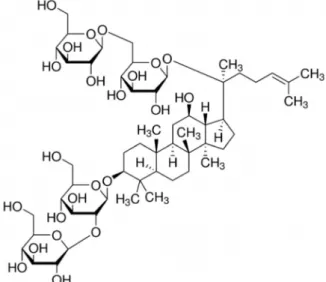
Fig. 1. The chemical structures of ginsenoside Rb1.
Table 1. Primer sequences for qPCR
Gene name Primer sequence
FAS Left Primer
Right Primer
5’-tggtgtcaatgaagccaaaa-3’
5’-tgccaattacgaagcagttg-3’
PLA2G4A Left Primer Right Primer
5’-tacaaggctccaggtgttcc-3’
5’-tggtgattctgggtcatcaa-3’
포로부터 TRIzol를 이용하여 제공된 매뉴얼에 따라 total RNA 를 추출하여 정량하고 순도를 측정하였다. cDNA 합성은 2- step으로 진행하였다. AccuPower® RT PreMix K-2041 (Bio- neer, Korea)를 이용하여 Total RNA 1 μg을 주형으로 Oligo dT (Bioneer) 100 pmole을 primer로 사용하였으며 MyGenie 96 Thermal block (Bioneer)를 이용하여 RT-1(Primer anneal- ing)은 70℃에서 5분 조건으로 RT-2 (cDNA synthesis and RTase inactivation)는 42℃ 60분 85℃ 5분 조건으로 수행하여 cDNA를 합성하였다. 합성된 cDNA는 RNase free water (Sigma-aldrich)로 희석한 후 PCR template으로 사용하였다.
유전자의 발현분석은 AriaMX system (Agillent Technolo- gies)을 이용하여 유전자 특이적인 oligo primer (Table 1) 1 pmole, cDNA 5 ng, AccuPower® 2X Greenstar qPCR Master, K-6253 (Bioneer) 10 ul, Rnase Free water (Sigma-aldrich) 4 ul로 각 well 당 20 ul로 혼합하는 조건으로 qPCR을 수행하였 다. PCR 조건은 enzyme activation 95℃ 3분, denature 95℃
15초, annealing/extension 20초 조건으로 40 cycle을 수행하 였다. 유전자 상대정량 분석은 HmGAPDH의 발현량을 기준 으로 normalize를 수행하였으며 상대정량은 ΔΔCT 방법 (Applied Biosystems, 2004)을 사용하여 Excel (Microsoft, USA)에서 계산하였다.
siRNA transfection
HaCaT 세포를 60 mm dish에 접종 후, 약 24시간 뒤에 lip- ofectamine 2000 reagent (Invitrogen, USA)를 이용하여 제조 사의 방법에 따라 siRNA를 transfection하였다. 대조구로 사용 한 negative control (NC)-siRNA (Bioneer)와 STAT3-siRNA (sense, 5'-CUU CAG ACC CGU CAA ATT-3'; antisense, 5'- UUU GUU GAC GGG UCU GAA GTT-3) (Bioneer) con- struct을 serum free DMEM 배지 50 μl에 lipofectamin 10 μl와 6 μg의 siRNA를 각각 첨가한 뒤 혼합하여 transfection 시켰 다. 4시간 후 FBS가 첨가된 DMEM 배지로 교환한 후, 24시간 동안 추가 배양한 다음 10 μg/ml의 진세노사이드 Rb1를 24시 간 동안 처리하고 세포를 수확하였다.
통계처리
모든 실험은 최소한 3회 이상 실험을 실시하였으며, 실험군 당 평균±표준편차로 표현하였다. 데이터의 통계처리는 SPSS program (ver. 12.0.01)을 이용하여 일원배치 분산분석(one- way ANOVA)을 시행하였으며, 신뢰수준 p<0.05에서 평균값 들에 대한 유의성을 검증하였다.
결과 및 고찰
HaCaT 세포에 대한 진세노사이드 Rb1의 세포독성 및 세 포보호 효과
사람유래 피부각질세포 HaCaT은 항노화, 항산화, 피부탄 력, 미백 등의 연구 및 그 작용기전 연구를 위하여 많은 연구자 들이 흔히 사용하고 있는 세포 주이다[8, 17, 32]. 합성된 진세 노사이드 Rb1의 활성여부를 HaCaT 세포의 독성(cytotoxicity) 및 독성에 대한 세포보호 효과로 확인하였다. 진세노사이드 Rb1의 각 농도(0.01, 0.1, 1 및 10 μg/ml)를 1×10⁴ cells/well의 HaCaT 세포에 처리하여 24시간 동안 배양하여 세포 생존율 (viability)을 MTT assay로 확인한 결과, 시료의 농도에 따른 HaCaT 세포의 생존율에는 큰 차이가 없는 것으로 보아 세포 증식에 영향을 미치지 않았다(Fig. 2). 한편 산화적 스트레스에 의해 세포 생존율을 감소시키는 것으로 알려져 있는 H2O2를 50%의 세포사멸을 보이는 산화 스트레스의 조건(IC50)인 500 μM [19]에서 HaCaT 세포보호 효과를 측정하였다(Fig. 3).
H2O2 처리군의 세포 생존율은 대조군인 100%(±1.77)에 비하 여 37.57%(±0.79)로 나타났다. 반면 농도 별 진세노사이드 Rb1 를 각각 HaCaT 세포에 처리하였을 때 세포 생존율은 농도의 존적으로 증가한 것으로 나타났으며(p<0.05), 1, 10 μg/ml의 진세노사이드 Rb1에서 각각 76.85%(±0.40), 91.40%(±4.16)의 세포보호 효과를 나타내었다. 산화 스트레스에 의한 세포 손 상은 과도한 활성산소종(reactive oxygen specis, ROS)에 의해 매개되는데[9], 인체 내 DNA나 단백질 및 지질과 같은 다양한 생물학적 고분자를 공격하여 세포 기능을 파괴하여 세포사멸
Fig. 2. Effects of ginsenoside Rb1 on cell viability in HaCaT cells. Cells were treated with different concentration of ginsenoside Rb1 as indicated in material and methods.
Cell viability was analyzed using the MTT assay. The date indicated the mean ± SD for triplicate experiments.
Fig. 3. Cytoprotective effects of ginsenoside Rb1 against H2O2- induced oxidative stress in HaCaT cells. Cells were pre-treated for 24 hr with various concentrations of gin- senoside Rb1. The cells were then treated with 500 μM H2O2 (IC50) for 4 hr. No H2O2 treated cells were used as negative control. Viable cells were determined using the MTT assay. The data is represented as the mean ± SD obtained from three independent experiments con- ducted in triplicate. *p<0.05 vs. the control.
을 초래한다[7]. 기존 연구들에 의하면 H2O2로 인해 유도되는 세포사멸은 미토콘드리아의 기능적 손상에 따른 내인성 세포 사멸(intrinsic apoptosis) 경로의 활성과 직접적인 관련이 있 는 것으로 알려져 있다[4, 29, 35]. 본 연구의 진세노사이드 Rb1 은 H2O2로부터 유도된 산화적 스트레스에 대한 손상으로부터 HaCaT 세포를 보호하여 세포 생존율을 높여 주는 것으로 사 료되나, 이러한 진세노사이드 Rb1의 세포보호 효과가 세포사 멸 유발 억제와 연관성 및 미토콘드리아 기능 손상의 연계 여부는 향후 보다 구체적인 기전 규명이 필요할 것이다.
유전자 발현 양상
HaCaT 세포에서 진세노사이드 Rb1의 작용기전을 규명하
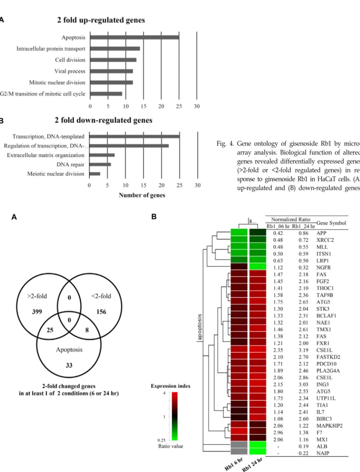
기 위하여 유전체 분석(microarray analysis: 전체 34,127 genes)를 실시하였다. 진세노사이드 Rb1를 HaCaT 세포에 6 또는 24시간 처리한 시료(control 시료 대비)에서 2 배 이상 발현이 증가(424 genes) 및 감소(164 genes)된 유전자들로부터 DAVID gene ontology를 분석한 결과, 발현이 증가된 유전자 들은 세포사멸(apoptosis), 유사분열 세포주기의 G2/M 전이 (G2/M transition of mitotic cell cycle), 세포분열(cell divi- sion), 유사 핵분열(mitotic nuclear division), 세포내 단백질 수송(intracellular protein transport) 등의 유전자였으며(Fig.
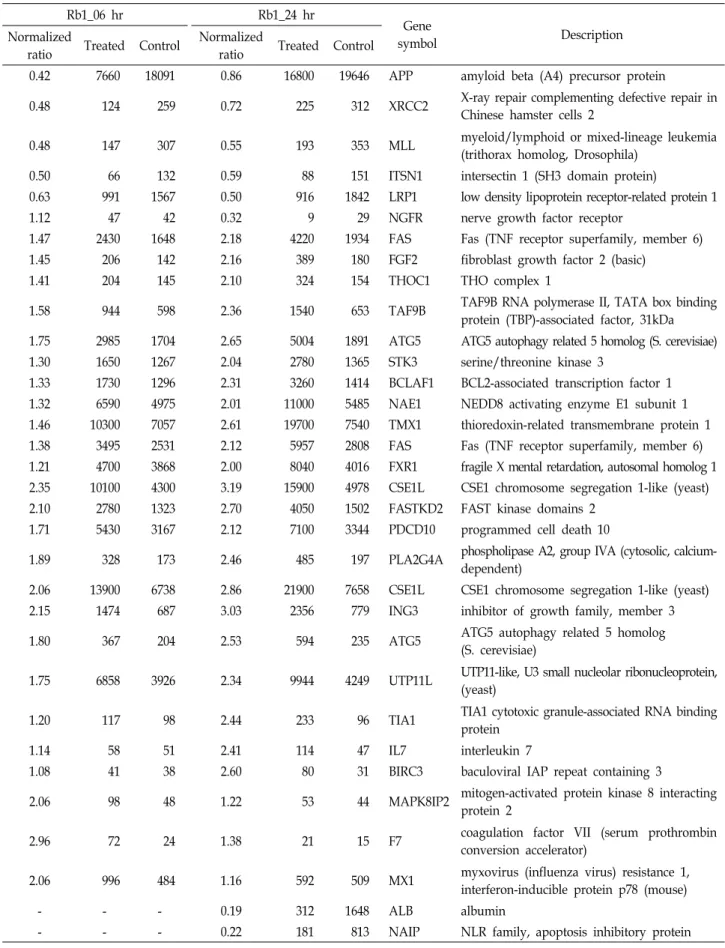
4A), 발현이 감소된 유전자들은 DNA 수선(DNA repair), 감수 핵분열(meiotic nuclear division), 세포 외 기질 체제(extracel- lular matrix organization) 등의 유전자였다(Fig. 4B). 진세노 사이드 Rb1에 대한 기존 연구로 B16-BL6 melanoma 세포의 사멸 야기[33], 쥐의 전두엽에서 일시적인 허혈에 의한 신경세 포의 사멸 지연 효과[1], C6 glioma 세포의 사멸 유도[26] 등이 있다. 따라서 진세노사이드 Rb1에 의해 발현이 증가된 세포사 멸 신호전달에 작용한 유전자들을 venn diagram으로 표현하 였다(Fig. 5A). 두 배 이상 발현이 증가된 유전자는 25개, 2 배 이상 발현이 감소된 유전자는 8개 이었다. 또한 세포사멸 신호전달에 작용한 33개 유전자들의 시간대 별 발현 양상을 hierarchical clustering으로 확인하였다(Fig. 5B). 대부분의 유 전자들은 10 μg/ml의 진세노사이드 Rb1를 6시간 처리한 시료 보다 24시간 처리한 시료에서 보다 현저하게 발현(2 배 이상) 된 것으로 나타났다(Table 2).
세포사멸 반응 경유 유전자의 확인
상기 진세노사이드 Rb1에 의한 다양한 유전자의 발현 변화 에서 세포사멸 신호전달에 작용한 33개 유전자들 간의 온톨로 지-관련 상호작용 네트워크 및 상위 조절자를 IPA로 분석하였 다. 진세노사이드 Rb1에 의한 세포사멸 반응은 TNF receptor superfamily에 속하는 FAS (TNF receptor superfamily, mem- ber 6)와 Phospholipase인 PLA2G4A [phospholipase A2, group IVA (cytosolic, calcium-dependent)]를 경유하는 것으 로 나타났으며(Fig. 6), 이들의 상위 조절자로 STAT3 (signal transducer and activator of transcription 3)에 의하여 조절되 었을 가능성이 높은 것으로 예측되었다. 세포사멸은 발생 또 는 다양한 스트레스의 상황에서 세포가 스스로 죽는 현상으로 정상세포가 비정상적으로 되는 것을 막는다[12]. 특정 유전자 의 변이가 있는 암세포는 세포사멸에 저항하며 끊임없이 증식 한다. 세포사멸은 다양한 원인에 의해 활성화되는데, 크게 세 포 외부에 존재하는 인자로부터 시작되는 외인성 세포사멸과 세포 내부의 요인으로 시작되는 내인성 세포사멸로 분류된다 [11]. 외인성 세포사멸을 유도하는 수용체 결합 인자(TNFα, TRAIL, FasL)가 수용체(TNFR, DR4, FAS 등)에 결합하면 수 용체 연결 단백질인 FADD (fas-associated protein with death domain)와 카스파제-8 (procaspase-8)가 함께 결합해 세포사멸
A
B
Fig. 4. Gene ontology of gisenoside Rb1 by micro- array analysis. Biological function of altered genes revealed differentially expressed genes (>2-fold or <2-fold regulated genes) in re- sponse to ginsenoside Rb1 in HaCaT cells. (A) up-regulated and (B) down-regulated genes.
A B
Fig. 5. Gene expression profiles of the genes related to the apoptosis signaling by ginsenoside Rb1. (A) Venn diagram showed 45 genes related to apoptosis signaling in HaCaT cells treated with ginsenoside Rb1 for 6 or 24 hr and (B) A gene set representing >2-fold changes in at least one treatment is presented by hierarchical clustering analysis (red, up-regulated genes; green, down-regulated genes). The clustering is represented using tools provided by the GeneSpring GX7.3 software.
Table 2. Gene lists in apoptosis signaling response by Rb1 in the HaCaT cells Rb1_06 hr
Rb1_24 hr
Gene
symbol Description
Normalized
ratio Treated Control Normalized
ratio Treated Control
0.42 7660 18091 0.86 16800 19646 APP amyloid beta (A4) precursor protein
0.48 124 259 0.72 225 312 XRCC2 X-ray repair complementing defective repair in Chinese hamster cells 2
0.48 147 307 0.55 193 353 MLL myeloid/lymphoid or mixed-lineage leukemia
(trithorax homolog, Drosophila)
0.50 66 132 0.59 88 151 ITSN1 intersectin 1 (SH3 domain protein)
0.63 991 1567 0.50 916 1842 LRP1 low density lipoprotein receptor-related protein 1
1.12 47 42 0.32 9 29 NGFR nerve growth factor receptor
1.47 2430 1648 2.18 4220 1934 FAS Fas (TNF receptor superfamily, member 6)
1.45 206 142 2.16 389 180 FGF2 fibroblast growth factor 2 (basic)
1.41 204 145 2.10 324 154 THOC1 THO complex 1
1.58 944 598 2.36 1540 653 TAF9B TAF9B RNA polymerase II, TATA box binding
protein (TBP)-associated factor, 31kDa 1.75 2985 1704 2.65 5004 1891 ATG5 ATG5 autophagy related 5 homolog (S. cerevisiae)
1.30 1650 1267 2.04 2780 1365 STK3 serine/threonine kinase 3
1.33 1730 1296 2.31 3260 1414 BCLAF1 BCL2-associated transcription factor 1
1.32 6590 4975 2.01 11000 5485 NAE1 NEDD8 activating enzyme E1 subunit 1
1.46 10300 7057 2.61 19700 7540 TMX1 thioredoxin-related transmembrane protein 1
1.38 3495 2531 2.12 5957 2808 FAS Fas (TNF receptor superfamily, member 6)
1.21 4700 3868 2.00 8040 4016 FXR1 fragile X mental retardation, autosomal homolog 1 2.35 10100 4300 3.19 15900 4978 CSE1L CSE1 chromosome segregation 1-like (yeast)
2.10 2780 1323 2.70 4050 1502 FASTKD2 FAST kinase domains 2
1.71 5430 3167 2.12 7100 3344 PDCD10 programmed cell death 10
1.89 328 173 2.46 485 197 PLA2G4A phospholipase A2, group IVA (cytosolic, calcium- dependent)
2.06 13900 6738 2.86 21900 7658 CSE1L CSE1 chromosome segregation 1-like (yeast)
2.15 1474 687 3.03 2356 779 ING3 inhibitor of growth family, member 3
1.80 367 204 2.53 594 235 ATG5 ATG5 autophagy related 5 homolog
(S. cerevisiae)
1.75 6858 3926 2.34 9944 4249 UTP11L UTP11-like, U3 small nucleolar ribonucleoprotein, (yeast)
1.20 117 98 2.44 233 96 TIA1 TIA1 cytotoxic granule-associated RNA binding
protein
1.14 58 51 2.41 114 47 IL7 interleukin 7
1.08 41 38 2.60 80 31 BIRC3 baculoviral IAP repeat containing 3
2.06 98 48 1.22 53 44 MAPK8IP2 mitogen-activated protein kinase 8 interacting
protein 2
2.96 72 24 1.38 21 15 F7 coagulation factor VII (serum prothrombin
conversion accelerator)
2.06 996 484 1.16 592 509 MX1 myxovirus (influenza virus) resistance 1,
interferon-inducible protein p78 (mouse)
- - - 0.19 312 1648 ALB albumin
- - - 0.22 181 813 NAIP NLR family, apoptosis inhibitory protein
The relative expression ratio was calculated by dividing the test channel intensity (Rb1 treated) by the control channel intensity.
Fig. 6. Signaling network of the genes related to the apoptosis signaling. Nodes col- ored by using IPA are the genes in the apoptosis signaling network in giseno- side Rb1-treated HaCaT cells (red, upre- gulated genes; green, downregulated genes), showing the interactions of pro- tein-protein or protein-DNA.
A B
Fig. 7. Effect of ginsenoside Rb1 on the gene expressions of FAS (A) and PLA2G4A (B) in HaCaT cells. The cells were treated with the indicated concentration of gisenoside Rb1 and incubated for 6 or 24 hr. The relative levels of gene expression were determined by qPCR analysis. Each value represents the mean ± SD of triplicate experiments. *p<0.05, **p<0.01 vs.
the control (NT, no treatment).
유도 신호 복합체(death-inducing signaling complex, DISC) 가 형성되고 카스파제-8의 활성화를 통해 세포사멸이 일어난 다[6, 19]. 진세노사이드 Rb1에 의한 이러한 세포사멸 반응이 FAS와 PLA2G4A를 경유하는지를 qPCR로 확인하였다(Fig.
8). 10 μg/ml의 진세노사이드 Rb1를 24시간 동안 처리하였을 경우 FAS 유전자는 약 2 배로 유의성 있게 발현이 증가되었다 (p<0.01). PLA2G4A는 6시간 처리부터 약 2 배로 증가되었고 (p<0.05), 24시간 동안 처리시 2 배 이상의 유전자 발현이 증가 되었다(p<0.01). 이와 같은 유전자 활성은 상기 microarray 분 석 결과와 일치하였다.
상위 조절자 STAT3의 확인
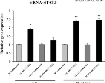
IPA 분석에서 진세노사이드 Rb1에 의한 세포사멸 작용 유 전자들이 상위 조절자 STAT3의 조절을 받아 FAS와 PLA2G4A 를 경유하는지를 확인하기 위하여 STAT3-siRNA를 이용하여 검증하였다. 즉 HaCaT 세포에 대조구인 NC-siRNA와 STAT3- siRNA를 transfection한 후, 24시간 후에 진세노사이드 Rb1을 10 μg/ml의 농도로 처리 혹은 DMSO를 처리하였고 qPCR로 분석하였다(Fig. 7). NC-siRNA를 transfection 한 후 진세노사 이드 Rb1을 처리한 경우 FAS 유전자의 발현이 약 2 배 증가되 었고(p<0.05), STAT3-siRNA를 transfection 후 진세노사이드 Rb1을 처리한 경우 FAS 유전자의 발현은 감소됨을 확인하였
Fig. 8. Effects of knock-down of STAT3 on expression of FAS and PLA2G4A. The HaCaT cells were transfected with STAT3-siRNA or control siRNA, after which they were treated with ginsenoside Rb1 (10 μg/ml) for 24 hr. The expression levels of FAS and PLA2G4A from harvested cells were measured by qPCR as indicated in material and methods. The data are presented as the mean ± SD of three independent experiments performed in triplicate.
*p<0.05, **p<0.01 vs. the control.
다. 반면 NC-siRNA 및 STAT3-siRNA를 transfection 한 후 진세노사이드 Rb1을 처리한 경우 PLA2G4A 유전자의 발현이 동일하게 증가되어(p<0.01), 상위 조절자 STAT3와 PLA2G4A 는 서로 다른 작용점을 나타내었다. 이러한 결과는 진세노사 이드 Rb1의 처리에 의해 상위 조절자인 STAT3는 FAS를 경유 하여 세포사멸을 유도하는 것임을 추측할 수 있다. 한편 STAT3는 7 family STAT 중 하나로 세포질 내에서 비활성형으 로 존재하다가 사이토카인(cytokine), 성장인자(growth fac- tor), 그리고 호르몬의 자극 등에 의해 인산화가 일어난 뒤 이 합체(dimer)를 형성한 후 핵 내로 이동하여 신호를 전달하는 전사조절 인자(transcription factor)이다[13, 16, 28]. STAT3는 다양한 종류의 유전자 발현을 조절함으로써 세포의 분화, 생 존, 증식, 사멸 등을 유발시키며, 특히 많은 종양세포에서 발현 이 증가하는 것으로 알려져 있다[24]. STAT3의 활성은 STAT3 인산화를 촉진하는 STAT의 upstream kinase인 Janus kinase 2 (JAK2)의 인산화와 JAK2와 결합하여 활성을 억제하는 SOCS3 (suppressor of cytokine signaling 3) 단백질 발현 정도 와 tyrosine phosphatase인 SHP-1 (SH2 domain-containing protein tyrosine phosphatase)의 활성에 의해서 조절 된다[2].
따라서 진세노사이드 Rb1에 의한 외인성 세포사멸 신호전달 은 JAK2와 결합된 전사조절 인자 STAT3의 조절을 받으며 FAS를 경유하는 작용기전임을 예상할 수 있다.
References
1. Attele, A. S., Wu, J. A. and Yuan, C. S. 1999. Ginseng phar- macology: multiple constituents and multiple actions.
Biochem. Pharmacol. 58, 1685-1693.
2. Chim, C. S., Fung, T. K., Cheung, W. C., Liang, R. and Kwong, Y. L. 2004. SOCS1 and SHP1 hypermethylation in multiple myeloma: implications for epigenetic activation of the JAK/STAT pathway. Blood 103, 4630-4635.
3. Cho, W. J., Yoon, H. S., Kim, Y. H., Kim, J. M., Yoo, I. J., Han, M. D. and Bang, I. S. 2013. Cytoprotective effects and gene expression patterns observed based on the antioxidant activity of Lonicera japonica extract. J. Life Sci. 23, 989-997.
4. Choi, E. O., Kwon, D. H., Hwang, H. J., Kim, K. J., Lee, D. H. and Choi, Y. H. 2018. Antioxidant and cytoprotective effects of socheongja and socheong 2, Korean black seed coat soybean varieties, against hydrogen peroxide-induced oxidative damage in HaCaT human skin keratinocytes. J.
Life Sci. 28, 454-464.
5. Choo, M. K., Sakurai. H., Kim. D. H. and Saiki, I. 2008. A ginseng saponin metabolite suppresses tumor necrosis fac- tor-alpha-promoted metastasis by suppressing nuclear fac- tor-kappaB signaling in murine colon cancer cells. Oncol.
Rep. 19, 595-600.
6. Degterev, A., Boyce, M. and Yuan, J. 2003. A decade of caspases. Oncogene 22, 8543-8567.
7. Deng, G., Su, J. H., Ivins, K. J., Van Houten, B. and Cotman, C. W. 1999. Bcl-2 facilitates recovery from DNA damage af- ter oxidative stress. Exp. Neurol. 159, 309-318.
8. Fung, C. K., Xi, N., Yang, R., Seiffert-Sinha, K. K., Lai, W.
and Sinha, A. A. 2011. Quantitative analysis of human kera- tinocyte cell elasticity using atomic force microscopy (AFM).
IEEE Trans. Nanobioscience 10, 9-15.
9. Halliwell, B., Gutteridge, J. M. C. and Cross, C. E. 1992. Free radicals, antioxidants, and human disease: Where are we now? J. Lab. Clin. Med. 119, 598-620.
10. Hasegawa, H. 2004. Proof of the mysterious efficacy of gin- seng: basic and clinical trials: metabolic activation of ginse- noside: deglycosylation by intestinal bacteria and ester- ification with fatty acid. J. Pharmacol. Sci. 95, 153-157.
11. Hengartner, M. O. 2000. The biochemistry of apoptosis.
Nature 407, 770-776.
12. Im, J. Y., Kim, B. K., Lee, J. Y., Park, S. H., Ban, H .S., Jung, K. E. and Won, M. 2018. DDIAS suppresses TRAIL-medi- ated apoptosis by inhibiting DISC formation and destabiliz- ing caspase-8 in cancer cells. Oncogene 37, 1251-1262.
13. Inghirami, G., Chiarle, R., Simmons, W. J., Piva, R., Schles- singer, K. and Levy, D. E. 2005. New and old functions of STAT3: a pivotal target for individualized treatment of cancer. Cell Cycle 4, 1131-1133.
14. Kim, B. G., Choi, S. Y., Kim, M. R., Suh, H. J. and Park, H. J. 2010. Changes of ginsenosides in korean red ginseng (Panax ginseng) fermented by Lactobacillus plantarum M1.
Process Biochem. 45, 1319-1324.
15. Kim, G., Sumiyoshi, M., Sakanaka, M. and Kimura, Y. 2009.
Effects of ginseng saponins isolated from red ginseng on
ultraviolet B-induced skin aging in hairless mice. Eur. J.
Pharmacol. 602, 148-156.
16. Kim, H., Suh, J. M., Hwang, E. S., Kim, D. W., Chung, H.
K., Song, J. H., Hwang, J. H., Park, K. C., Ro, H. K., Jo, E. K., Chang, J. S., Lee, T. H., Lee, M. S., Kohn, L. D. and Shong, M. 2003. Thyrotropin-mediated repression of class II trans-activator expression in thyroid cells: involvement of STAT3 and suppressor of cytokine signaling. J. Immunol.
171, 616-627.
17. Kimura, Y., Sumiyoshi, M. and Sakanaka, M. 2012. Effects of ginsenoside Rb1 on skin changes. J. Biomed. Biotechnol.
2012, 1-11.
18. Krämer, A., Green, J., Pollard, J. Jr. and Tugendreich, S.
2014. Causal analysis approaches in ingenuity pathway analysis. Bioinformatics 30, 523-530.
19. Krammer, P. H., Arnold, R. and Lavrik, I. N. 2007. Life and death in peripheral T cells. Nat. Rev. Immunol. 7, 532-542.
20. Lee, C. H. and Kim, J. H. 2014. A review on the medicinal potentials of ginseng and ginsenosides on cardiovascular diseases. J. Ginseng Res. 38, 161-166.
21. Lee, J. O., Choi, E., Shin, K. K., Hong, Y. H., Kim, H. G., Jeong, D., Hossain, M. A., Kim, H. S., Yi, Y. S., Kim, D., Kim, E. and Cho, J. Y. 2019. Compound K, a ginsenoside metabolite, plays an antiinflammatory role in macrophages by targeting the AKT1-mediated signaling pathway. J.
Ginseng Res. 43, 154-160.
22. Lee, Y. M., Yoon, H., Park, H. M., Song, B. C. and Yeum, K. J. 2017. Implications of red Panax ginseng in oxidative stress associated chronic diseases. J. Ginseng Res. 41, 11311- 119.
23. Lü, J. M., Yao, Q. and Chen, C. 2009. Ginseng compounds:
an update on their molecular mechanisms and medical applications. Curr. Vasc. Pharmacol. 7, 293-302.
24. Mitchell, T. J. and John, S. 2005. Signal transducer and acti- vator of transcription (STAT) signalling and T-cell lympho- mas. Immunology 114, 301-312.
25. Namgoong, S., Lee, H., Han, S. K., Lee, H. W., Jeong, S.
H. and Dhong, E. S. 2019. Effect of Panax ginseng extract on the activity of diabetic fibroblasts in vitro. Int. Wound J. 16, 737-745.
26. Park, J. H., Lee, Y. H., Kang, K. S., Lee, S. K., Kim, S. Z., Park, J. Y., Kwak, E. K. and Sohn, Y. K. 2004. The effects of ginsenoside Rb1 on the apoptosis and the production of nitric oxide in Rat C6 glioma cells. Kor. J. Pathol. 38, 1-7.
27. Park, S .E., Na, C. S., Yoo, S. A., Seo, S. H. and Son, H.
S. 2017. Biotransformation of major ginsenosides in ginseno-
side model culture by lactic acid bacteria. J. Ginseng Res.
41, 36-42.
28. Park, Y. J., Park, E. S., Kim, M. S., Kim, T. Y., Lee, H. S., Lee, S., Jang, I. S., Shong, M., Park, D. J. and Cho, B. Y.
2002. Involvement of the protein kinase C pathway in thyro- tropin-induced STAT3 activation in FRTL-5 thyroid cells.
Mol. Cell Endocrinol. 194, 77-84.
29. Richter, C., Gogvadze, V., Laffranchi, R., Schlapbach, R., Schweizer, M., Suter, M., Walter, P. and Yaffee, M. 1995.
Oxidants in mitochondria: from physiology to diseases.
Biochim. Biophys. Acta. 1271, 67-74.
30. Shibata, S. 2001. Chemistry and cancer preventing activities of ginseng saponins and some related triterpenoid com- pounds. J. Kor. Med. Sci. 16, 28-37.
31. Shin, J. E., Park, E. K., Kim, E. J., Hong, Y. H., Lee, K. T.
and Kim, D. H. 2003. Cytotoxicity of compound K (IH-901) and ginsenoside Rh2, main biotransformants of ginseng sap- onins by bifidobacteria, against some tumor cells. J. Ginseng Res. 27, 129-134.
32. Simmler, C., Antheaume, C. and Lobstein, A. 2010. Antiox- idant biomarkers from vanda coerulea stems reduce irradi- ated HaCaT PGE-2 production as a result of COX-2 inhibition. PLoS One 5, 1-9.
33. Wakabayashi, C., Murakami, K., Hasegawa, H., Murata, J.
and Saiki, I. 1998. An intestinal bacterial merabolite of gin- seng protopanaxadiol saponins has the ability to induce apoptosis in tumor cells. Biochem. Biophys. Res. Commun. 246, 725-730.
34. Yayeh, T, Jung, K. H., Jeong, H. Y., Park, J. H., Song, Y.
B., Kwak, Y. S., Kang, H. S., Cho, J. Y., Oh, J. W. and Kim, S. K., et al. 2012. Korean red ginseng saponin fraction down regulates proinflammatory mediators in LPS stimulated RAW264.7 cells and protects mice against endotoxic shock.
J. Ginseng Res. 36, 263-269.
35. Yoon, J. J., Jeong, J. W., Choi, E. O., Kim, M. J., Hwang-Bo, H., Kim, H. J., Hong, S. H., Park, C., Lee, D. H. and Choi, Y. H. 2017. Protective effects of Scutellaria baicalensis Georgi against hydrogen peroxide-induced DNA damage and apoptosis in HaCaT human skin keratinocytes. EXCLI J. 16, 426-438.
36. Zhang, G., Xia, F., Zhang, Y., Zhang, X., Cao, Y., Wang, L., Liu, X., Zhao, G. and Shi, M. 2016. Ginsenoside Rd is effica- cious against acute ischemic stroke by suppressing micro- glial proteasome-mediated inflammation. Mol. Neurobiol. 53, 2529-2540.
초록:피부각질세포 HaCaT에서 진세노사이드 Rb1에 의한 유전자 발현 양상
이동우1․김정민2․방인석1*
(1호서대학교 생명과학과 및 기초과학연구소, 2제노플랜코리아㈜ 유전체분석팀)
인삼(Panax ginseng C. A. Meyer)의 사포닌 진세노사이드 Rb1이 처리된 인간 피부각질세포 HaCaT에서 micro- array 분석 및 발현이 증가된 세포사멸 반응에 대한 작용기전을 연구하였다. HaCaT 세포에 진세노사이드 Rb1의 처리로 세포사멸, 유사분열 세포주기의 G2/M 전이, 세포분열, 핵분열, 그리고 단백질 수송 등의 작용기전에 관여 하는 유전자들이 2 배 이상 발현이 증가된 것으로 나타났으며, DNA 수선, 감수 핵분열, 그리고 세포외기질 체계 등의 작용기전에 관여하는 유전자들은 2 배 이상 발현이 감소된 것으로 나타났다. 특히 세포사멸 신호전달은 FAS 와 PLA2G4A를 경유하는 것으로 나타났으며, 이들 유전자의 상위 조절자로 STAT3가 예측되었다. 세포사멸 반응 경유 유전자 FAS와 PLA2G4A의 활성을 qPCR로 확인한 결과, FAS 유전자는 10 μg/ml의 진세노사이드 Rb1를 24시간 동안 처리하였을 경우 약 2 배의 발현 증가와, PLA2G4A 유전자는 6시간 처리부터 약 2 배로 증가되어 24시간 동안 처리시 2 배 이상의 유전자 발현이 증가되었다. 한편 STAT3-siRNA를 이용한 knock-down 실험에서 FAS의 발현 감소와 PLA2G4A의 발현 증가로 상위 조절자 STAT3로부터 FAS 만을 경유하는 것을 알 수 있었다.
이상의 결과 진세노사이드 Rb1의 처리에 의해 상위 조절자인 STAT3는 FAS를 경유하여 세포사멸을 유도하는 것 임을 알 수 있다.