• 검색 결과가 없습니다.

The Effect of Codium fragile (Chlorophyta) Extract on Hepatic Dysfunction and Hyperlipidemia in Rats

N/A
N/A
Protected

Academic year: 2021

Share "The Effect of Codium fragile (Chlorophyta) Extract on Hepatic Dysfunction and Hyperlipidemia in Rats"

Copied!
7
0
0

로드 중.... (전체 텍스트 보기)

전체 글

(1)

INTRODUCTION

Recently, due to the diversification and westernization of dietary habits, the consumption of high calorie food and meats have increased, and consequently, obesity, stroke, arteriosclerosis, hypertension, diabetes, and other life style diseases are on the increase. According to the national nutri- tion survey by the health and welfare department (Korea), in Korean diet, the fat consumption rate per day is continuously increasing from 16.9 g in the year of 1969 to 36.8 g in 2007 (Annual report 2008). Such increase of fat consumption has led to an increase in the incidence and mortality of circula- tory diseases by increasing the lipid content in the body.

Among diseases of the circulatory system, hyperlipidemia refers to the condition wherein plasma cholesterol or trigly- ceride levels are abnormally elevated. In the development of cardiovascular diseases, the concentration of cholesterol has been known to act as an important contributing factor.

Blood cholesterol concentration is controlled in vivo and maintained constantly. Nonetheless if it were consumed excessively for a long period of time, its blood concentration becomes accumulated and induces hyperlipidemia, arterio- sclerosis, coronary artery diseases and other cardiovascular diseases (Lusis 1988).

Chronic high calorie food intake can impair the function of liver cells, which play an important role in the metabolism of nutrients. Hepatic injury can result in severe damage to the serum level of AST, ALT and cholesterol (LDL. Chol and HDL. chol.) (Lee et al. 2005). The degradation of nor-

─ 79 ─

The Effect of Codium fragile (Chlorophyta) Extract on Hepatic Dysfunction and Hyperlipidemia in Rats

Kap Joo Park, Eun Kyoung Hwang

1

, Chan Sun Park

2

, Myung Hwan Cho and Jae Seok Lee*

Department of Biological Sciences, Konkuk University, Seoul 143-701, Korea

1

Seaweed Research Center, NFRDI, Jeonnam 530-831, Korea

2

Department of Marine Resources, Mokpo National University, Jeonnam 534-729, Korea

Abstract - - To examine the effect of Codium fragile on blood cholesterol and lipid metabolism, hyperlipidemia was induced in experimental animal rats through the administration of a hyper- cholesterolemic diet. Codium fragile powder was then administered to the rats for 5 weeks, after which, blood biochemical changes such as blood cholesterol, Aspartate Aminotransferase (AST:

serum SGOT) and Alanine Aminotransferase (ALT: serum SGPT) enzyme activity, etc. were determined. And histological changes in liver cells were examined using an electron microscope.

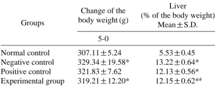
Codium fragile treatment resulted in a significant reduction of the levels of total cholesterol, blood triglyceride and low-density cholesterol (LDL. Chol) compared to the control rats. In contrast the expression levels of high-density cholesterol (HDL. chol.) were increased. The AST value of the Codium fragile administration group was significantly reduced and the blood ALT value of the Codium fragile group showed a significant decrease in comparison to the negative control group.

In summary, this study demonstrated the beneficial possibilities of Codium fragile in improving the abnormality of lipid metabolism caused by liver cell damage and hyperlipidemia.

Key words : hyperlipidemia, Codium fragile, rat liver cell, lipid metabolism

* Corresponding author: Jae Seok Lee, Tel. 02-447-5018,

Fax. 02-3436-5432, E-mail. [email protected]

(2)

mal liver functions is indicated by the increased levels of serum AST and ALT enzymes that are normally concentrat- ed in the hepatic tissue. Increased levels of AST and ALT enzymes in the serum can be caused by fatty liver or expo- sure to by-products, resulting in metabolic problems of the liver and hepatocyte death. And high levels of cholesterol are major causal factors in the development of atherosclero- sis and subsequent cardiovascular disease (Park et al. 2002).

Recently, drugs suppressing 3-hydroxy-3-methylglutaryl CoA reductase (HMG-CoA reductase), which is an enzyme required for the synthesis of cholesterol in hepatocytes, have been reported to be most effective among drugs for the treatment of hypercholesterolemia (Samuel and McNanara 1983). In addition, cholestyramine, clofibrate, gemfivrozil, nicotinic acid, probucol, etc. have been developed as drugs to decrease blood lipid concentration. However, the effect of these drugs are not constant between various individuals and furthermore, various side effects of these drugs have recently been revealed (Lim et al. 2005).

Therefore, to resolve the safety problem of the long-term intake of lipid-depression drugs currently supplied by clinics, the appropriate consumption of natural food types has been recommended for the prevention and treatment of cardiova- scular diseases (Lee et al. 1997). Interests in natural diet therapy has heightened as the physiological approach to lowering blood cholesterol level. Studies on the prevention and improvement of hyperlipidemia by applying functional materials extracted from natural food types are also genera- ting widespread interest (Harris et al. 1993; Cameron et al.

1997).

A type of green algae, Codium fragile is used widely as food in Korea, China, Japan as well as Philippines, Hawaii, Africa, and other countries (Champman 1962; Oh et al.

1990). It has been used as a helminthic in folk medicine as well as for urinary diseases and obesity treatments (Tseng and Zhang 1984). The extract of Codium fragile contains acrylic acid that has antibiotic activity, anticoagulation acti- vation materials, agglutinin and also anticancer as well as anti-mutation and immune activity (Cho et al. 1990). Thus it is a useful marine plant that could be applied in the field of pharmacology and medicine and has the potential to be a candidate material for the treatment of hyperlipidemia as well as obesity (Rogers et al. 1990; Rogers et al. 1991).

This study was therefore conducted to elucidate the effects of Codium fragile on hepatic function damaged by hyperli-

pidemia and elevated blood lipid concentrations in rats.

Codium fragile extract was administered to rats that were previously maintained on a high-fat diet so as to induce hyperlipidemia. Aspartate Aminotransferase (AST: serum SGOT) and Alanine Aminotransferase (ALT: serum SGPT) activities were measured. Also, the serum major lipid com- ponents, i.e. triglycerides, total cholesterol (T. chol.), low- density cholesterol (LDL. chol.), and high-density cholesterol (HDL. chol.) activity were measured.

MATERIALS AND METHODS

1. Experiment animals and diet

As experiment animals, 32 Sprague Dawley male rats, 4 weeks of age, with an average weight of 79.29 1.73 g were obtained from Daehan Biolink Co., Ltd. (Seoul). The ani- mals were allowed to adapt to the animal facility for 1 week.

The temperature and humidity of the animal room were maintained as 22±2� C and 55±5%, respectively. The rats were kept on the 12 h light/dark cycle and acclimatized to the housing situation. Under a free environment, basal food (Superspeed, Co. Seoul) (Table 1), food inducing fatty liver (Table 2), and drinking water were freely supplied.

2. Classification of experiment groups

After one week of the adaptation period, the rats were divided into 4 groups (n= =8) as follows: (i) normal control rats administered with a basic diet

++

distilled water, (ii) nega- tive control rats administered fatty liver-inducing feed

++

dis- tilled water, (iii) positive control rats administered fatty liver- inducing feed

++

blood circulation promotion Solution (BCPS), (iv) experimental control rats administered fatty liver-induc- ing feed

++

Codium fragile extract (Table 3) (Kang et al. 2003).

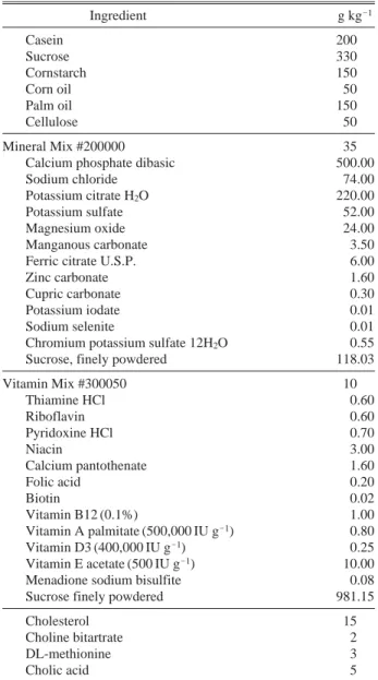
Each feed was prepared by 200 g fatty liver feed (Dyets Table 1. Composition of basal diet

Ingredients Contents (%)

1)

Crude protein 22.1

Crude fat 3.5

Fiber 5.0

Crude ash 8.0

Calcium 0.6

Phosphate 0.4

1)

per 200 g

(3)

INC., New York, USA, DYET# 101865 Custom, AIN-76A Based Purfied Rat Diet With 15% Palm Oil, 1.5% Chole- sterol, and 0.5% Cholic Acid), and 200 g each diet was allowed to be consumed freely for 5 weeks. Distilled water, blood circulation promotion Solution and Codium fragile extract was administered orally using a syringe everyday at a constant time.

3. Preparation of Codium fragile extract and BCPS

Boiling water extracts of Codium fragile were prepared from the dried Codium fragile. 25 g of Codium fragile was added into 1,000 mL of sterilized water and boiled for 150 min using a herbal and medicinal boiling pot (Daewoong

Co., Ltd., Seoul). After centrifugation at 6,000 ×g for 15 min, aqueous extracts from the sample were filtered through 3 mm filter papers (Whatman, England), and the final volume was adjusted to around 400 mL in order to prepare an appro- priate volume for administration (about 1.6 g kg

-1

body weight/day) (Lee et al. 2005). BCPS was mixture of 120 mg Ginkgo biloba extract and 60 mg mL

-1

sodium benxoate (Cho-A Pharmaceutical Co., Ltd., Seoul), and the dose was calculated by the average weight of experimental animals based on humans (60 mL kg

-1

daily) and administered.

4. Animal autopsy

On the last day of the experiment, the animals were fasted for 16 hours, the abdomen was resected under weakly anesthetized condition with ethyl ether and 3~4 mL blood was collected from the venae cavae and distributed into an EDTA-free test tube. Immediately after blood collection, the liver was extracted, washed with cold saline and the weight was measured.

5. Biochemical analysis (AST, ALT, HDL-cholesterol, LDL-cholesterol, Total cholesterol, Triglyceride)

The blood collected from each experiment group was kept at room temperature for approximately 30 minutes. The serum was separated by centrifugation (6,000 ×g, 15 min) and used for serum biochemical tests. Aspartate transami- nase (AST), a marker enzyme of the deterioration of liver function was measured using an AST (GOT) kit (Bayer Co.

Ltd., Frankfurt, German) while alanine transaminase (ALT) was measured using an ALT (GPT) Reagents kit (Bayer Co.

Ltd., Frankfurt, German). The total cholesterol concentration was measured using a Cholesterol Reagents kit (Bayer Co.

Table 2. Composition of hyperlipidemic diet

Ingredient g kg

-1

Casein 200

Sucrose 330

Cornstarch 150

Corn oil 50

Palm oil 150

Cellulose 50

Mineral Mix #200000 35

Calcium phosphate dibasic 500.00

Sodium chloride 74.00

Potassium citrate H

2

O 220.00

Potassium sulfate 52.00

Magnesium oxide 24.00

Manganous carbonate 3.50

Ferric citrate U.S.P. 6.00

Zinc carbonate 1.60

Cupric carbonate 0.30

Potassium iodate 0.01

Sodium selenite 0.01

Chromium potassium sulfate 12H

2

O 0.55

Sucrose, finely powdered 118.03

Vitamin Mix #300050 10

Thiamine HCl 0.60

Riboflavin 0.60

Pyridoxine HCl 0.70

Niacin 3.00

Calcium pantothenate 1.60

Folic acid 0.20

Biotin 0.02

Vitamin B12 (0.1%) 1.00

Vitamin A palmitate (500,000 IU g

-1

) 0.80

Vitamin D3 (400,000 IU g

-1

) 0.25

Vitamin E acetate (500 IU g

-1

) 10.00

Menadione sodium bisulfite 0.08

Sucrose finely powdered 981.15

Cholesterol 15

Choline bitartrate 2

DL-methionine 3

Cholic acid 5

Table 3. Experimental designs

Groups No. of rats Composition of treatments Normal control group 8 basic diet

++

distilled water Negative control group 8 fatty liver inducing feed

++

distilled water

Positive control group 8 fatty liver inducing feed

++

BCPS*

Experiment group 8 fatty liver inducing feed

++

Salicornia herbacea extract

*BCPS: Blood Circulation Promotion Solution (mixture of Ginkgo biloba

extract 120 mg and sodium benxoate 60 mg mL

-1

, Cho-A Pharmaceutical

Co., Ltd., Seoul)

(4)

Ltd., Frankfurt, German) by an automated biochemical analyzer (ADVIA 1650/2400, Bayer Co. Ltd., Frankfurt, German), high-density lipoprotein (HDL) was measured using a Direct HDL-Cholesterol Kit (Bayer Co. Ltd., Frank- furt, German) and low-density lipoprotein (LDL)-cholesterol was measured using a LDL-cholesterol kit (Daiichi Co.

Ltd., Tokyo, Japan) by an automated biochemical analyzer (ADVIA 1650, Bayer Co. Ltd., Frankfurt, German). Trigly- ceride (TG) concentration was measured using a Triglyceri- des reagents (Bayer Co. Ltd., Frankfurt, German) by an auto- mated biochemical analyzer (ADVIA 1650/2400, Bayer Co.

Ltd., Frankfurt, German) (Park et al. 2004).

6. Statistical analysis

All results were shown as mean±standard deviation.

Statistical evaluation of data was performed by Duncan’s multiple range test to make comparisons between groups.

RESULTS AND DISCUSSION

1. Weight gain and ratio of liver weight to body weight

The weight change of experiment animals that were admi- nistered with a high fat diet for 5 weeks is shown in Table 4. The final weight change of the normal group and the negative control group was shown to be 307.11±5.24 g and 329.34±19.58 g, respectively. The final weight change of the positive control group was 321.83±7.62 g, while the Codium fragile administration group was shown to be 319.21

±12.20 g. It was also found that in the negative control group, the overweight phenomenon was shown in compari- son to the normal group. The Codium fragile administration group showed a significant weight change similar to the normal group. Thus the effect of the negation of weight gain caused by the high fat diet in the Codium fragile administra- tion group was confirmed.

The ratio of total weight of experiment animals and the ratio of body weight to liver weight obtained from our ex- periment is shown in Table 4. After consumption of the high fat diet for a long period, the liver was enlarged because of fatty liver and hepatic fibrosis. Several research results show- ed that the liver weight of all high fat diet groups was in- creased by the accumulation of lipid in hepatic tissues due

to consumption of a high fat diet (Samuel and McNanara 1983). In the present study, a similar trend to previously reported studies was shown. The ratio of body weight and the liver of the negative control group was 13.22±0.64%, and it was significantly high in comparison to the normal group of 5.53±0.45%. Thus it was found that fatty liver and liver fibrosis had already progressed; this was in agreement with previous reports showing that the liver became enlarged upon administration of high fat diet for a long period of time.

On the other hand, the positive group was 12.13±0.56%, and a slightly reduced value was shown, and the ratio of body weight and the liver in the experimental group was shown to be 12.15±0.62%, and a significant decreased was shown in comparison with the normal group and the negative control group.

2. Activities of AST and ALT

Changes in AST and ALT activity of each group is pre- sented in Table 5. AST and ALT levels both increased with increased high fat diet intake. These enzymes are well-docu- mented indicators of hepatic disfunction, with increased Table 4. Body weight gain and ratio of liver weight to body weight

administered for 5 weeks

Change of the Liver Groups body weight (g) (% of the body weight)

Mean±S.D.

5-0

Normal control 307.11±5.24 5.53±0.45 Negative control 329.34±19.58* 13.22±0.64*

Positive control 321.83±7.62 12.13±0.56*

Experimental group 319.21±12.20* 12.15±0.62*

#

Each value was represented as mean

±standard deviation for 8 rats. Means

with different superscripts within a column are significantly different from the normal control at *p⁄0.05, and significantly different from negative control at

#

p⁄0.05.

Table 5. Serum levels of AST and ALT Groups

AST ALT

Means±S.D Means±S.D Normal control group 131.50 ±24.80 42.25 ±10.31 Negative control group 951.75 ±204.80* 180.00±17.72*

Positive control group 787.00 ±162.45*

#

154.00 ±75.77

Experiment treatment group 662.25 ±233.70* 143.50±61.21

#

Each value was represented as mean±standard deviation for 8 rats. Means

with different superscripts within a column are significantly different from

the normal control at *p⁄0.05, and significantly different from negative

control at

#

p⁄0.05.

(5)

AST and ALT levels reflecting impaired liver function (Lim et al. 2005). In the normal group, the AST value was shown to be 131.50±24.80 U L

-1

, the value of the negative group showed a rapid increase to 951.75±204.80 U L

-1

, and thus the liver damage caused by the consumption of high fat diet for a long time could be confirmed.

In the positive group, the AST value was 787.00±162.45 U L

-1

, which showed a slight decrease compared to negative group. The AST value of the Codium fragile administration group was 662.25±233.70 U L

-1

. It was measured to be lower than the AST value of the negative control group.

The ALT level of the normal group was shown to be 42.25

±10.31 U L

-1

, the negative control group was 180.00±

17.72 U L

-1

, and it was shown to be markedly higher. This also confirmed the presence of liver damage due to the con- sumption of the high fat diet for the period of time. The ALT value of the positive control group was shown to be 154.00 ±75.77 U L

-1

, the ALT value of the Codium fragile administration group was shown to be 143.50±61.21 U L

-1

. In this study, the Codium fragile administration group exhi- bited significantly reduced AST and ALT levels compared to the negative control group. These data suggest the possi- bility of Codium fragile being an excellent candidate to ame- liorate the effect of hepatocytes and anti-hyperlipidemia from high fat diet-mediated damage in the rat.

3. Total cholesterol and triglyceride levels

Serum total cholesterol and triglyceride concentrations are presented in Table 6. Total cholesterol concentration of the normal group was 55.25±10.94 mg dL

-1

, while that of the negative control group was increased greatly to 191.50

±63.98 mg dL

-1

. Total cholesterol levels in the positive control group and the Codium fragile administration groups were shown to be 130.00±14.54 mg dL

-1

and 130.22±

18.23 mg dL

-1

, respectively. Serum triglyceride concentra- tion of the normal group, the negative control group, the positive control group, and the Codium fragile administration group were 54.75±10.87 mg dL

-1

, 61.75±5.19 mg dL

-1

, 46.20±6.45 mg dL

-1

, and 55.00±10.30 mg dL

-1

, respec- tively. In comparison to the negative control group, the Codi- um fragile administration group showed a significantly low total cholesterol and triglyceride content. Many reports indi- cated that a high fat diet intake significantly increased both serum and hepatic triglyceride (TG) levels resulting in hyper-

triglyceridemia and fatty liver (Lim et al. 2005). Data sum- marized in Table 6 indicates that the administration of Codium fragile extract have markedly beneficial effects on the serum lipid levels. Based on reports. Indicating that an elevated blood cholesterol level is one of the main causes of vascular disease in the heart and circulatory system (Kang et al. 2003), a number of drugs have been developed to lower plasma cholesterol concentrations, such as cholesty- ramine, probucol and statins. However, little work has been done in developing natural materials to prevent hyperlipide- mia. In this context, this report suggests that Codium fragilel may represent an alternative therapeutic agent to assist in the prevention and treatment of hyperlipidemia

4. HDL-cholesterol and LDL-cholesterol levels

Serum HDL-cholesterol and LDL-cholesterol concen- trations are presented in Table 7. HDL-cholesterol has the effect of improving arteriosclerosis and angiosclerosis. LDL- cholesterol accumulates in the artery blood vessel wall pri- marily and may cause arteriosclerosis, and thus it has been known to be an important risk factor in the development of arteriorsclerosis and cardiovascular diseases (Kannel et al.

1964). In our experiments, the HDL-cholesterol level of the Table 6. Serum levels of total cholesterol and triglyceride

Groups Total cholesterol Triglyceride Means ±S.D Means ±S.D Normal control group 55.25±10.94 54.75±10.87 Negative control group 191.50±63.98* 61.75±5.19*

Positive control group 130.00±14.54** 46.25±6.45*

Experiment treatment group 130.22±18.23

#

55.00±10.30**

#

Each value was represented as mean

±standard deviation for 8 rats. Means

with different superscripts within a column are significantly different from the normal control at *p⁄0.05, **p⁄0.01, and significantly different from negative control at

#

p⁄0.05.

Table 7. Serum levels of HDL-cholesterol and LDL-cholesterol

Groups HDL LDL

Means±S.D Means±S.D Normal control group 24.25±2.63 9.50±1.29 Negative control group 13.50±1.29* 100.25±27.44*

Positive control group 20.00±1.83** 55.50±9.98*

#

Experiment treatment group 17.50±3.11*

#

68.75±12.53*

#

Each value was represented as mean±standard deviation for 8 rats. Means

with different superscripts within a column are significantly different from

the normal control at *p⁄0.05, **p⁄0.01, and significantly different from

negative control at

#

p⁄0.05.

(6)

normal group was 24.25±2.63 mg dL

-1

, while the negative control group and the positive control group was shown to be 13.50±1.29 mg dL

-1

and 20.00±1.83 mg dL

-1

, respec- tively. That of the experiment group was shown to be 17.50

±3.11 mg dL

-1

, and in comparison to the normal group, it was significantly low by more than 30%. The LDL-chole- sterol of the normal group (9.50±1.29 mg dL

-1

) and the negative group (100.25±27.44 mg dL

-1

) was noticeably different. Comparing the positive control group (55.50±

9.98 mg dL

-1

) and the experimental group (68.75±12.53 mg dL

-1

) with the negative control group, the value of LDL- cholesterol was significantly decreased. It was found that Codium fragile directly lowered the blood LDL-cholesterol value and increased HDL-cholesterol value.

In the American NIH standard, for the determination of the risk level of cardiovascular diseases, the arteriosclerosis index and cardiac risk index have been applied (Goldstein and Brown 1975). In our experiments, the ratio of HDL- cholesterol and total-cholesterol (HTR: HDL-cholesterol/

total-cholesterol), the fluctuation of arteriosclerosis index (A.I., atherogenic index: total-cholesterol - HDL-cholesterol/

HDL-cholesterol), and cardiac risk factor (C.R.F., cardiac risk factor: total-cholesterol/HDL-cholesterol) was calculated.

The HTR of the Codium fragile administration group was 2 times higher than that of the negative control group, and thus it was found to reduce the risk of the development of car- diovascular diseases. The arteriosclerosis index of the nega- tive control group showed a value reaching almost 2 times higher than that of the Codium fragile administration group.

Its cardiac risk index showed a pattern similar to arterio- sclerosis index (Goldstein and Brown 1975).

Hyperlipidemia is an index of arteriosclerosis symptoms.

It has been reported to increase the synthesis of triglycerides in the small intestine. The secretion of chylomicron increases the synthesis of triglycerides in the liver, thereby increasing the synthesis and secretion of VLDL-cholesterol and LDL- cholesterol, decrease HDL-cholesterol synthesis and decreas- ing lipase activity. A decrease in the lipase activity induces the reduction of the removal of triglycerides in peripheral tissues. In our study, similar results were demonstrated in rats wherein an induced dietary hyperlipidemia allowed for the noticeable elevation of the content of total-cholesterol and LDL-cholesterol.

In summary, a high concentration of total-cholesterol and LDL-cholesterol acts as a major factor in the induction of

artherosclerosis and cardiovascular diseases, and thus, nu- merous studies and efforts have been made to develop drugs and functional materials. It is anticipated that if drugs or functional materials were developed from natural materials with less side effects, its value would be great. From such a point of view, the results of our study suggest that Codium fragile could be tested as a candidate substance for natural therapeutic against hyperlipidemia.

ACKNOWLEDGEMENT

This research was supported by a grant from the Korea Ministry of Agriculture and Forest and by Priority Research Centers Program through the National Research Foundation of Korea (NRF) funded by the Ministry of Education, Sci- ence and Technology (2009-0093828).

REFERENCES

Korea national statistical office. 2008. Annual report of the cause of death statistics.

Cameron-Smith D, R Habito, M Barnett and GR Collier. 1997.

Dietary guar gum improves insulin sensitivity in streptozo- tocin-induced diabetic rats. J. Nutr. 127:359-364.

Champman VJ. 1962. The algae. St. Martin’s Press. New York.

pp. 94-104.

Cho KJ, YS Lee and BH Ryu. 1990. Antitumor effect and im- munology activity of seaweeds toward sarcoma-180. Bull.

Kor. Fish. Soc. 23:345-352.

Cho SH and TY Ha. 2004. In vitro and in vivo effects of pro- somillet and sorghum on cholesterol metabolism. Food Sci.

Biotechnol. 12:485-490.

Goldstein JL and MS Brown. 1975. Familial hypercholesterole- mia. A genetic regulatory defect in cholesterol metabolism.

Am. J. Med. 58:147-152.

Harris PJ, AM Roberton, ME Watson, CM Triggs and LR Fer- guson. 1993. The effects of soluble-fiver polysaccharides on the adsorption of a hydrophobic carcinogen to an in- soluble dietary fiber. Nutr. Cancer. 19:43-54.

Kang SM, JY Shim, SJ Hwang, SG Hong, HE Jang and MH Park. 2003. Effect of saengshik supplementation on health improvement in diet-induced hypercholesterolemic rats. J.

Kor. Soc. Food Sci. Nutr. 32:906-912.

Kannel WB, TR Dawber and GD Frieman. 1964. Risk factor in

coronary heart disease continous and grade; Primary screens

of multiple risk factor intervention trial (MEFIT). J AMA.

(7)

256:2853-2870.

Lee JS, KH Ahn and KJ Park. 2005. Ameliorative effects of pine needle oil on liver protection and lipid metabolism of alcohol-fed Rats. Food Sci. Biotechnol. 14:99-101.

Lee JS, HY Kim, KJ Park and HH Lee. 2005. Effects of soybean embryo on liver protection and lipid metabolism of alcohol- fed rats. Food Sci. Biotechnol. 14:102-107.

Lee JS, GS Lee and HK Shin. 1997. Effects of chicory extract on the serum glucose and lipid metabolism in streptozotocin - induced diabetic rats. Korean J. Nutr. 30:781-788.

Lim HJ, KH Chol and RW Cho. 2005. The effects of functional tea supplement with medical nutrition therapy on the blood lipid levels and antioxidant status in subjects with hyper- lipidemia. J. Korean Soc. Food Sci. Nutr. 34:42-56.

Lusis AJ. 1988. Genetic factors affecting blood lipoproteins:

The candidate gene approach. J. Lipid Res. 29:397-429.

Oh YS, YP Lee and IK Lee. 1990. A taxonomic study on the genus Codium chlorophyta, in Cheju island. Korean J. Phy- col. 2:61-72.

Park KJ, HY Kim, BJ Chang and HH Lee. 2004. Ameliorative effects of soy 11s protein on liver damage and hyperlipide-

mia in alcohol-fed rats. Bio. Pharm. Bull. 27:1636-1641.

Park KJ, MJ Lee, H Kang and HH Lee. 2002. A medicinal herb complex, protects liver cell damage induced by alcohol.

Bio. Pharm. Bull. 25:451-1455.

Rogers DJ, KM Jurd, S Blunden and F Zanetti. 1990. Anticoa- gulant activity of a proteoglycan in extracts of Codium fra- gile sp. atlanticum. J. Appl. Phycol. 2:357-361.

Rogers DJ and RW Loveless. 1991. Electron microscopy of human erythrocytes agglutinated by lectin from Codium fragile sp. tomentosoides and pseudohaemagglutinin from Ascophyllum nodosum. J. Appl. Phycol. 3:83-86.

Samuel P and DJ McNanara. 1983. Dfferential absorption of exogeneous and endogeneous cholesterol in man. J. Lipid Res. 24:265-269.

Tseng CK and JF Zhang. 1984. Chinese seaweeds in herbal medicine. Proc. Int’l. Seaweed Symp. pp. 62-65.

Manuscript Received: March 18, 2010

Revision Accepted: May 20, 2010

Responsible Editor: Sun Il Kwon

수치

Table 3. Experimental designs
Table 5. Serum levels of AST and ALT  Groups
Table 7. Serum levels of HDL-cholesterol and LDL-cholesterol

참조

관련 문서

Therefore, the present study aimed to (i) investigate the effect of surfactant on the solubility of valsartan and (ii) improve the dissolution profiles of valsartan

The purpose and necessity of this study was to investigate the effect of the 8-week GX complex exercise program on the stress index, blood lipid and APG

Methods: Eighty ASA physical status I and II pediatric patients (1~7 years) were randomly divided into four groups: Group I (Lactated Ringer`s solution, n=20), Group II

I examined ticks that collected by Flagging and Dragging method in 5 regions of Gwangju suburb and captured rats. nipponensis ) I classified ticks

Contents of total lipid in the epididymal adipose of rats fed high fat-high cholesterol diet containing Stachys sieboldii Miq root powder for 6 weeks ···78 Fig.. Contents of

Effect of caloric restriction on the expression of PGC-1 and PPARs mRNA in liver of Otsuka Long-Evans Tokushima Fatty Rats.. Long-lived growth hormone

Mean arterial plasma concentration-time profiles of 4-hydroxytamoxifen after an oral administration of tamoxifen (10 mg/kg) to rats in the presence or absence

Contents of total cholesterol in the epididymal adipose tissue of STZ-induced diabetic rats containing barley leaf powder for 4 weeks ··· 147 Fig.. The change of growth rate