LPS로 유도된 RAW264.7 염증모델에서 MAPK 조절에 의한 羊乳의 항염증효과
김범회1․이용태․강경화*
동의대학교 한의과대학 생리학교실 & 한의학 연구소, 1 : 해부학교실 & 한의학 연구소
Codonopsis Lanceolata Inhibits Inflammation through Regulation of MAPK in LPS-stimulated RAW264.7 cells
Bum Hoi Kim1, Yong Tae Lee, Kyung Hwa Kang*
Department of Oriental Physiology,
1 : Department of Anatomy, College of Oriental Medicine and Research Institute of Oriental Medicine, Dong-Eui University
Codonopsis Lanceolata (CL) has been widely used in Oriental medicine for treatment of chronic inflammatory diseases, such as bronchitis, cough, and spasm; however, the mechanism of its anti-inflammatory activity has not been clarified. In this study, therefore, we investigated the inhibitory effect of CL on LPS-induced inflammation. The effect of CL was analyzed by ELISA, RT-PCR and Western blotting in LPS-stimulated RAW264.7 cells. We found that CL suppressed not only the mRNA expression of pre-inflammatory cytokines, inducible nitric oxide synthase (iNOS), and cyclooxygenase (COX)-2, but also the phosphorylation of ERK1/2, JNK1/2 and p38 MAPK. These results suggest that CL exerts an anti-inflammatory effect through the regulation of the mitogen-activated protein kinases (MAPK) pathway, thereby decreasing production of pre-inflammatory cytokines, NO, and PGE2.
Key words : codonopsis anceolata (CL), inflammation, mitogen-activated protein kinases (MAPK), RAW264.7
* 교신저자 : 강경화, 부산시 부산진구 양정2동 동의대학교 한의과대학
․E-mail : [email protected], ․Tel : 051-850-7423
․접수 : 2010/01/12 ․수정 : 2010/01/19 ․채택 : 2010/01/28
서 론
염증은 조직손상과 병원균의 침입으로부터 인체를 방어하는 가장 중요한 기전중의 하나로, 정상적인 염증반응은 전구 염증단 백질의 발현을 감소시키고 항염증 단백질의 발현을 증가시키며 초기 면역세포 보충을 촉진시키기 위한 혈관의 변화를 복구하는 등의 조절이 일어난다 1,2) . 그러나 만성염증반응은 궁극적으로 만 성피부질환, 류마티스 관절염, 기관지염, 위염, 다발성 경화증, 염 증성 장질환 등의 염증질환으로 발전할 수 있으므로 적절한 치 료가 요구된다 2) .
대식 세포는 혈액 단핵세포로부터 분화한 조직 세포로 염증 반응에서 방어적 역할을 담당한다. Lipopolysaccharide (LPS)는 대식세포 표면의 TLR4를 자극하여 하부 세포신호전달경로인 mitogen-activated protein kinase (MAPK)의 활성화를 유도한다
3) . 활성화된 신호전달경로는 pro-inflammatory cytokines, nitric
oxide (NO), prostaglandin (PG)s와 같은 여러 가지 염증성 매개 인자들의 발현을 유도한다 4) .
양유(Codonopsis Lanceolata, CL)는 더덕의 뿌리로 한국, 일 본 및 중국에서 기관지염, 기침, 경련 및 대식 세포에 기인한 염 증 치료에 널리 사용되어져 왔다 5) . 최근 연구에서 양유는 알콜성 지방간 6) , 스트레스성 간손상 7) , 대장염 8) 에 억제효과가 보고되었 으며, 항산화, 항균, 및 항돌연변이 효과 9) 도 알려졌다. CL의 항염 증기전에 대한 연구에서 Nuclear Factor kappa B (NF-kB)를 통 한 항염증발현에 대한 연구는 있었으나 8,10) 또 다른 중요한 항염 증기전인 MAPK에 대한 연구는 아직 보고되지 않았다.
이에 저자는 LPS로 유도된 RAW264.7 cells의 염증반응에서 CL의 항염증 효과와 세포내 MAPK를 통한 염증발현기전에 미 치는 영향을 조사하여 유의한 결과를 얻었기에 보고하는 바이다.
재료 및 방법
1. 약재의 추출
본 실험에 사용된 CL은 자연으로(제주, 한국)에서 구입한 국
내산 더덕 300 g을 증류수로 水洗하여 1 L의 증류수를 加하여 3 시간 동안 가열 추출하였다. 추출된 용액을 원심분리를 통하여 상층액을 분리하고, 0.2 mm 여과지에서 여과하여 rotary evaporator (N-1100V, EYELA, JAPAN)로 200 ml이 되도록 감압 농축한 후 냉동 건조하여 7.38 g의 분말을 얻었다. CL의 수득율 은 2.46%로 -20℃에 보관하였다가 실험직전 생리식염수나 배지 에 희석하여 0.2 mm syringe filtering 후 실험에 사용하였다.
2. 시약
본 실험에 사용된 LPS (Escherichia coli O55:B5), HEPES, Igepal CA-630, Sodium deoxycholate, NaCl, Tris-HCl, Sodium pyrophosphate, NaVO 4 , NaF, Leupeptin, Phenylmethylsulfonyl fluoride는 Sigma (St Louis, MO, USA)로부터, FBS, penicillin, streptomycin, DMEM 및 trypsin-EDTA solution은 Gibco BRL (NY, USA)로부터, MTT, TRizol 및 DMSO는 Amresco (Cochran Road Solon, OH USA)로부터, reverse transcription polymerase chain reaction (RT-PCR) kit (AccuPower RT/PCR PreMix)는 Bioneer (Daejeon, Korea)로부터, anti-phospho-ERK, anti-ERK, anti-phospho-JNK, anti-JNK, anti-phospho-p38, anti-p38 및 anti-β-actin primary antibody는 Cell Signaling Technology (Beverly, MA, USA)으로부터, HRP-conjugated secondary antibody는 KPL (Gaithersburg, MD, USA)로 부터 구입하여 사 용하였다.
3. 세포 배양
RAW264.7 cells는 한국세포주은행 (Korea Cell Line Bank, KCLB, No. 40071) (Seoul, Korea)에서 분양받았으며 세포의 배양 을 위하여 10% heat-inactivated FBS (Gibco BRL, USA)과 1%
penicillin 및 streptomycin (Gibco BRL, USA)을 포함한 DMEM (Gibco BRL, USA) 배양액에서 배양하였다. 세포는 37℃, 5%
CO 2 조건하에서 배양하였고, 2일마다 배지를 교환하였으며, 세 포의 증식에 따른 과밀도 현상을 해소하기 위하여 계대 배양하 였다.
4. MTT assay
RAW264.7 cells를 10% FBS를 포함한 DMEM에 현탁시킨 후 96 well plate (Corning, USA)에 2 × 10 4 cells/ml의 세포수가 되도록 200 ml씩 분주하여 37℃ 5% CO 2 incubator에서 24시간 배양한 후 CL을 농도별 (0, 0.25, 0.5, 1, 2 및 4 mg/ml)로 처리한 후 4시간동안 배양하였다. 배양액을 제거 한 후 5 mg/ml의 MTT를 각 well에 넣고 잘 섞어 준 후 4시간동안 37℃ incubator 에서 배양한 후 tetrazolium bromide salt를 제거하고 DMSO를 200 ml씩 분주하여 well에 생성된 formazan이 잘 녹을 수 있게 충분히 흔들어 모두 녹인 후 microplate reader (Molecular Devices, USA)를 사용하여 540 nm에서 흡광도를 측정하였으며, 3 회의 측정으로 그에 대한 평균값과 표준 편차를 구하였다.
5. RT-PCR
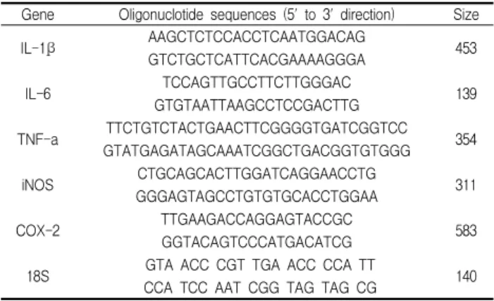
RAW264.7 cells를 10% FBS를 포함한 DMEM에 현탁시킨 후 6 well plate (Corning, USA)에 5 × 10 5 cells/ml의 세포수가 되도록 3 ml씩 분주하여 37℃ 5% CO 2 incubator에서 24 시간 배 양하였다. 새로운 DMEM배지로 교환한 후 CL (2 mg/ml)로 세 포에 처리하여 1시간 동안 배양한 후 자극제 LPS (1 mg/ml)를 처리하고 3시간 또는 24시간동안 배양하였다. 상층액을 제거한 후 1 ml의 TRizol을 넣고 2분 동안 방치한 후 chloroform을 넣고 10초 동안 vortexing하고 12,000 rpm에서 15분 동안 원심분리한 후, 상층액을 취하여 동량의 isopropanol을 혼합하여 흔들어 주 었다. 12,000 rpm에서 10분 동안 원심분리하여 상층액을 제거하 고 pellet은 DEPC (diethyl pyrocarbonate)-DW 20 ml에 녹여 RT-PCR에 사용하였다. RT-PCR kit (Bioneer, Korea)를 사용하여 45℃에서 30분, 94℃에서 5분 동안 반응시킨 후 94℃에서 30초 동안 denaturation시키고, 55~62℃에서 30초 동안 annealing시 킨 다음, 72℃에서 1분동안 extension시키는 cycle을 30~35회 반 복한 뒤, 마지막 extension은 72℃에서 5분 동안 PCR machine (GeneAmp, PCR system 9700, USA)에서 수행하였다. 각 PCR products는 2% agarose gel에 loading하여 100 V 조건에서 30분 동안 전기영동을 통하여 분석하였다. 각각의 primer의 염기서열 은 다음과 같다(Table 1).
Table 1. The primers for RT-PCR analysis
Gene Oligonuclotide sequences (5' to 3' direction) Size IL-1β AAGCTCTCCACCTCAATGGACAG
GTCTGCTCATTCACGAAAAGGGA 453
IL-6 TCCAGTTGCCTTCTTGGGAC GTGTAATTAAGCCTCCGACTTG 139
TNF-a TTCTGTCTACTGAACTTCGGGGTGATCGGTCC GTATGAGATAGCAAATCGGCTGACGGTGTGGG 354
iNOS CTGCAGCACTTGGATCAGGAACCTG GGGAGTAGCCTGTGTGCACCTGGAA 311
COX-2 TTGAAGACCAGGAGTACCGC GGTACAGTCCCATGACATCG 583
18S GTA ACC CGT TGA ACC CCA TT CCA TCC AAT CGG TAG TAG CG 140
6. Western blot
RAW264.7 cells를 10% FBS를 포함한 DMEM에 현탁시킨 후 100 mm cell culture dishes (Corning, USA)에 5×10 5 cells/ml 의 세포수가 되도록 10 ml씩 분주하여 37℃ 5% CO 2 incubator에 서 24시간 배양하였다. 새로운 DMEM배지로 교환한 후 CL (2 mg/ml)로 세포에 처리하여 1시간 동안 배양한 후 자극제 LPS를 처리하고 배양하였다. MAPK 활성을 측정하기 위하여 LPS (1 mg/ml)에 10분 동안 expose하였다. 반응이 종료된 후 배지를 제 거하고 Cold PBS로 세척한 후 cell lysates는 lysis buffer (10 mM pH 7.4 Tris-HCl, 5 mM NaF, 1 mM Na 3 VO 4 , 1 mM EDTA abd 1 mM EGTA)를 첨가하여 protein을 추출하였다. Protein content를 Bradford법으로 정량하여 20-50 mg의 단백질을 l0%
sodium dodecyl sulfate-polyacrylamide gel electrophoresis
(SDS-PAGE)로 분리하고, Hypond-PVDF membrane
(Amersham, Little Chalfont, UK) 으로 transfer하였다. Transfer
된 membrane은 Tris-buffered saline Tween-20 (TBST) (20 mM Tris, pH 7.6, 136 mM NaCl, 0.1% Tween 20)에 용해된 5% skim milk에 1시간동안 실온에서 blocking한 후 anti-phospho-ERK와 anti-ERK, anti-phospho-JNK와 anti-JNK, anti-phospho-p38과 anti-p38 MAP kinase와 β-actin primary antibody (1 : 1000 dilution)로 4℃에서 overnight 반응한 후 TBST로 3회 washing하 고, HRP-conjugated secondary antibody (1 : 1000 dilution)로 1 시간동안 실온에서 반응시켰다. TBST로 3회 세척한 후 면역반응 성 단백질 밴드는 X-ray films에서 enhanced chemiuminescence regents (ECL) (Amersham, Little Chalfont, UK)을 이용하여 검 출하였다.
7. mRNA 및 단백질 밴드의 정량
mRNA 발현 및 면역반응성 단백질 밴드는 Scion Image Release Beta3b software (Maryland, USA)를 사용하여 정량하였 다. 각 밴드영역에서 측정된 pixels의 수를 정량하여 LPS 비자극 군에 대한 비율로 나타내었다.
8. 統計 분석
成績은 SPSS 17.0 K for Windows 통계 프로그램 패키지를 사용하여 평균치 ± 표준오차로 나타내었고 유의수준은 P<0.05로 하였다. 각 실험군 간의 통계학적 분석은 one way-ANOVA와 Dunett test 검정을 실시하였다.
결 과
1. 세포생존률에 미치는 영향
RAW264.7 cells의 생존률에 미치는 영향을 알아보기 위하여 MTT assay를 실시하였다. 아무런 처리를 하지 않은 대조군에서 3.14 ± 0.14의 흡광도를 나타내었으며, CL (0.25, 0.5, 1, 2 and 4 mg/ml)의 농도를 처리한 세포에서 각각 3.35 ± 0.07, 3.21 ± 0.29, 3.41 ± 0.03, 3.30 ± 0.15 및 3.29 ± 0.14의 흡광도를 나타내 어 모든 농도에서 정상세포에 비해 유의한 영향을 보이지 않았 다(Fig. 1).
Fig. 1. Effects of CL on the cell viability in RAW264.7 cells.
The cells were treated with the indicated concentrations (0, 0.25, 0.5, 1, 2, and 4 mg/ml) of CL for 4 h. Cell viability was evaluated using a colorimetric assay based on MTT assay. Data represent the mean± S.D. of three independent experiments.2. 전구 염증 매개 사이토카인 유전자 발현에 미치는 영향
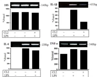
RAW264.7 cells에서 전구 염증 매개 사이토카인 유전자의 발현에 대해 조사한 결과, LPS로 자극한 세포에서 IL-1b, IL-6와 TNF-a mRNA 발현량이 현저하게 증가하였으며, CL (2 mg/ml) 을 전처리한 세포에서 유전자의 발현이 감소하는 것으로 나타났 다(Fig. 2).
Fig. 2. Effects of CL in the expression of IL-1β, IL-6, and TNF-α gene in the LPS stimulated RAW 264.7 cells.
The cells were incubated for 1 h in the presence or absence of CL (2 mg/ml) prior to LPS stimulation (3 h). Expression of pro-inflammatory cytokine and enzyme genes was assayed by RT-PCR. 18S was used as internal control genes.3. 전구 염증 매개 사이토카인과 효소 유전자 발현에 미치는 영향 RAW264.7 cells에서 전구 염증 매개 효소 유전자의 발현에 대해 조사한 결과, LPS로 자극한 세포에서 iNOS와 COX-2 mRNA의 발현량이 현저하게 증가하였으며, CL (2 mg/ml)을 전 처리한 세포에서 유전자의 발현이 감소하는 것으로 나타났다 (Fig. 3).
Fig. 3. Effects of CL in the expression of iNOS and COX-2 gene in the LPS stimulated RAW 264.7 cells.
The cells were incubated for 1 h in the presence or absence of CL (2 mg/ml) prior to LPS stimulation (24 h).Expression of pro-inflammatory cytokine and enzyme genes was assayed by RT-PCR. 18S was used as internal control genes.
4. MAPK 활성화에 미치는 영향
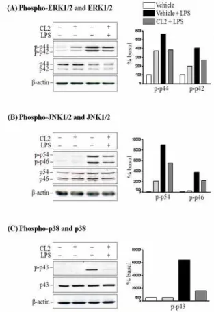
LPS로 자극한 RAW264.7 cells에서 ERK1/2, JNK1/2와 p38
의 인산화에 미치는 영향을 살펴본 결과 LPS로 자극한 세포에서
phospho-ERK1/2, phospho-JNK1/2와 phospho-p38의 발현이
현저히 증가하였고 CL (2 mg/ml)을 전처리한 세포에서 발현을
현저히 억제하는 것으로 보아 CL이 ERK1/2, JNK1/2와 p38의 인산화를 억제하는 것으로 나타났다(Fig. 4).
Fig. 4. Effects of CL on the activation of MAPK in LPS stimulated RAW264.7 cells.
The cells were incubated for 1 h in the presence or absence of CL (2 mg/ml) prior to LPS stimulation (10 min), and the phosphorylation of ERK1/2(A), JNK1/2(B) and p38(C) was assayed by Western blot.고 찰
본 연구는 RAW264.7, murine macrophage cell line을 사용 하여 CL의 항염증효과와 MAPK를 통한 조절 기전을 밝히기 위 해 실시하였다.
염증의 병리기전은 사이토카인 상호작용과 많은 전구 염증 유전자의 유도에 의해 조절되는 복잡한 과정이다. Macrophages 는 인체전반에 걸쳐 분포하고 있고, 염증과정에서 백혈구 유주에 앞서 병원체에 대항하는 즉각적인 방어를 제공함으로써 중요한 역할을 담당하며, LPS는 macrophages에서 interleukins (ILs), TNF-a, iNOS, COX-2와 같은 염증매개인자의 생산을 촉진한다
11,12) . LPS로 유도된 염증 매개인자들의 약리학적 감소는
macrophages의 활성화에 의해 야기되는 염증반응을 포함하는 여러 가지 증상을 완화시키는 효과적인 방법으로 간주되고 있다.
본 연구에 사용된 RAW 264.7 cells는 항염증 약물의 효능을 검증하고, 전구 염증 매개인자들의 유도를 주도하는 신호전달경 로의 억제효능을 가진 약물을 평가하기위한 실험모델로 가장 일 반적으로 사용되고 있다.
본 연구에서 RAW264.7 cells의 생존률과 증식을 저해하지
않는 CL의 농도를 조사한 결과 4 mg/ml 이하의 농도에서 CL이 정상세포와 유의한 차이를 보이지 않았다(Fig. 1). 따라서 이 농도 범위 내의 CL (2 mg/ml)를 사용하여 항 염증효과를 조사하였다.
본 연구에서 전구 염증 매개 사이토카인 유전자의 발현에 미치는 영향을 살펴본 결과 CL의 전처리가 LPS 자극에 의해 증 가된 IL-1b, IL-6와 TNF-a mRNA 발현을 모두 억제하는 것을 알 수 있었다(Fig. 2).
전구염증매개 효소에는 inducible NOS (iNOS)과 Cyclooxygenase (COX)가 대표적인데, iNOS는 주로 자극된 macrophages에 의해 생산되어 NO의 염증 조건을 제공한다. 특 히 macrophages에서 과도한 NO 생산이 세포독성, 염증, 암 발 생, 자가면역질환들을 초래할 수 있다는 점에서 NO 생산과 iNOS 발현의 억제가 염증질환을 예방할 수 있다는 것을 뒷받침 하고 있다 13,14) . COX family는 prostacyclin과 thromboxanes 뿐만 아니라 PGs의 형성에 역할을 담당하고 있다. COX는 arachidonic acid를 PGs로 전환하는 효소로 COX-1과 COX-2로 존재하는데 COX-2의 억제는 염증과 통증의 증상을 감소할 수 있다 15) . COX-2는 정상 생리조건에서 거의 발현되지 않으나 전구염증매 개인자에 의해 일시적으로 빨리 유도되어 PGs의 생합성을 자극 하여 염증반응을 일으킨다 16) .
본 연구에서 전구 염증 매개 효소 유전자의 발현에 미치는 영향을 살펴본 결과 CL의 전처리가 iNOS와 COX-2 mRNA 발현 을 억제하는 것을 알 수 있었다(Fig. 3).
MAPK는 세포 성장과 분화의 조절, 사이토카인과 stress에 대한 세포 반응의 조절에 중요한 역할을 한다 17) . MAPK 신호전 달경로는 진화적으로 보존되어왔고 세포외 환경으로부터 세포질 을 통해 핵으로 정보를 전달하는데 중요한 역할을 하고 있으며, 최소 3가지의 신호전달경로가 존재한다 18) .
ERK 신호전달경로에서 활성화된 ERK는 다양한 전사인자를 인산화할 수 있다. ERK가 자극인자에 의해 광범위하게 활성화되 는 반면, p38과 JNK는 stress 반응경로의 부분을 구성하며 염증 성 사이토카인같은 인자에 의해 유도된 세포스트레스에 의해 활 성화된다 18) . JNK 신호전달경로는 LPS와 TNF-a와 같은 면역 염 증자극에 반응하는 세포에서 활성화되고, 세포의 형태적 증대와 사이토카인 전사에 포함된다 20,21) .
본 연구에서 MAPK 신호전달경로에 미치는 영향을 조사한 결과 CL이 RAW264.7 cells에서 LPS로 유도된 ERK1/2, JNK1/2와 p38의 인산화를 억제하는 것을 관찰하였다(Fig. 4).
이러한 결과는 CL가 ERK1/2, JNK1/2와 p38 활성화와 관련된 MAPK 신호전달경로의 억제를 통해 항염증 작용을 발휘하는 것으로 보인다.
연구결과를 요약하면 CL가 LPS로 자극한 RAW264.7 cells에
서 세포내 ERK1/2, JNK1/2와 p38 활성화와 관련된 MAPK 신
호전달경로를 억제하고 전구 염증 매개인자들의 유전자 발현을
억제함으로써 염증반응을 촉발하는 전구 염증 매개인자들의 생
산을 감소시킬 것으로 사료된다. 이는 CL이 임상에서 염증질환
을 예방하고 초기단계에 효과적으로 제어할 수 있을 것으로 사
료된다.
감사의 글
본 연구는 2008년도 동의대학교 교내연구비에 의해 지원되 었음
참고문헌