Antioxidant and Cytoprotective Effects of Socheongja and Socheong 2, Korean Black Seed Coat Soybean Varieties, against Hydrogen Peroxide-induced
Oxidative Damage in HaCaT Human Skin Keratinocytes
Eun Ok Choi1,2, Da Hye Kwon1,2, Hye-Jin Hwang3, Kook Jin Kim4, Dong Hee Lee4 and Yung Hyun Choi1,2*
1Open Laboratory for Muscular & Skeletal Disease Control and Department of Biochemistry, Dongeui University College of Korean Medicine, Busan 47227, Korea
2Anti-Aging Research Center, Dongeui University, Busan 47340, Korea
3Department of Food and Nutrition, College of Natural Sciences and Human Ecology, Dongeui University, Busan 47340, Korea
4Genomine Advanced Biotechnology Research Institute, Genomine Inc., Pohang 37668, Korea Received October 31, 2017 /Revised November 15, 2017 /Accepted November 18, 2017
Black soybeans are used as food sources as well as for traditional medicines because they contain an abundance of natural phenolic compounds. In this study, total phenolic contents (TPCs) of Korean black seed coat soybean varieties Socheongja (SCJ), Socheong 2 (SC2) and Cheongja 2 (CJ2) as well as their antioxidant capacities were investigated. Among them, TPCs were abundantly present in the order of CJ2<SCJ<SC2, and we confirmed that SCJ and SC2 have a higher antioxidant potential than CJ2. Based on these results, we investigated the cytoprotective effects of SCJ and SC2 in H2O2- stimu- lated HaCaT human keratinocytes. Our results revealed that treatment with SCJ and SC2 prior to H2O2 exposure significantly increases the viability of HaCaT cells, indicating that the exposure of HaCaT cells to SCJ and SC2 conferred a protective effect against oxidative stress. SCJ and SC2 also effectively inhibited H2O2-induced apoptotic cell death through the blocking of mitochondrial dysfunction.
SCJ and SC2 also attenuated the phosphorylation of Histone H2AX. Furthermore, they effectively in- duced the levels of thioredoxin reductase (TrxR) 1, a potent antioxidant enzyme, which is associated with the induction of nuclear transcription factor erythroid-2-like factor 2 (Nrf2); however, the protective effects of SCJ and SC2 were significantly reversed by Auranofin, a TrxR inhibitor. These results indicate that they have protective activity through the blocking of cellular damage related to oxidative stress via the Nrf2 signaling pathway. In conclusion, our study indicated that SCJ and SC2 might potentially serve as novel agents for the treatment and prevention of skin disorders caused by oxidative stress.
Key words : Antioxidant, HaCaT cells, Nrf2, Socheongja, Socheong 2
*Corresponding author
*Tel : +82-51-850-7413, Fax : +82-51-853-4036
*E-mail : [email protected]
This is an Open-Access article distributed under the terms of the Creative Commons Attribution Non-Commercial License (http://creativecommons.org/licenses/by-nc/3.0) which permits unrestricted non-commercial use, distribution, and reproduction in any medium, provided the original work is properly cited.
Journal of Life Science 2018 Vol. 28. No. 4. 454~464 DOI : https://doi.org/10.5352/JLS.2018.28.4.454
서 론
활성산소종(reactive oxygen species, ROS)은 스트레스, 음 주, 흡연, 자외선 및 유해 화학물질 노출 등의 요인으로 인해 과도하게 생성되거나 항산화 시스템의 기능이 저하되어 체내 산화-환원 조절 장애가 발생할 경우 증가한다[2, 29]. 과도한 ROS의 생성은 다양한 질병의 발생과 진행을 촉진하는데 그 중 피부 세포의 항산화 방어체계를 손상시키기도 하며[39, 41], 지질과산화, 단백질산화, 피부의 염증반응 유발, 피부 면역기 능 억제, 세포성분의 손상을 야기시켜 광노화를 촉진시키기도
한다[12, 18, 30, 31]. 따라서 피부건강과 노화방지를 위한 항산 화 소재 탐색 중요성이 강조되고 있으며, 특히 인체에 유해한 기능성 천연소재를 대상으로 효능 평가가 광범위하게 이루어 지고 있다.
식용작물인 콩은 단백질과 다양한 isoflavone, 각종 mineral 및 vitamin 등의 성분이 풍부한 완전식품으로 널리 애용되고 있으며, 최근 다양한 유효 성분 추출법의 개발로 새롭게 관심 을 모으고 있다[24, 38]. 특히, 검은콩은 다량의 단백질 외에도 lecithin, phytoestrogen, 불포화지방산, anthocyanins, iso- flavone, saponin, carotene 등이 함유되어 있어, 항산화, 항암, 항비만, 면역 증강 등의 다양한 약리 작용을 나타낸다[13, 27, 37, 43]. 또한 검은콩 추출물과 유효성분들은 혈전 생성 억제를 통한 혈액 순환 개선효과를 가지며[21, 46], 강력한 항산화 및 tyrosinase 활성 저해효과를 가져 피부 미백효과를 가지는 것 으로 알려진 바 있다[15]. 또한 고 콜레스테롤 및 고 지방식이 로 유도된 비 알코올성 지방간 모델에서 검은콩 추출물이 간 의 콜레스테롤 축적을 억제시켰다고 보고된 바도 있다[6, 14, 16]. 아울러 검은콩의 생리학적 활성을 높이기 위한 우수한
품종의 발굴에도 많은 진전이 있어 왔으며, 그 중 소청자 (Socheongja), 소청2호(Socheong 2) 및 청자2호(Cheongja 2)는 isoflavone 함량을 높여 기능성 강화를 목적으로 개발된 품종 들이다[23, 36, 42]. 그러나 이들 품종에 대한 생리학적 효능에 대한 연구는 상대적으로 미비한 실정이므로 본 연구에서는 이들에 대한 항산화 활성을 비교 하였으며, 산화적인 스트레 스로 유도된 in vitro 피부세포 모델에서 항산화 효능 및 관련 기전 연구를 시도하였다.
재료 및 방법 검은콩 추출물의 준비
본 실험에 사용된 3가지 검은콩 품종(Korean black seed coat soybean varieties)은 모두 국립식량과학원(National In- stitute of Crop Science, Rural Development Administration, Republic of Korea)으로부터 제공받았으며, 2시간 동안 저온 처리된 종자를 발아시켜 콩나물 제조기를 이용하여 실온, 암 실 조건에서 6일 동안 재배하여 시료로 사용하였다. 저온용출 추출물을 얻기 위하여 ethanol에 소독한 후 물로 충분히 세척 하고 물기를 제거한 뒤 정제수와 1:1의 비율로 섞어서 homog- enizer로 잘게 분쇄하여 저온(4℃)에서 2시간 동안 교반하여 원료의 유효성분이 정제수로 우러나오게 하였다. 제조된 추출 물들은 4-5겹의 mesh로 여과한 뒤 5,000 rpm에 15분 원심분리 하여 상층액만 분리하여 찌꺼기를 제거하고 50 mg DW (dry weight)/ml 농도로 만든 다음 효능성분의 손실이 없도록 -80
℃에 보관하였으며, 실험에 사용시 적정 농도로 배지에 희석 하여 처리하였다.
총 페놀 함량
각 검은콩 추출물들에 함유된 총 페놀 함량(total phenoloic content, TPC)은 Li et al. [25]의 방법에 준하여 측정하였으며, gallic acid (Sigma-Aldrich Chemical Co., St. Louis, MO, USA) 를 표준물질로 사용하였다. 이를 위하여 적정 실험 농도로 희 석한 시료 50 μl에 2%의 Na2CO3 1 ml을 첨가하고 2분간 실온 에 방치한 후 50% Folin-Ciocalteu reagent (Sigma-Aldrich Chemical Co.)를 50 μl를 가하고 실온에서 30분간 반응시킨 다음 750 nm에서 흡광도를 측정하였다. Gallic acid를 이용하 여 검량선을 작성하였으며, 총 페놀 함량은 mg gallic acid equivalents (GAE)/g of sample dry weight (DW)으로 나타내 었다.
Ferric reducing antioxidant power (FRAP) 활성 측정 이 방법은 산성 pH 영역에서 ferric tripyridyltrizaine (Fe3+- TPTZ) 복합체가 환원성 물질에 의해 청색의 ferrous tripyr- idytriazine (Fe2+-TPTZ)으로 환원되어 593 nm에서 흡광도가 증가 하는 원리[3]를 이용한 것으로 대부분의 항산화제가 환원
력을 가지고 있다는 점에 착안하여 고안된 실험방법이다.
FRAP에 의한 검은콩 추출물들의 항산화력을 측정하기 위하 여 300 mM acetate buffer (pH 3.6), 40 mM HCl에 용해한 10 mM 2,4,6-tripyridyl-s-triazine (TPTZ, Sigma-Aldrich Chemical Co.) 및 20 mM FeCl3·6H2O를 각각 10:1:1(v/v/v)의 비율로 혼합하여 FRAP 시약을 제조하였다. 이어서 적정 농도 로 희석된 검은콩 추출물 시료액 10 μl과 240 μl의 FRAP 시약 을 혼합하고 37℃에서 5분간 반응시킨 후 593 nm에서 흡광도 를 측정하였다. 양성 대조군(positive control)은 trolox (Sigma- Aldrich Chemical Co.)를 사용하여 검량선을 작성하였으며, 결 과는 mg trolox/g of sample dry weight (DW)로 나타내었다.
2,2'-diphenyl-1-picrylhydrazyl (DPPH) radical 소거능 검은콩 추출물들의 항산화력을 측정하기 위한 2,2-diphenyl- 1-picrylhydrazl (DPPH) radical에 대한 전자공여능은 Blios의 방법[4]에 준하여 조사하였다. 이 방법은 DPPH radical에 전 자를 공여함으로써 radical을 소거하는 효과를 측정하는 방법 으로 0.4 mM의 DPPH (Sigma-Aldrich Chemical Co.) 용액 150 μl에 검은콩 추출물이 적정 농도로 희석된 시료 100 μl을 첨가하여 37℃에서 30분간 반응시킨 후 518 nm에서 흡광도를 측정하였다. 그리고 반응물에 대한 흡광도 값을 대조군에 대 한 검은콩 추출물 시료의 DPPH radical 소거 활성으로 항산화 활성도를 나타내었으며, 양성 대조군으로 trolox를 사용하였 고 음성 대조군으로는 0.4 mM DPPH 용액 대신 메탄올을 이 용한 아래 식에 준하여 산출하였다.
DPPH radical scavenging activity (%) = 100-[(OD of sample/ OD of control) ×100]
2,2'-azinobis(3-ethylbenzothiazoline- 6-sulfonate) cationic (ABTS・+) 소거능
ABTS・+ 소거능을 이용한 검은콩 추출물들의 항산화능 측 정은 Potassium persulfate와의 반응에 의해 생성된 ABTS 유 리 radical이 제거되어 radical 특유의 색인 청록색으로 탈색되 는 원리에 근거하여 Re et al. [33]의 방법에 준하여 조사하였 다. 즉 7 mM의 ABTS (Sigma-Aldrich Chemical Co.)와 2.45 mM potassium persulfate를 1:1로 혼합하여 실온, 암실에서 24시간 동안 방치하여 radical을 형성시킨 다음 실험 직전에 ABTS 용액을 734 nm에서 흡광도가 0.7±0.02가 되도록 phos- phate buffer saline (PBS, pH 7.4)으로 희석하였다. ABTS radi- cal cation solution 190 μl에 실험 농도로 희석한 시료 10 μl를 가하여 실온에서 6분간 반응시켜 734 nm에서 흡광도를 측정 하였으며 trolox를 양성 대조군으로 하여 다음 식에 준하여 항산화 효능을 비교하였다.
ABTS radical scavenging activity (%) = 100-[(OD of sam- ple/ OD of control) ×100]
세포 배양
본 연구에 사용된 세포들 중에서 HEI 193 인체 신경초종세 포(House Ear Institute, Los Angeles, California, USA)를 제외 한 RAW 264.7 대식세포, RT4-D6P2T 신경집세포, C2C12 근아 세포, V79-4 폐섬유아세포 및 HaCaT 피부각질세포는 Amer- ican Type Culture Collection (ATCC, Manassas, VA, USA)에 서 분양받았으며, 10% fetal bovine serum (FBS, Welgene, Daegu, Republic of Korea)과 1%의 penicillin/streptomycin 이 포함된 Dulbecco's modified Eagle's medium (DMEM, Welgene)을 사용하여 37℃, 5% CO2 조건하에서 배양하였다.
세포의 증식에 따른 과밀도 현상을 해소하기 위하여 매 48 시간마다 0.05% trypsin-0.02% ethylene-diaminetetracetic acid (EDTA)를 이용하여 세포를 부유시킨 후 적정수의 세포를 유 지하였다.
검은콩 추출물의 세포독성 및 세포보호 효과 평가
다양한 세포에서 검은콩 추출물의 세포독성 여부와 H2O2
처리에 따른 산화적 스트레스에 대한 세포보호 효과를 확인하 기 위하여 3-(4,5-dimethyl-2-thiazolyl)-2,5-diphnyl-2H-tetra- zolium bromide (MTT) assay를 이용하였다. 이를 위하여 세 포 배양용 6 well plate에 HaCaT, C2C12 및 V79-4 세포를 1×10⁵ cells/well로, RAW 264.7 세포는 3×10⁵ cells/well로, RT4-D6P2T 세포는 5×10⁴ cells/well로, HEI 193 세포는 2×10⁵ cells/well로 각각 분주하여 안정화시킨 후 각 검은콩 추출물 을 적정농도로 처리하였다. 산화적 스트레스에 의한 세포 손 상 보호효과를 확인하기 위하여 세포 배양용 6 well plate에 적정 세포를 분주하고 24시간 동안 안정화시킨 후 검은콩 추 출물 및 H2O2 (Sigma-Aldrich Chemical Co.)를 단독 또는 검 은콩 추출물을 1시간 전처리한 후 H2O2 처리하여 추가 배양하 였다. 24시간 후, 각 세포들의 형태 변화를 도립현미경(inver- ted microscope, Carl Zeiss, Oberkochen, Germany)하에서 관 찰하고, MTT (Sigma-Aldrich Chemical Co.)를 0.5 mg/ml 농 도로 희석하여 200 μl씩 분주하고 37oC에서 2시간 동안 다시 반응시켰다. 반응이 끝난 다음 MTT 시약을 제거하고 dimethyl sulfoxide (DMSO)를 2 ml씩 분주하여 각 well에 생성된 for- mazan을 모두 녹인 후 96 well plate에 200 μl씩 옮겨서 en- zyme-linked immunosorbent assay (ELISA) reader (Molecu- lar Devices, Sunnyvale, CA, USA)로 540 nm에서 흡광도를 측정하였다. 각 세포에 대한 독성은 각각의 대조군의 평균 흡 광도 값을 구하여 평균 흡광도 값에 대한 백분율로 나타내었 다.
Annexin V-FITC 염색에 의한 apoptosis 유발의 정량적 측정
H2O2 처리에 의한 apoptosis 유발 및 검은콩 추출물에 의한 차단 효과의 정량적 분석을 위해 준비된 세포들을 모은 다음
2,000 rpm으로 5분간 원심분리하여 상층액을 제거하였다. 이 들 세포를 phosphate-buffered saline (PBS)으로 2~3회 정도 세척하고 10 mM HEPES/NaOH, pH 7.4, 140 mM NaCl 및 2.5 mM CaCl2가 포함된 annexin V binding buffer (Becton Dickinson, San Jose, CA, USA)에 부유시킨 다음 annexin V-fluorescein isothiocyanate (FITC, Becton Dickinson) 및 propidium iodide (PI; Sigma-Aldrich Chemical Co.)를 처리 하여 암실에서 15분 동안 반응을 시켰다. 반응이 끝난 후 35- mm mesh를 이용하여 단일세포로 분리하고 flow cytometer (FACS Calibur, Becton Dickinson, San Jose, CA, USA)를 적용 시켜 apoptosis가 유발된 세포(V+/PI–)를 형광반응에 따라 분 석하였다.
Mitochondrial membrane potential (MMP, Δψm) 변 화의 측정
세포 내 미토콘드리아 기능 손상의 여부를 확인하기 위하여 MMP 값 변화 정도를 측정하였다. 이를 위하여 다양한 조건에 서 배양된 세포들에 10 μM의 5,5’,6,6’-tetrachloro-1,1’,3,3’- tet- raethyl-imidacarbocyanine iodide (JC-1, Sigma-Aldrich Chem- ical Co.) 용액을 처리하여 암실, 상온에서 20분 동안 반응시켰 다. 반응이 끝난 후 상층액을 제거하고 PBS를 첨가하여 세포 를 부유시킨 다음 flow cytometer에 적용시켜 MMP의 변화를 측정하였다.
단백질의 분리, 전기영동 및 Western blot analysis 준비된 세포에 적당량의 lysis buffer [25 mM Tris-Cl (pH 7.5), 250 mM NaCl, 5 mM EDTA, 1% Nonidet-P40 (NP-40), 1 mM phenymethylsulfonyl fluoride (PMSF), 5 mM dithio- threitol (DTT)]를 첨가하여 4℃에서 1시간 이상 반응시킨 후, 14,000 rpm으로 30분간 원심 분리하여 상층액에 있는 단백질 을 분리하였다. 상층액의 단백질 농도는 Bio-Rad 단백질 정량 시약(Bio-Rad Lab., Hercules, CA, USA)과 그 사용방법에 따 라 정량 한 다음 동량의 Laemmli sample buffer와 혼합하여 sodium dodecyl sulfate (SDS)-polyacrylamide gel을 이용하 여 전기영동을 실시하였다. 이를 다시 nitrocellulose mem- brane (Schleicher and Schuell, Keene, NH, USA)으로 electro- blotting에 의해 전이시키고 5% skim milk를 1시간 처리하여 비특이적인 단백질들에 대한 blocking을 실시하였다. 그리고 적정 1차 항체를 처리하여 상온에서 2시간 이상 또는 4℃에서 over night 반응시킨 다음 PBS-T (PBS with Tween 20)로 세척 하고 1차 항체에 맞는 2차 항체를 사용하여 상온에서 1시간 정도 반응시켰다. 반응이 끝난 후 암실에서 enhanced chem- iluminoesence (ECL) solution (Santa Cruz Biotechnology Inc., Santa Cruz, CA, USA)을 적용시킨 다음 X-ray film에 감 광시켜 특정 단백질의 발현 변화를 분석하였다. 본 실험에서 단백질 분석을 위하여 사용된 항체들은 Santa Cruz Biotech-
Table 1. Total phenolic contents (GAE, gallic acid equivalent) and ferric reducing antioxidant power (Trolox, Trolox equivalent) of water extracts of SCJ, SC2 and CJ2
Total phenolic contents FRAP
Samples mgGAE/g Samples mgTrolox/g
SCJ SC2 CJ2
447.8±4.57 524.2±5.24 437.6±3.66
SCJ SC2 CJ2
86.4±2.42 87.2±1.72 75.2±0.78
*Values are presented as the mean ± SD.
A
B
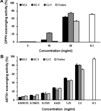
Fig. 1. Effects of SCJ, SC2 and CJ2 extracts on DPPH and ABTS radical scavenging activities. The absorbances of only DPPH and ABTS solution at 517 nm and 734 nm were 0.98±0.032 and 0.71±0.032 (experimental control), re- spectively. Trolox was used as the positive control. Each point represents the mean ± SD of three independent experiments.
nology Inc. 및 Abcam (Danvers, MA, USA)에서 구입하였다.
Immunoblotting을 위해 2차 항체로 사용된 horseradish per- oxidase (HRP)-conjugated anti-mouse 및 anti-rabbit 항체는 Santa Cruz Biotechnology Inc.에서 구입하였다.
통계 분석
실험 결과들의 유의성을 검정하기 위하여 분산분석(ANOVA) 을 실시한 후 p<0.05 수준에서 Duncan's multiple range tests 를 실시하였으며, 그 결과는 평균(mean) ± 표준편차(stan- dard deviation, SD)로 표시하였다.
결 과 총 페놀 함량 비교
본 연구에서 선정한 3가지 검은콩 추출물의 총 페놀 함량 (TPC)을 비교하기 위하여 gallic acid를 표준용액으로 선정하 여 작성한 검정곡선으로부터 각 추출물의 페놀 함량을 비교하 여 Table 1에 나타내었다. 3가지 검은콩 추출물의 총 페놀 함량 은 437.6±3.66~524.2±5.24 mg GAE/g DW의 범위로 나타났으 며, 추출물의 페놀 함량은 소청2호(SC2) > 소청자(SCJ) > 청자 2호(CJ2) 순으로 높게 나타났다.
항산화 활성 비교
3가지의 검은콩 추출물의 항산화능을 비교하기 위하여 먼 저 FRAP법을 적용한 결과, 가장 높은 환원력을 보인 것은 소 청2호(SC2, 87.2±1.72 mg Trolox/g)였으며, 청자2호(CJ2)에서 가장 낮은 값(75.2±0.78 mg Trolox/g)을 나타내었다(Table 1).
DPPH radical 소거능을 이용한 항산화능의 결과는, Fig. 1A에 나타낸 바와 같이, 소청2호(SC2)가 74.5%, 소청자(SCJ)가 64.6
%, 청자2호(CJ2)가 53.9%로 나타났다. ABTS free radical에 대 한 소거 활성의 경우는 소청자(SCJ)가 80.7%, 소청2호(SC2)가 80%, 청자2호(CJ2)가 69.8%로 나타나, 소청자(SCJ)와 소청2호 (SC2)가 유사한 활성을 보였다(Fig. 1B). 따라서 실험법에 따른 약간의 차이가 있었으나, 각 추출물의 항산화력은 그들 시료 에 함유되어 있는 총 페놀 함량이 증가할수록 높게 나타나는 경향을 보여주었다.
HaCaT 세포에서 산화적 스트레스로 유도된 생존율 저하에 대한 검은콩 추출물의 보호 효과
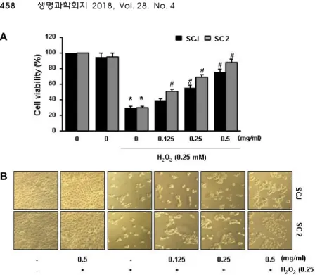
3가지 검은콩 추출물 중 항산화 활성이 상대적으로 높았던 소청자(SCJ)와 소청2호(SC2)를 선정하여 HaCaT 세포를 대상 으로 산화적 세포손상에 대한 보호효과를 조사하였다. 이를 위하여 적정 농도의 추출물(0.125~0.5 mg/ml)을 1시간 전처리 한 후 산화적 스트레스를 유발하기 위하여 H2O2 (0.25 mM)를 처리하고 24시간 동안 배양한 후 세포 생존율을 확인한 결과, H2O2를 단독 처리 하였을 때 감소하였던 세포 생존율과 형태 적 변형이 소청자(SCJ)와 소청2호(SC2)의 전처리군에서 처리 농도의존적으로 회복되었음을 확인하였다(Fig. 2).
HaCaT 세포에서 산화적 스트레스에 의한 apoptosis 유 발에 미치는 검은콩 추출물의 영향
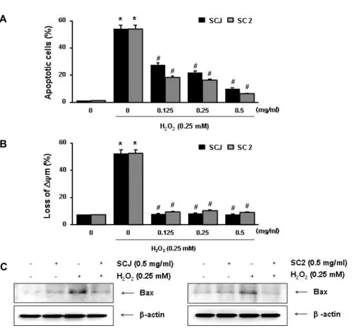
산화적 스트레스에 대한 소청자(SCJ)와 소청2호(SC2)의 세 포보호효과가 apoptosis 유발 억제와 연관성이 있는지를 확인 하기 위하여 annexin V-FITC/PI 염색을 이용한 flow cytome- ter 분석을 실시하였다. Fig. 3A의 결과에서 알 수 있듯이, H2O2 단독 처리된 배지에서 배양된 HaCaT 세포에서는 약 54.13%에 해당되는 세포에서 apoptosis가 유발되었으나, 소청 자(SCJ)와 소청2호(SC2)의 전처리 농도가 증가할수록 감소되
A
B
Fig. 2. Effects of SCJ and SC2 on the H2O2- induced growth inhibition in HaCaT cells. Cells were pre-treated with or without SCJ (0.125~0.5 mg/ml) or SC2 (0.125~0.5 mg/ml) for 1 hr, then in- duced with H2O2 (0.25 mM). (A) After 24 hr, the cell viability was measured by MTT assay. The results are the mean ± SD values obtained in three independent experiments (*p<0.05 com- pared with the untreated control group;
#p<0.05 compared with H2O2treated group). (B) Cell morphology was vi- sualized by light microscopy (Magnifi- cation, ×200).
어 0.5 mg/ml 농도 조건에서는 각각 10.24%와 4.51% 정도로 나타나 소청자(SCJ)와 소청2호(SC2)의 산화적 스트레스 보호 효과가 apoptosis 억제와 직접 연관성이 있음을 알 수 있었다.
HaCaT 세포에서 산화적 스트레스에 의해 유도된 미토콘 드리아 기능 손상에 미치는 검은콩 추출물의 영향
산화적 스트레스에 의한 HaCaT 세포의 apoptosis 유도에 미토콘드리아 기능 손상이 연계되어 있는지, 그리고 소청자 (SCJ)와 소청2호(SC2)가 이를 차단할 수 있는지의 여부를 조사 하였다. 그 결과, H2O2 단독 처리된 배지에서 배양된 HaCaT 세포에서는 미토콘드리아 기능 손상을 의미하는 MMP 소실 이 매우 증가(52.56%)되었으나, 소청자(SCJ)와 소청2호(SC2) 에 의해 이를 유의적으로 차단하였다(Fig. 3B). 아울러 H2O2의 처리에 의하여 증가되었던 미토콘드리아 기능 손상을 유발하 는 pro-apoptotic protein인 Bax의 발현이 증가되었으나 소청 자(SCJ)와 소청2호(SC2)의 전처리에 의해서는 발현이 대조군 수준으로 감소되었다(Fig. 3C). 이러한 결과들은 소청자(SCJ) 와 소청2호(SC2)가 H2O2에 의한 Bcl-2 family 관련 유전자의 발현 변화를 억제함으로서 미토콘드리아 기능 손상을 차단시 켜 apoptosis 유도를 억제하였음을 의미한다.
HaCaT 세포에서 산화적 스트레스에 의해 유도된 DNA 손 상에 미치는 검은콩 추출물의 영향
다음은 HaCaT 세포에서 산화적 스트레스에 의한 apopto- sis 차단에 미치는 소청자(SCJ) 및 소청2호(SC2)의 효과가 DNA 손상 차단 억제와 연관성이 있는지를 조사하였다. 이를 위하여 대표적인 DNA 손상 마크인 γH2A.X 단백질의 인산화 (p-γH2A.X) 여부를 조사하였다. Fig. 4의 Western blot 분석 결과에 따르면, H2O2에 노출된 HaCaT 세포에서는 증가되었
던 인산화형의 γH2A.X 발현이 소청자(SCJ)와 소청2호(SC2)의 전처리에 의해 대조군 수준으로 억제되었다. 이는 산화적 스 트레스에 대한 소청자(SCJ)와 소청2호(SC2)의 HaCaT 세포보 호 효과가 DNA 손상 차단과도 직접적인 연관성이 있음을 알 수 있었다.
HaCaT 세포에서 Nrf2/TrxR1 경로 활성화에 미치는 검 은콩 추출물의 영향
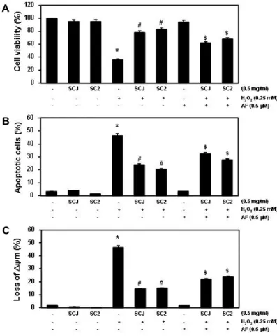
이상에서 관찰된 HaCaT 세포에서의 소청자(SCJ)와 소청2 호(SC2)의 항산화 효능에 nuclear factor erythroid 2-related factor 2 (Nrf2) 신호계가 관여하는지를 조사하였다. Fig. 5의 결과에 의하면 소청자(SCJ)와 소청2호(SC2)의 처리에 따라 Nrf2의 발현은 농도 의존적으로 증가된 반면 Nrf2의 활성 억 제에 관여하는 Kelch-like ECH-associated protein-1 (Keap1) 의 발현은 감소되었다. 아울러 Nrf2의 전사활성에 의하여 조 절되는 항산화 관련 유전자 중에서 thioredoxin reductase-1 (TrxR1)의 발현이 소청자(SCJ) 및 소청2호(SC2)의 처리 농도가 증가할수록 증대되었다. 소청자(SCJ)와 소청2호(SC2)에 의한 TrxR 발현 증가가 그들의 항산화 활성과 직접 연관성이 있는 지의 여부를 조사하기 위하여 TrxR1 활성 억제제인 auranofin 이 동시 처리되었을 경우 나타나는 현상을 조사하였다. Fig.
6에 나타낸 바와 같이, auranofin을 소청자(SCJ) 및 소청2호 (SC2)와 함께 처리하였을 경우, 산화적 스트레스에 대응하여 소청자(SCJ)와 소청2호(SC2)에 의해 증가되었던 세포생존율 과 억제되었던 apoptosis 및 MMP 소실 유의적으로 상쇄되었 다. 이러한 결과는 소청자(SCJ)와 소청2호(SC2)의 HaCaT 세 포에서의 세포보호 효과에 Nrf2/TrxR1 경로가 직접적으로 관 여할 가능성이 있음을 의미하는 것이다.
A
B
C
Fig. 3. Effects of SCJ and SC2 on the H2O2-induced apoptosis and MMP loss in HaCaT cells. Cells were pretreated with or without SCJ (0.125~0.5 mg/ml) or SC2 (0.125~0.5 mg/ml) for 1 hr, then induced with or without H2O2 (0.25 mM) for 24 hr. (A) The cells were stained with FITC-conjugated Annexin-V and PI for flow cytometry analysis. The percentages of apoptotic cells were determined by counting the percentage of Annexin V-positive cells. (B) The cells were stained with 10 μM JC-1 for 20 min at 37˚C in the dark. Cells were washed once with PBS, and MMP loss was then evaluated using a flow cytometer analysis. Data are presented as mean ± SD values obtained from three independent experiments (*p<0.05 compared with the untreated control group; # p<0.05 compared with H2O2treated group). (C) The cells were lysed and then equal amounts of cell lysates were separated on SDS-polyacrylamide gels and transferred to membranes. The membranes were probed with specific antibody against Bax, and the proteins were visualized using an ECL detection system. β-actin was used as an internal control.
A
B
Fig. 4. Effects of SCJ and SC2 on H2O2-induced phosphorylation of γH2A.X in HaCaT cells. Cells were pretreated with or without SCJ (0.5 mg/ml) or SC2 (0.5 mg/ml) for 1 hr, then induced with or without H2O2 (0.25 mM) for 24 hr. (A and B) The cells were lysed and then equal amounts of cell lysates were separated on SDS-polyacrylamide gels and transferred to membranes. The membranes were probed with specific antibodies against p-γH2A.X, and the proteins were visualized using an ECL detection system. β-actin was used as an loading control.
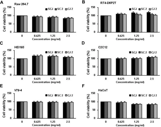
다양한 세포주의 생존율에 미치는 검은콩 추출물의 영향 HaCaT 세포에서 산화적인 스트레스로 인해 유도되는 손상 에 대해 유의적인 보호효과를 나타낸 소청자(SCJ), 소청2호 (SC2)를 비롯하여 청자2호(CJ2)가 다양한 다른 기관 유래 세포
의 생존율에 미치는 영향을 추가로 조사하였다. 이를 위하여 검은콩 추출물이 함유된 배지에서 24시간 동안 배양된 세포들 을 대상으로 MTT assay를 실시하였다. Fig. 7의 결과에서 알 수 있듯이 HaCaT 세포에서 사용된 농도(0.5 mg/ml) 전후에
A
B
Fig. 5. Effects of SCJ and SC2 on the levels of Nrf2, Keap1 and TrxR1 in HaCaT cells. The cells were incubated with various concentrations of SCJ (A) and SC2 (B) for 6 hr. Cellular proteins were separated on SDS-polyacrylamide gels and transferred to membranes. The membranes were probed with specific antibodies against Nrf2, Keap1 and TrxR1. β-actin was used as the loading control.
A
B
C
Fig. 6. Effects of a TrxR1 inhibitor on the SCJ or SC2- mediated attenuation of growth inhibition, apo- ptosis induction and MMP loss by H2O2 in HaCaT cells. The cells were pretreated for 1 hr with 0.5 mg/ml SCJ or SC2 and then treated for 24 hr rwith or without 0.25 mM H2O2 in the absence or presence of 0.5 μM auranofin. (A) Cell viability was assessed using an MTT reduction assay. (B) To quantify the degree of apoptosis, the cells were stained with FITC-conjugated Annexin V and propidium iodide (PI) for flow cytometry analysis.
(C) After incubation with 10 μM JC-1 for 20 min, JC-1 fluorescence intensity was detected using a flow cytometer. Data are presented as mean ± SD values obtained from three independent experi- ments (*p<0.05 compared with the untreated con- trol group; # p<0.05 compared with H2O2treated group; $ p<0.05 compared with H2O2 and SCJ or SC 2-treated group).
서 6가지 세포주들 모두 유의적인 세포증식 억제효과가 관찰 되지 않아 검은콩 추출물들이 세포독성에는 효과가 없음을 알 수 있었다.
고 찰
과도한 ROS의 생성은 다양한 질병의 발생과 진행을 촉진할 뿐만 아니라 피부의 항산화 방어체계를 손상시키고[39, 41], 피부의 염증반응 유발, 피부면역기능 억제, 세포성분의 손상 을 야기 시키고 광노화를 촉진시키기도 한다[12, 18, 30, 31].
본 연구에서 사용된 소청자, 소청2호 및 청자2호는 isoflavone 의 함량이 증대된 우리나라에서 개발된 검은콩 육성품종들이 며, 이들의 추출물을 이용한 산화적 스트레스에 대한 피부세 포 보호 효과를 조사하였다. 이를 위하여 다양한 약리성분이 파괴될 수 있는 기존의 열수추출법의 단점을 보완하기 위하여 저온용출 추출법을 적용하였다.
본 연구에 사용된 3가지 검은콩 품종의 기본적인 항산화 활성 평가를 위하여 먼저 총 페놀 함량(TPC)을 조사한 결과 소청2호(SC2) > 소청자(SCJ) > 청자2호(CJ2)의 순으로 나타났 다(Table 1). 페놀 복합물은 유해한 radical을 제거할 수 있는
A B
C D
E F
Fig. 7. Effects of SCJ or SC2 on the cell viability in various normal cell lines. (A) RAW 264.7 (A), RT4-D6P2T (B), HEI193 (C), C2C12 (D), V79-4 (E) and HaCaT (F) cells were treated with the indicated concentrations of SCJ, SC2 and CJ2 (0.625~2.5 mg/ml). After 24 hr, cell viability was assessed by MTT assays and the results are expressed as mean ± SD values obtained from three independent experiments.
phenolic hydroxyl기를 가지고 있어서 강력한 항산화 활성을 나타내기 때문에[22, 25] 이를 기준으로 3가지 방법으로 항산 화능을 평가하였다. 그 결과, FRAP법에서는 소청2호(SC2)가 가장 높은 항산화력으로 보였으며, 청자2호(CJ2)에서 가장 낮 게 나타났다. DPPH radical 소거 활성능에서도 소청2호(SC2) 가 가장 높았으며, 청자2호(CJ2)에서 가장 낮았고, ABTS radi- cal 소거 활성은 소청자(SCJ)에서 가장 높게 나타났으며, 청자2 호(CJ2)에서 상대적으로 낮은 항산화 활성을 보여 주었다.
이상의 cell-free system에서 소청자(SCJ)와 소청2호(SC2)가 전반적으로 청자2호(CJ2)에 비하여 항산화 활성이 높았기 때 문에 이 두 품종 추출물을 대상으로 HaCaT 피부 각질세포에 서 산화적 스트레스로 인해 유도되는 산화적 세포손상에 대한 보호효과를 조사하였다. 본 연구의 결과에 의하면, H2O2를 단 독 처리 하였을 때 감소하였던 HaCaT 세포의 생존율과 형태 학적인 변화가 소청자(SCJ)와 소청2호(SC2)가 함유된 배지에 서는 이러한 처리 농도 의존적으로 회복이 되어 0.5 mg/ml 처리군에서는 대조군과 유사한 세포 생존율을 보였으며, 두 품종 사이에서 그 효과가 유사하게 나타났다. 이러한 두 품종 추출물의 세포보호 효과는 산화적 스트레스에 대한 apoptosis 차단에 의한 것이었음을 알 수 있었다.
선행 연구들에 의하면 H2O2로 인해 유도되는 apoptosis는
미토콘드리아의 기능적 손상에 따른 intrinsic apoptosis 경로 의 활성과 직접적인 관련이 있는 것으로 알려져 있다[35, 45].
따라서 산화적 스트레스에 의한 HaCaT 세포의 apoptosis 유 도에 미토콘드리아 기능 손상이 연계되어 있는지, 그리고 소 청자(SCJ)와 소청2호(SC2)가 이를 차단할 수 있는지의 여부를 조사한 결과, H2O2 단독 처리시 증가되었던 MMP의 소실이 소청자(SCJ)와 소청2호(SC2)가 전처리된 세포에서는 거의 대 조군 수준으로 유지되었다. 한편, 미토콘드리아 기능 손상과 연계된 apoptosis 유도 조절에 Bcl-2 family 유전자군이 가장 중요하게 작용하며, Bcl-2 family에 속하는 단백질은 apopto- sis를 유도하거나 억제하는 인자들로 구성되어 있다[9, 26, 34].
Bcl-2 family 구성인자 중 대표적인 pro-apoptotic 단백질인 Bax의 발현을 확인한 결과, H2O2 단독 처리에 의하여 증가되 었던 Bax의 발현이 두 품종 추출물의 전처리에 의해서는 현저 히 감소되었다. 이러한 결과는 소청자(SCJ)와 소청2호(SC2)가 H2O2에 의한 Bax의 발현 증가를 차단함으로서 미토콘드리아 기능 손상을 보호하여 apoptosis 유도를 억제하였음을 의미한 다.
한편, 산화적 스트레스에 의한 DNA의 손상은 돌연변이의 유발뿐만 아니라 해당 세포의 죽음을 유도할 수 있으며[7, 32], γH2A.X 단백질은 DNA 이중 나선이 산화적 스트레스에 의하
여 절단되었을 경우 인산화형의 발현이 증가하기 때문에 DNA 손상 마커로 활용된다[8, 10, 28]. 소청자(SCJ)와 소청2호(SC2) 에 의한 HaCaT 세포의 산화적 스트레스에 의한 보호 효과가 DNA 손상 차단 효과와 연관성이 있는지를 조사한 결과, HaCaT 세포에 H2O2를 단독 처리하였을 경우 증가되었던 인산화형의 γH2A.X의 발현이 소청자(SCJ)와 소청2호(SC2)의 전처리에 의 해 효과적으로 억제되었다. 따라서 산화적 스트레스에 대한 HaCaT 세포에서 소청자(SCJ)와 소청2호(SC2)의 보호 효과는 DNA 손상 차단과 직접적인 연관성이 있음을 알 수 있었다.
최근 항산화와 관련해서 가장 큰 연구 대상인 Nrf2 신호 경로의 활성은 산화적 스트레스에 대응하는 주요한 방어 기전 이다. 일반적인 상황에서 Nrf2는 Keap1이라 불리는 Nrf2 억제 단백질과 결합된 채로 세포질에 존재하다가 외부의 자극이나 산화적 스트레스에 의해서 Keap1 단백질에서 떨어져 나와 핵 으로 이동한다[20, 44]. 핵에서 Nrf2는 small Maf 단백질과 이 합체를 형성하여 antioxidant response element (ARE)에 결합 해 항산화 유전자들의 발현을 촉진함으로써 생체 방어기전을 활성화 시킨다[11, 20, 44]. 이러한 Nrf2 신호전달을 통해 생성 되는 항산화 효소는 지질의 과산화, 단백질산화 및 핵산 손상 등에 대한 방어 작용 등 세포 내 산화적 스트레스 제거와 세포 방어 기작에 중요한 역할을 하고 있다[1, 19]. 따라서 Nrf2 신호 전달을 통해 생성되는 항산화 효소들의 발현 증가가 검은콩 추출물의 항산화력과 연관성이 있는지의 여부를 조사한 결과, 소청자(SCJ)와 소청2호(SC2) 추출물의 처리 농도 증가에 따라 Keap1의 발현 감소와 함께 Nrf2의 발현이 증가되었다. 아울러 Nrf2의 downstream 유전자들 중 산화적 스트레스에 대한 항 상성 유지에 필수 유전자 중의 하나인 TrxR1의 발현이 유의적 으로 증가되었다[5, 17, 40]. 이러한 소청자(SCJ)와 소청2호 (SC2) 추출물에 의한 TrxR1의 발현 증가가 산화적 스트레스에 대한 보호효과에 직접적으로 관여하는지의 여부를 확인하기 위하여 TrxR1 활성 억제제인 auranofin을 동시에 처리하여 TrxR1의 활성을 억제하였을 때 소청자(SCJ)와 소청2호(SC2) 추출물 처리에 의하여 화복되었던 세포생존율과 억제되었던 apoptosis 및 MMP 손상이 유의적으로 상쇄되었음을 확인할 수 있었다. 이러한 결과는 소청자(SCJ)와 소청2호(SC2)의 세포 보호 효과에 Nrf2/TrxR1 경로가 직접적으로 관여하고 있음을 의미하는 것이다.
추가적으로 HaCaT 세포에서 산화적인 스트레스로 인해 유 도되는 손상에 대해 보호효과를 나타낸 소청자(SCJ)와 소청2 호(SC2)를 비롯하여 청자2호(CJ2) 추출물이 다양한 세포주들 에서의 세포생존율에 미치는 영향을 통해 추출물의 안전성을 확인한 결과, 추가로 조사된 다양한 세포들에서도 유의적인 세포독성은 관찰되지 않았다. 비록 본 연구에서 사용된 추출 물에 대한 정확한 구성 성분의 분석과 각 구성성분들에 대한 추가적인 효능 검증이 있어야겠지만, 본 연구의 결과는 검은 콩 품종의 항산화력을 기초로 한 기능성 평가를 위한 자료로
서 활용될 수 있을 것으로 생각한다.
감사의 글
본 연구는 산업통상자원부와 한국산업기술진흥원의 “지역 특화산업육성사업(KIAT, R0005567)”의 지원을 받아 수행된 연구임.
References
1. Ahmed, S. M., Luo, L., Namani, A., Wang, X. J. and Tang, X. 2017. Nrf2 signaling pathway: Pivotal roles in inflamma- tion. Biochim. Biophys. Acta. 1863, 585-597.
2. Alan Mitteer, R., Wang, Y., Shah, J., Gordon, S., Fager, M., Butter, P. P., Jun, K. H., Guardiola-Salmeron, C., Carabe- Fernandez, A. and Fan, Y. 2015. Proton beam radiation in- duces DNA damage and cell apoptosis in glioma stem cells through reactive oxygen species. Sci. Rep. 5, 13961.
3. Benzie, I. F. and Strain, J. J. 1996. The ferric reducing ability of plasma (FRAP) as a measure of “antioxidant power”: the FRAP assay. Anal. Biochem. 239, 70-76.
4. Blios, M. S. 1958. Antioxidant determination by the use of a stable free radical. Nature 26, 1158-1200.
5. Cebula, M., Schmidt, E. E. and Arnér, E. S. 2015. TrxR1 as a potent regulator of the Nrf2-Keap1 response system.
Antioxid. Redox. Signal. 23, 823-853.
6. Ceni, E., Mello, T. and Galli, A. 2014. Pathogenesis of alco- holic liver disease: role of oxidative metabolism. World J.
Gastroenterol. 20, 17756-17772.
7. Chen, Z., Wang, D., Liu, X., Pei, W., Li, J., Cao, Y., Zhang, J., An, Y., Nie, J. and Tong, J. 2015. Oxidative DNA damage is involved in cigarette smoke-induced lung injury in rats.
Environ. Health Prev. Med. 20, 318-324.
8. Cha, H., Lowe, J. M., Li, H., Lee, J. S., Belova, G. I., Bulavin, D. V. and Fornave, A. J. Jr. 2010. Wip1 directly dephosphor- ylates gamma-H2AX and attenuates the DNA damage response. Cancer Res. 70, 4112-4122.
9. Choi, Y. H., Kim, M. J., Lee, S. Y., Lee, Y. N., Chi, G. Y., Eom, H. S., Kim, N. D. and Choi, B. T. 2002. Phosphoryla- tion of p53, induction of Bax and activation of caspases dur- ing β-lapachone-mediated apoptosis in human prostate epi- thelial cells. Int. J. Oncol. 21, 1293-1299.
10. Chowdhury, D., Keogh, M. C., Ishii, H., Peterson, C. L., Buratowski, S. and Lieberman, J. 2005. gamma-H2AX de- phosphorylation by protein phosphatase 2A facilitates DNA double-strand break repair. Mol. Cell. 20, 801-809.
11. Espinosa-Diez, C., Miguel, V., Mennerich, D., Kietzmann, T., Sánchez-Pérez, P., Cadenas, S. and Lamas, S. 2015. Antiox- idant responses and cellular adjustments to oxidative stress.
Redox Biol. 6, 183-197.
12. Fu, P. P., Xia, Q., Sun, X. and Yu, H. 2012. Phototoxicity and environmental transformation of polycyclic aromatic hydrocarbons (PAHs)-light-induced reactive oxygen spe- cies, lipid peroxidation, and DNA damage. J. Environ. Sci.
Health C Environ. Carcinog. Ecotoxicol. Rev. 30, 1-41.
13. Jang, E. H., Moon, J. S., Ko, J. H., Ahn, C. W., Lee, H. H., Shin, J. K., Park, C. S. and Kang, J. H. 2008. Novel black soy peptides with antiobesity effects: activation of leptin-like signaling and AMP-activated protein kinase. Int. J. Obes (Lond). 32, 1161-1170.
14. Jang, Y. S. and Jeong, J. M. 2001. Anti-obesity effects of black bean Chungkugjang extract in 3T3-L1 adipocytes and obese mice induced by high fat diet. J. Kor. Soc. Food Sci. Nutr.
40, 1235-1243.
15. Jhan, J. K., Chung, Y. C., Chen, G. H., Chang, C. H., Lu, Y. C. and Hsu, C. K. 2016. Anthocyanin contents in the seed coat of black soya bean and their anti-human tyrosinase ac- tivity and antioxidative activity. Int. J. Cosmet. Sci. 38, 319- 324.
16. Jung, J. H. and Kim, H. S. 2013. The inhibitory effect of black soybean on hepatic cholesterol accumulation in high cholesterol and high fat diet-induced non-alcoholic fatty liv- er disease. Food Chem. Toxicol. 60, 404-412.
17. Kang, J. S., Kim, G. Y., Kim, B. W. and Choi, Y. H. 2017.
Antioxidative effects of diallyl trisulfide on hydrogen per- oxide-induced cytotoxicity through regulation of nuclear factor-E2-related factor-mediated thioredoxin reductase 1 expression in C2C12 skeletal muscle myoblast cells. Gen.
Physiol. Biophys. 36, 129-139.
18. Kammeyer, A. and Luiten, R. M. 2015. Oxidation events and skin aging. Ageing Res. Rev. 21, 16-29.
19. Kensler, T. W., Wakabayashi, N. and Biswal, S. 2007. Cell survival responses to environmental stresses via the Keap1- Nrf2-ARE pathway. Annu. Rev. Pharmacol. Toxicol. 47, 89- 116.
20. Keum, Y. S. and Choi, B. Y. 2014. Molecular and chemical regulation of the Keap1-Nrf2 signaling pathway. Molecules 19, 10074-10089.
21. Kim, K., Lim, K. M., Kim, C. W., Shin, H. J., Seo, D. B., Lee, S. J., Noh, J. Y., Bae, O. N., Shin, S. and Chung, J. H. 2011.
Black soybean extract can attenuate thrombosis through in- hibition of collagen-induced platelet activation. J. Nutr.
Biochem. 22, 964-970.
22. Kim, S. C., Kwon, H. S., Kim, C. H., Kim, H. S., Lee, C.
Y. and Cho, S. J. 2016. Comparison of antioxidant activities of pileus and stipe from white beech mushrooms (Hypsizy- gus marmoreus). J Life Sci. 26, 928-935.
23. Korea Seed & Variety Service, https://www.seed.go.kr 24. Lee, M. J., Chung, I. M., Kim, H. and Jung, M. Y. 2015.
High resolution LC-ESI-TOF-mass spectrometry method for fast separation, identification, and quantification of 12 iso- flavones in soybeans and soybean products. Food Chem. 176, 254-262.
25. Li, X., Wu, X. and Huang, L. 2009. Correlation between anti- oxidant activities and phenolic contents of radix Angelicae sinensis (Danggui). Molecules 14, 5349-5361.
26. Loh, K. P., Huang, S. H., De Silva, R., Tan, B. K. and Zhu, Y. Z. 2006. Oxidative stress: apoptosis in neuronal injury.
Curr. Alzheimer. Res. 3, 327-337.
27. Magalhães, M. S., Fechine, F. V., Macedo, R. N., Monteiro,
D. L., Oliveira, C. C., Brito, G. A., Moraes, M. E. and Moraes, M. O. 2008. Effect of a combination of medium chain trigly- cerides, linoleic acid, soy lecithin and vitamins A and E on wound healing in rats. Acta. Cir. Bras. 23, 262-269.
28. Park, Y. and Gerson, S. L. 2005. DNA repair defects in stem cell function and aging. Annu. Rev. Med. 56, 495-508.
29. Patwardhan, J. and Bhatt, P. 2016. Flavonoids derived from Abelmoschus esculentus attenuates UV-B induced cell damage in human dermal fibroblasts through Nrf2-ARE pathway.
Pharmacogn. Mag. 12, S129-138.
30. Piao, M. J., Lee, N. H., Chae, S. and Hyun, J. W. 2012. Eckol inhibits ultraviolet B-induced cell damage in human kerati- nocytes via a decrease in oxidative stress. Biol. Pharm. Bull.
35, 873-880.
31. Poljšak, B. and Dahmane, R. 2012. Free radicals and extrinsic skin aging. Dermatol. Res. Pract. 2012, 135206.
32. Punchard, N. A. and Kelly, F. J. 1996. Free radicals. A prac- tical approach. Oxford University Press, Oxford. 1-8.
33. Re, R., Pellegrini, N., Proteggente, A., Pannala, A., Yang, M. and Rice-Evans, C. 1999. Antioxidant activity applying an improved ABTS radical cation decolorization assay. Free Radic. Biol. Med. 26, 231-1237.
34. Reed, J. C. 2000. Mechanisms of apoptosis. Am. J. Pathol.
157, 1415-1430.
35. Richter, C., Gogvadze, V., Laffranchi, R., Schlapbach, R., Schweizer, M., Suter, M., Walter, P. and Yaffee, M. 1995.
Oxidants in mitochondria: from physiology to diseases.
Biochim. Biophys. Acta. 1271, 67-74.
36. Rural Development Administration National Institute of Crop Science, Republic of Korea, www.nics.go.kr 37. Sehitoglu, M. H., Farooqi, A. A., Qureshi, M. Z., Butt, G.
and Aras, A. 2014. Anthocyanins: targeting of signaling net- works in cancer cells. Asian Pac. J. Cancer Prev. 15, 2379-2381.
38. Slavin, M. and Yu, L. L. 2012. A single extraction and HPLC procedure for simultaneous analysis of phytosterols, toco- pherols and lutein in soybeans. Food Chem. 135, 2789-2795.
39. Trouba, K. J., Hamadeh, H. K., Amin, R. P. and Germolec, D. R. 2002. Oxidative stress and its role in skin disease.
Antioxid. Redox. Signal. 4, 665-673.
40. Wei, Y. S., Wung, B. S., Lin, Y. C. and Hsieh, C. W. 2009.
Isolating a cytoprotective compound from Ganoderma tsugae:
effects on induction of Nrf-2-related genes in endothelial cells. Biosci. Biotechnol. Biochem. 73, 1757-1763.
41. Wölfle, U., Seelinger, G., Bauer, G., Meinke, M. C., Lade- mann, J. and Schempp, C. M. 2014. Reactive molecule spe- cies and antioxidative mechanisms in normal skin and skin aging. Skin Pharmacol. Physiol. 27, 316-332.
42. www.nongsaro.go.kr
43. Xu, B. and Chang, S. K. 2008. Antioxidant capacity of seed coat, dehulled bean, and whole black soybeans in relation to their distributions of total phenolics, phenolic acids, an- thocyanins, and isoflavones. J. Agric. Food Chem. 56, 8365- 8373.
44. Yates, M. S. and Kensler, T. W. 2007. Chemopreventive promise of targeting the Nrf2 pathway. Drug News Perspect.
20, 109-117.
초록:HaCaT 인간 피부 각질세포에서 과산화수소 유도 산화 손상에 대한 소청자 및 소총2호의 항산화 및 세포보호 효능
최은옥1,2․권다혜2․황혜진3․김국진4․이동희4․최영현1,2*
(1동의대학교 한의과대학 생화학교실 및 근ˑ골격계 질환제어 융합연구실, 2동의대학교 항노화연구소, 3동의대학교
의료․보건․생활대학 식품영양학과, 4제노마인㈜ 첨단생명공학연구소)
식품 원료로도 널리 애용되는 검은콩은 풍부한 천연 페놀 화합물을 함유하고 있기 때문에 기능성 소재로서의 개발에도 매우 유용한 자원이다. 본 연구에서는 3가지 검은콩 품종[소청자(SCJ), 소청2호(SC2) 및 청자2호(CJ2)]을 대상으로 TPCs과 항산화 능을 조사하였다. 그 중에서도 TPCs는 CJ2 <SCJ <SC2의 순서로 풍부하게 존재하였으 며, DPPH, ABTS 및 FRAP 분석을 통하여 SCJ와 SC2가 CJ2 보다 높은 항산화능을 가지고 있음을 확인하였다.
이러한 결과를 토대로 우리는 산화적 스트레스에 대한 SCJ와 SC2의 세포보호 효과를 HaCaT 인간 각질세포를 대상으로 조사하였다. 본 연구의 결과에 의하면, SCJ와 SC2는 H2O2 처리에 의한 HaCaT 세포의 생존력 감소를 현저히 억제하여 산화적 스트레스에 대한 보호 효과가 있음을 알 수 있었다. SCJ와 SC2 전처리는 또한 HaCaT 세포에서 mitochondrial dysfunction의 차단과 pro-apoptotic Bax의 발현 변화의 정상화를 통해 H2O2에 의하여 유도된 apoptosis를 효과적으로 억제하였으며, DNA 손상에 대한 보호 효과와 연관성이 있었다. 또한 SCJ와 SC2 는 Nrf2와 연관된 TrxR1의 발현을 효과적으로 유도하였으나, 산화적 스트레스에 대한 SCJ와 SC2의 보호 효과는 TrxR 억제제에 의하여 상쇄되었다. 이러한 결과는 SCJ와 SC2가 Nrf2 신호전달 경로 활성을 통하여 산화적 스트레 스와 관련된 세포 손상을 차단함으로써 세포 보호 활성을 갖는다는 것을 의미한다. 결론적으로, SCJ와 SC2는 산화 스트레스로 인한 피부 질환의 치료와 예방을 위한 응용 가능성이 높음을 보여주었다.
45. Yoon, J. J., Jeong, J. W., Choi, E. O., Kim, M. J., Hwang-Bo, H., Kim, H. J., Hong, S. H., Park, C., Lee, D. H. and Choi, Y. H. 2017. Protective effects of Scutellaria baicalensis Georgi against hydrogen peroxide-induced DNA damage and apoptosis in HaCaT human skin keratinocytes. EXCLI J. 16, 426-438.
46. Ziberna, L., Lunder, M., Moze, S., Vanzo, A., Tramer, F., Passamonti, S. and Drevensek, G. 2010. Acute cardiopro- tective and cardiotoxic effects of bilberry anthocyanins in ischemia-reperfusion injury: beyond concentration-depend- ent antioxidant activity. Cardiovasc Toxicol. 10, 283-294.