Inhibition of Proliferation and Neurogenesis of Mouse Subventricular Zone Neural Stem Cells by a Mitochondrial Inhibitor Rotenone
Ki-Youb Park1* and Man Su Kim2
1Korea Science Academy of KAIST 105-47 Baegyanggwanmun-ro, Busanjin-Gu, Busan 614-100, Korea
2College of Pharmacy, Inje University, Gimhae 50834, Korea
Received July 24, 2018 /Revised August 27, 2018 /Accepted August 27, 2018
Mitochondria have multiple functions in cells: providing chemical energy, storing cellular Ca2+, gen- erating reactive oxygen species, and regulating apoptosis. Through these functions, mitochondria are also involved in the maintenance, proliferation, and differentiation of stem/progenitor cells. In the brain, the subventricular zone (SVZ) is one of the neurogenic regions that contains neural stem cells (NSCs) throughout a lifetime. However, reports on the role of mitochondria in SVZ NSCs are scarce.
Here, we show that rotenone, a complex I inhibitor of mitochondria, inhibits the proliferation and dif- ferentiation of SVZ NSCs in different ways. In proliferating NSCs, rotenone decreases mitosis as meas- ured through phosphorylated histone H3 detection; moreover, apoptosis is not induced by rotenone at 50 nM. In differentiating NSCs, rotenone blocks neurogenesis and oligodendrogenesis while glial fibrillary acidic protein-positive astrocytes are not affected. Interestingly, in this study there were more cells in the differentiating NSCs treated with rotenone for 4-6 days than in the vehicle control group which was a different effect from the reduced number of cells in the proliferating NSCs. We examined both apoptosis and mitosis and found that rotenone decreased apoptosis as detected by staining cleaved caspase-3 but did not affect mitosis. Our results suggest that functional mitochondria are nec- essary in both the proliferation and differentiation of SVZ NSCs. Furthermore, mitochondria might be involved in the mitosis and apoptosis that occur during those processes.
Key words : Mitochondria, neural stem cells (NSCs), neuronal differentiation, rotenone, subventricular zone (SVZ)
*Corresponding author
*Tel : +82-51-606-2225, Fax : +82-51-606-2226
*E-mail : [email protected]
This is an Open-Access article distributed under the terms of the Creative Commons Attribution Non-Commercial License (http://creativecommons.org/licenses/by-nc/3.0) which permits unrestricted non-commercial use, distribution, and reproduction in any medium, provided the original work is properly cited.
Journal of Life Science 2018 Vol. 28. No. 12. 1397~1405 DOI : https://doi.org/10.5352/JLS.2018.28.12.1397
Introduction
Energy metabolism is versatile and switches among dif- ferent modes to best meet the requirements of the cells under certain conditions. For example, stem cells, progenitor cells, and differentiated cells also utilize energy metabolism differently. Quiescent stem cells depend on glycolysis for ATP generation, instead of oxidative phosphorylation occur- ring in mitochondria [10]. This probably helps cells to mini- mize damages upon genomic DNA that can be caused by reactive oxygen species generated during oxidative phos- phorylation. Proliferating pluripotent stem cells also utilize glycolysis and intermediates from the glycolytic pathway might provide building blocks for new cells such as nucleic
acids, proteins, and lipids [16]. In these pluripotent stem cells, mitochondria are functionally and morphologically im- mature [32]. When pluripotent stem cells undergo differ- entiation, mitochondria become mature and oxidative phos- phorylation increases to support energy demand of differ- entiated cells [9, 32]. Since neurons are well-known ATP con- sumers, energy metabolism using mitochondria might need to be constantly surveyed by cells.
Functional mitochondria are necessary in proliferation and differentiation processes of various kinds of stem cells.
Differentiation of embryoid bodies to cardiomyocytes was disrupted by mitochondrial respiratory chain blockers, anti- mycin and rotenone [5]. Mitochondrial function was also necessary in proliferation and early onset of differentiation of embryonic stem cells [17]. In addition, mitochondrial res- piratory chain inhibitors, potassium cyanide (KCN) or rote- none blocked generation of dopaminergic neurons from hu- man induced pluripotent stem cells (iPSCs) [8]. Cell pro- liferation and neurogenesis of hippocampal neural stem cells (NSCs) were perturbed by mitochondrial dysfunction [3].
When mitochondrial dysfunction was introduced through
genetic deletion of the membrane anchoring subunit D of succinate dehydrogenase, self-renewal capacity of sub- ventricular zone (SVZ) NSCs was reduced and neurons and oligodendrocytes differentiated from cultured NSCs did not survive [6]. However, underlying mechanisms how func- tional mitochondria are involved in fate determination of stem cells are unknown.
Besides ATP generation, mitochondria have other func- tions including generation of reactive oxygen species, stor- age of Ca2+, and signaling apoptosis. Especially, pro-apop- totic proteins act on mitochondria and cytochrome c is re- leased from mitochondria [29]. This leads to cleavage of pro- caspase-9 followed by activation of caspase-3/7 proteases that digest endogenous proteins and finally disrupt cells.
Rodent NSC cultures treated exposed with a toxicant ex- pressed multiple features of mitochondria-mediated apopto- sis including activation of caspase-3 [28]. Under physio- logical stimulation such as retinoic acid treatment, some fraction of cells of murine PCC7-Mz1 cell line underwent apoptosis along with normal differentiation to neural cells [7]. In addition, caspase-3 inhibitor increased neuronal dif- ferentiation of cultured NSCs from rat brain [14]. These re- ports suggest that mitochondria mediate apoptosis of NSC under either normal or toxic stimulation.
In this study, rotenone, a complex I inhibitor, was used to examine the roles of mitochondria in mouse SVZ NSCs.
There have been numerous reports about the effects of rote- none on neural tissue. Above all, rotenone has been ex- tensively used to model Parkinson’s disease due to its capa- bility to recapitulate multiple symptoms of the disease in animal model [31]. In human brain spheroid model, rote- none inhibited various aspects of neuronal development such as neurite outgrowth and synaptogenesis [18]. In PC12 neuronal cell line, rotenone acted as a neurotoxin by induc- ing apoptosis and oxidative stress [30]. Here, we have shown that rotenone blocked normal proliferation and differ- entiation of SVZ NSC cultures. Furthermore, underlying mechanisms involved in effects of rotenone were also examined.
Materials and Methods
Mouse SVZ NSC culture
To culture mouse SVZ NSCs, 5-7 day old mouse pups (CD1 (ICR) mice from Orient Bio, Sungnam, Korea) were used. Postnatal mice were euthanized using carbon dioxide
and decapitated to take out brains. Preparation of NSCs from SVZ tissue was performed as previously reported [20]. All the procedures related to animal work followed the national guidelines and approved by Inje University Animal Care and Use Committee (approval ID number: 2017-020).
Once the SVZ tissues were cut out from the brains, tissues were dissociated using trypsin. Cells were plated and in- cubated at 37℃ with 5% carbon dioxide for one week until they became confluent. Cells from SVZ tissues from 1 mouse pup were plated into one well of 6-well culture dish. Once cells were confluent, cells were passaged to new wells and they are passage 1. Cells were passaged at 1:2 or 1:3 ratio.
For this study, cells with passage 5 to 7 were used.
Culture medium (N5 medium) is identical as in the pre- vious report with DMEM/F12-GlutaMAXTMsupplement, 5%
fetal bovine serum (GenDEPOT, Texas, USA), 10% N2 sup- plement, 35 µg/ml bovine pituitary extract, 20 ng/ml epi- dermal growth factor (EGF), 20 ng/ml basic fibroblast growth factor (bFGF), and 10% antibiotic/antimycotic [19].
All the reagents except for fetal bovine serum were pur- chased from the same company (ThermoFisher, Waltham, MA, USA).
Cell proliferation assay
NSCs were maintained in N5 medium and plated on a dish in N5 medium containing dimethyl sulfoxide (DMSO) (Sigma, St. Louis, MO, USA) as a vehicle control or rotenone (Sigma, St. Louis, MO, USA). Rotenone stock solution (50 mM) was prepared by dissolving rotenone in 100% DMSO.
For dose-dependency experiments, 1,10, and 50 nM rotenone were applied to cells. For all the other experiments, 50 nM rotenone was used. For dose-dependency experiments, DMSO was added to N5 medium in the equivalent amount as in 50 nM rotenone. After 3 days of incubation, cells were obtained using trypsin and counted using EVE automatic cell counter (NanoEnTek, Seoul, Korea).
For time course experiments, NSCs were trypsinized and plated on a dish in N5 medium containing either DMSO or 50 nM rotenone. At 1, 2, or 3 days of incubation, cells were harvested and counted in the same way above.
Immunocytochemistry and imaging
For immunocytochemistry, confluent NSCs with passage 4 to 6 were plated onto laminin-coated 8-well Lab-Tek CC2 chamber slides (Nunc, ThermoFisher, Waltham, MA, USA).
Therefore, cells were passage 5 to 7 on the chamber slides.
Laminin (Invitrogen, ThermoFisher, Waltham, MA, USA) coating was done for 4 hr to overnight in the 37℃ incubator.
To stain differentiated NSCs, N5 medium was switched with the differentiation N6 medium (same components as in N5, but lacking EGF, bFGF, and fetal bovine serum) containing either DMSO or 50 nM rotenone at 1 day post plating. Cells were fixed at 4 to 6 days post differentiation.
Immunocytochemisry was performed similarly as pre- viously reported [19]. Once cells were confluent in the cham- ber slides, they were rinsed with phosphate-buffered saline (PBS), fixed with 4% paraform aldehyde, and stained with antibodies. All primary antibodies were diluted in blocking solution (10% normal goat serum (Cell Signaling, Danvers, MA, USA) and 0.1~0.3% triton X-100 (Sigma, St. Louis, MO, USA) in PBS) at various dilutions and secondary antibodies in PBS at 1:500 dilutions. Primary and secondary antibodies and their dilutions were as following: mouse anti-Tuj1 (BioLegend, San Diego, CA, USA) at 1:500, rabbit anti-Olig2 (Millipore, Billerica, MA, USA) at 1:500, rabbit anti-histone H3 phosphorylated at Ser10 (pH3) at 1:500 or 1:100 (Milli- pore, Billerica, MA, USA), mouse anti-glial fibrillary acidic protein (GFAP) (Millipore, Billerica, MA, USA) at 1:500, rab- bit anti-active caspase-3 (R&D systems, Minneapolis, MN, USA) at 1:50, Alexa-488–conjugated anti-mouse, Alexa-488- conjugated anti-rabbit, and Alexa-594–conjugated anti- mouse, and Alexa-594–conjugated anti-rabbit secondary antibodies. All secondary antibodies were from Jackson ImmunoResearch, West Grove, PA, USA. For nuclear stain- ing, 4',6-diamidino-2-phenylindole (DAPI, Sigma, St. Louis, MO, USA) was added along with secondary antibodies at 1:1,000 dilution.
Fluorescence imaging and analysis of images For imaging, a fluorescence microscope (Olympus, Tokyo, Japan) was used. For each well, at least 4~7 fields of view were randomly chosen and imaged. The total number of cells (DAPI staining), Olig2+ cells, and pH3+ cells were counted using the cell count macro in iSolution software (IMT i-Solution Inc., Vancouver, BC, Canada). Tuj1+ cells and active caspase-3+ cells were counted manually.
Results
Inhibition of cell proliferation by rotenone
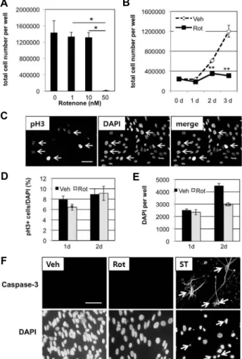
To examine the role of mitochondria in the proliferation of SVZ NSCs, cultured NSCs were treated with rotenone,
an inhibitor of complex I in the electron transport chain of mitochondria. When NSCs were incubated in N5 proliferat- ing medium containing 50 nM rotenone for 3 days, most of the cells were dead (Fig. 1A). However, 1 nM or 10 nM rotenone did not affect cell viability, resulting in similar numbers of cells as in 0 nM rotenone (DMSO-treated vehicle control). Therefore, all the following experiments using rote- none, 50 nM was used. Next, NSCs were treated with DMSO (vehicle control) or 50 nM rotenone for 0 to 3 days. The num- ber of DMSO-treated cells increased by 2.5 fold or 5 fold after 2 or 3 days, respectively, compared to 0 day treatment (Fig. 1B). However, the number of rotenone-treated cells in- creased by 1.5 fold (2d) or 1.3 fold (3d), compared to 0 day treatment. These less numbers of cells in rotenone-treated group than in control were statistically significant (p<0.01).
To investigate the mechanism how rotenone affected NSCs, immunocytochemistry was performed using anti- bodies against pH3, a hallmark of cell division (Fig. 1C).
After 1-day treatment with 50 nM rotenone, 6% of cells were positive for pH3, while 8 % in the control group (Fig. 1D).
This difference was statistically significant (p<0.05). After 2 days, there was no significant difference between control and rotenone-treated groups. Using DAPI staining and counting, total number of cells was compared. While the number of cells increased from 1 d to 2d of vehicle treatment by 1.8 fold, the number of cells increased only by 1.3 fold in rotenone-treated condition (Fig. 1E). Still, the rote- none-treated group had significantly less total number of cells than the control group at 2 d (p<0.01). Immunocyto- chemistry using antibodies against active caspase-3 was per- formed in NSCs treated with either vehicle or rotenone for 2 days. Both conditions resulted in negative for active cas- pase-3 staining, while 5 μM staurosporin treatment resulted in activation of caspase-3 (Fig. 1F). Combined with results from pH3 staining, theses suggest that rotenone probably blocked cell division and did not directly induce apoptosis, at least in the early phase of rotenone incubation.
Inhibition of neurogenesis and oligodendrogenesis by rotenone
Next, the effect of rotenone on differentiation of NSCs was examined. Proliferating NSCs are maintained in N5 pro- liferating medium and then, switched to N6 differentiation medium containing either DMSO (0 nM rotenone, vehicle control) or 1, 10, 50 nM rotenone. After 4 days, Tuj1 positive neurons and DAPI were counted (Fig. 2A ~ Fig. 2D). When
A B
C D
E
Fig. 2. Effect of rotenone on neurogenesis. A, B. SVZ NSCs of passage 7 were incubated in N6 differentiation medium containing different concentrations of rotenone for 4 days. Through immunocytochemistry and imaging, Tuj1- positive neurons and total number of cells (DAPI) were detected. C, D. Similar procedures were performed as in A and B except that SVZ NSCs of passage 5 were incubated in N6 medium for 5 days. A~D. Data are aver- age and standard deviation from three or four different wells of cells. One-way ANOVA test followed by post- hoc tests using Bonferroni corrected value was done and significantly different pairs of groups are indicated (*p<0.05). E. Images from experiments of C and D. Scale bar= 20 μm.
A B
C
D E
F
Fig. 1. Effect of rotenone on cell proliferation. A. SVZ NSCs were treated with different concentrations of rotenone for 3 days in N5 proliferating medium. Then, numbers of cells were measured. For 0 nM, DMSO was used as the same amount as in 50 nM rotenone. B. SVZ NSCs were treated with vehicle (Veh, DMSO) or 50 nM rotenone (Rot) in N5 medium. At 0, 1, 2, 3 days post treatment, cells were counted. C. Images from DMSO-treated cells for 1 day in N5 medium. Arrows indicate some of cells positive for histone H3 phosphorylated at serine 10 (pH3). Scale bar=10 μm. D and E. Proliferating NSCs were treated with either DMSO (Veh) or 50 nM rotenone (Rot) for 1 or 2 days. Fixed cells were stained to detect histone H3 phosphorylated at serine 10 (pH3) and nucleus (DAPI) as in C for counting. A, B, D and E. Data are average and standard deviation from three different wells of cells.
Statistical significance is shown, compared to 0d in B, or vehicle (Veh) treatment (D and E) (*p<0.05, **p<0.01, Student’s T-test). In A, one-way ANOVA followed by post-hoc tests with Bonferroni correction was performed and significant differences are indicated (*p<0.05). F.
Immunostaining with antibodies against active caspase-3 after treatment of proliferating NSCs with DMSO (Veh), 50 nM rotenone (Rot), or 5 μM stauosporin (ST) as a pos- itive control. Arrows indicate some of cells positive for active caspase-3. Scale bar=20 μm.
NSCs passaged 7 times (passage 7) before, about 3% of total cells were positive for Tuj1, a specific neuronal marker (Fig.
2A), while NSCs passaged 5 times (passage 5) before, about 32% were positive (Fig. 2C and upper lane of Fig. 2E).
However, NSCs incubated in N6 medium containing 50 nM rotenone had almost 0% in passage 7 cells (Fig. 2A) and 0.3%
in passage 5 cells (Fig. 2C and lower lane of Fig. 2E). Both 1 nM and 10 nM rotenone did not affect neurogenesis significantly. According to DAPI counting, more cells were present in 50 nM rotenone condition than control by 1.5 fold
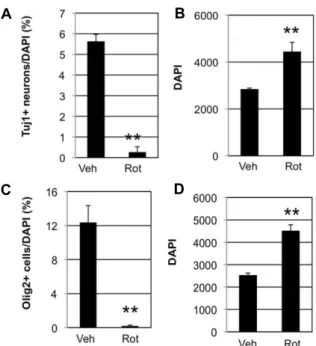
Fig. 3. Effects of rotenone on glial differentiation of SVZ NSCs.
Passage 6 SVZ NSCs were incubated in N6 differ- entiation medium containing DMSO (Veh) or 50 nM ro- tenone (Rot) for 6 days. Images from staining with Tuj1, Olig2, or GFAP are shown at the left panel under the columns, Veh or Rot. Merged images with DAPI staining are shown at the right panel under the columns, Veh or Rot. Scale bar= 20 μm.
A B
C D
Fig. 4. Effect of rotenone on differentiation of SVZ NSCs. A~D.
Images like in figure 3 were analyzed. For A and B, Tuj1 positive cells and total cell number (DAPI) were counted.
For C and D, Olig2 positive cells and DAPI were counted.
Data are average and standard deviation from three dif- ferent wells of cells. Statistical significance is shown, compared to vehicle control (**p<0.01, Student’s T-test).
(Fig 2B) or 1.4 fold (Fig. 2D). All these data indicate that rotenone blocked neurogenesis of NSCs.
Besides neurons, NSCs can become oligodendrocytes and astrocytes. To test the effects of rotenone on gliogenesis, sim- ilar experiments were done using antibodies against Olig2, a marker for oligodendrocytes, or GFAP, a marker for astrocytes. Passage 6 NSCs were incubated in N6 differ- entiation medium containing either DMSO (vehicle control) or 50 nM rotenone for 6 days. Representative images after immunocytochemistry are shown in figure 3. As expected, Tuj1+ cells were 0.2% in rotenone-treated cells and 6% in vehicle control (Fig. 4A). In addition, Olig2+ cells were 0.2%
in rotenone-treated cells and 12% in vehicle control (Fig. 4C).
Yet, GFAP+ cells was similar both in control and rote- none-treated cells (Fig. 3). These results indicate that rote- none inhibits NSCs from becoming either neurons or oligo- dendrocytes, while does not affect generation of astrocytes.
Effect of rotenone on cell proliferation during dif- ferentiation
One of the interesting phenomena in rotenone-treated NSCs during differentiation is that more cells were observed
than vehicle control (Fig. 2B, Fig. 2D, Fig. 4B, Fig. 4D). Since this result seems to be contrast to the inhibitory effect of rotenone on NSC proliferation, it invites more investigation.
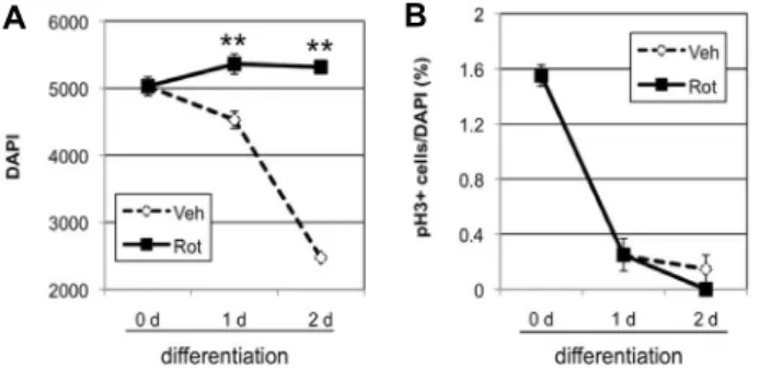
First, cell division was examined using pH3 staining. NSCs were incubated in N6 differentiation medium containing DMSO (vehicle control) or 50 nM rotenone for 0, 1, or 2 days. The number of cells in control decreased by 90% (1 day) and 50% (2 days), compared to 0-day treatment (Fig.
5A). In rotenone-treated group, the number of cells was maintained with 107% (1 day) and 106% (2 days), compared to 0-day. At 1 and 2 days, rotenone-treated groups con- sistently had more cells than control (Fig. 5A). However, pH3 staining was comparable in both vehicle control and rotenone-treated cells (Fig. 5B). In both groups, pH3+ cells decreased significantly from 1.6% (0 day) to 0.2% (1 day).
At 2 days of treatment, pH3+ cells were 0.1% in the control and 0% in rotenone-treated cells. Therefore, the maintained total cell number in rotenone does not seem to be caused by differential cell division.
As another possible mechanism, apoptosis was checked using antibodies against active caspase-3. NSCs were under- going differentiation in N6 medium for 12 hr and then, fur- ther incubated in N6 containing DMSO (vehicle control) or
A B
Fig. 5. Effect of rotenone on mitosis during neuronal differ- entiation. A, B. NSCs were incubated in N6 differ- entiation medium containing DMSO (Veh) or 50 nM ro- tenone (Rot) for 0, 1, or 2 days. Cells were fixed and stained with DAPI (A) and pH3 (B). Data are average and standard deviation from three different wells of cells. Statistical significance is shown, compared to Veh (**p<0.01, Student’s T-test).
A
B C
Fig. 6. Effect of rotenone on activation of caspase-3 during neu- ronal differentiation. A. NSCs were incubated in N6 for 42 hr including DMSO (Veh) or 50 nM rotenone (Rot) for 30 hr. Immunostaining with antibodies against active caspase-3 and DAPI staining are shown. Arrows indicate cells positive with active caspase-3. Scale bar= 10 μm.
B and C. Counting results are from three different wells of cells as shown in A with statistical significance (**p<
0.01, Student’s T-test).
50 nM rotenone for another 30h. In control, about 4% of total cells were positive for active caspase-3 (Fig. 6A, Fig. 6B).
In rotenone condition, about 2% of cells were active cas- pase-3+. This reduced number of active caspase-3+ cells by rotenone was statistically significant (p<0.01). This less stain- ing with active caspase-3 was consistent with more DAPI count in rotenone-treated cells (Fig. 6C).
Discussion
In our cultured NSCs obtained from neurogenic SVZ of mice, (1) rotenone inhibited cell proliferation, (2) it com- pletely blocked neurogenesis and oligodendrogenesis, (3) it increased a total number of cells in differentiation medium, and (4) it reduced activation of caspase-3 during differ- entiation. These results demonstrated important roles of mi- tochondria in proliferation and differentiation of SVZ NSCs and suggested possible involvement of apoptosis in fate de- termination of stem cells.
Rotenone inhibited cell proliferation in various cell types through cell cycle arrest [2, 12, 26]. Similarly, rotenone blocked cell proliferation of our cultured SVZ NSCs. While cytotoxicity was tested in earlier studies with μM concen- trations of rotenone, 50 nM was effective in our cultures.
Since other neural cells such as NSCs from mouse forebrain tissues, human iPSC-derived NSCs, or cultured neurons were sensitive to nM concentrations of rotenone [4, 23, 27, 33], neural cells might be more sensitive to rotenone than other cell types.
In proliferating condition, mitotic marker pH3 staining was less in the rotenone-treated NSCs than in the vehicle control. This difference was significant after 1-day incubation, but disappeared after 2-day incubation. Actual cell number difference was evident after 2-day incubation, according to both live cell counting and DAPI counting. However, there was no difference in activation of caspase-3, supporting that rotenone might have decreased cell number by reducing mi- tosis, rather than inducing apoptosis. Since rotenone in μM concentrations induced apoptosis in human NSCs in- dependently of caspase-9/3 activation [15], it is still possible that rotenone induced apoptosis without activating cas- pase-3 in our culture condition. Therefore, other methods of detecting apoptosis such as terminal deoxynucleotidyl transferase-mediated dUTP nick end labeling (TUNEL) staining and DNA fragmentation assay can be applied to test whether rotenone could induce apoptosis in proliferat- ing SVZ NSCs. Furthermore, increasing dose of rotenone up to μM concentrations can be tested.
In this report, cultured SVZ NSCs underwent neuronal differentiation when proliferating N5 medium was switched with N6 medium lacking growth factors and serum. Adding rotenone to N6 medium completely blocked neurogenesis and oligodendrogenesis. Previously, inhibitory effects of ro- tenone on the generation of dopaminergic neurons from hu-
man iPSC have been reported [8, 23]. To our knowledge, our study is the first report that showed inhibition of neuro- genesis and oligodendrogenesis in SVZ NSCs by rotenone.
Since both neurons and oligodendrocytes are generated form transient-amplifying cells (so-called type C cells) in SVZ [1], it is tempting to think that rotenone might have blocked dif- ferentiation of type C cells either to neuroblasts or oligoden- drocyte progenitor cells. To examine this hypothesis, the number of type C cells can be measured in rotenone- or ve- hicle-treated groups using marker staining such as Mash1+/
GFAP- or epidermal growth factor receptor (EGFR)+/GFAP- [21]. Additionally, rotenone treatment did not significantly affect GFAP staining. Since both type B NSCs and mature astrocytes express GFAP [13], more detailed identification of those GFAP+ cells should follow.
Interestingly, rotenone-treated group had more cells (DAPI+) than control cells after 1 or 2 days of incubation in differentiating N6 medium. This could have been either due to increased cell proliferation or decreased cell death.
Less staining of active caspase-3 was observed in the rote- none-treated group than in control, while pH3 staining was comparable between two groups. Therefore, it seems that presence of more cells in rotenone-treated groups might have been caused by a reduction of apoptotic cell death, not by an increase in mitosis. Since rotenone inhibited neuro- genesis, it is tempting to speculate that reduced apoptosis by rotenone treatment might be involved in perturbation of differentiation. Increased proliferation through genetic dele- tion coincided with increased neurogenesis [11, 24]. Here, more cells in the rotenone-treated group coincided rather with decreased neurogenesis. We think that disruption of probably normal apoptotic process during differentiation might have caused inhibition of differentiation, at least partly. Apoptosis has been known to be important in main- taining the constant size of olfactory bulb despite continual provision of neurons from SVZ [22, 25]. Yet, possible in- volvement of apoptosis in neuronal differentiation of cul- tured SVZ NSCs has not been known.
In summary, our study demonstrated that functional mi- tochondria are necessary in normal proliferation and neuro- nal differentiation of cultured SVZ NSCs. Unexpectedly, ro- tenone, a complex I inhibitor showed dual effects on cell proliferation depending culture conditions. While rotenone reduced mitosis in proliferating condition, it increased num- ber of cells by reducing apoptosis in non-proliferative differ- entiating condition. Furthermore, rotenone completely blocked
neurogenesis and oligodendrogenesis. Our data are the first report that suggests the possible involvement of apoptosis in neuronal differentiation. Finally, more detailed mecha- nisms how functional mitochondria take part in or signal to NSCs for differentiation are needed for further inves- tigation.
Acknowledgment
This work was supported by the Korea Science Academy of KAIST with funds from the Ministry of Science and ICT and by Basic Science Research Program through the National Research Foundation of Korea (NRF) funded by the Ministry of Science and ICT (2015R1C1A1A02037078).
References
1. Armada-Moreira, A., Ribeiro, F. F., Sebastião, A. M. and Xapelli, S. 2015. Neuroinflammatory modulators of oligo- dendrogenesis. Neuroimmunol. Neuroinflamm. 2, 263-273.
2. Armstrong, J. S., Hornung, B., Lecane, P., Jones, D. P. and Knox, S. J. 2001. Rotenone-induced g2/m cell cycle arrest and apoptosis in a human b lymphoma cell line pw. Bio- chem. Biophys. Res. Commun. 289, 973-978.
3. Beckervordersandforth, R., Ebert, B., Schaffner, I., Moss, J., Fiebig, C., Shin, J., Moore, D. L., Ghosh, L., Trinchero, M.
F., Stockburger, C., Friedland, K., Steib, K., von Wittgen- stein, J., Keiner, S., Redecker, C., Holter, S. M., Xiang, W., Wurst, W., Jagasia, R., Schinder, A. F., Ming, G. L., Toni, N., Jessberger, S., Song, H. and Lie, D. C. 2017. Role of mi- tochondrial metabolism in the control of early lineage pro- gression and aging phenotypes in adult hippocampal neurogenesis. Neuron 93, 560-573 e566.
4. Choi, W. S., Kruse, S. E., Palmiter, R. D. and Xia, Z. 2008.
Mitochondrial complex i inhibition is not required for dop- aminergic neuron death induced by rotenone, mpp+, or paraquat. Proc. Natl. Acad. Sci. USA. 105, 15136-15141.
5. Chung, S., Dzeja, P. P., Faustino, R. S., Perez-Terzic, C., Behfar, A. and Terzic, A. 2007. Mitochondrial oxidative me- tabolism is required for the cardiac differentiation of stem cells. Nat. Clin. Pract. Cardiovasc. Med. 4, S60-67.
6. Diaz-Castro, B., Pardal, R., Garcia-Flores, P., Sobrino, V., Duran, R., Piruat, J. I. and Lopez-Barneo, J. 2015. Resistance of glia-like central and peripheral neural stem cells to genet- ically induced mitochondrial dysfunction--differential ef- fects on neurogenesis. EMBO Rep. 16, 1511-1519.
7. Esdar, C., Milasta, S., Maelicke, A. and Herget, T. 2001.
Differentiation-associated apoptosis of neural stem cells is effected by bcl-2 overexpression: Impact on cell lineage determination. Eur. J. Cell Biol. 80, 539-553.
8. Fang, D., Qing, Y., Yan, S., Chen, D. and Yan, S. S. 2016.
Development and dynamic regulation of mitochondrial net- work in human midbrain dopaminergic neurons differ-
entiated from ipscs. Stem Cell Rep. 7, 678-692.
9. Folmes, C. D., Dzeja, P. P., Nelson, T. J. and Terzic, A. 2012.
Metabolic plasticity in stem cell homeostasis and differ- entiation. Cell Stem Cell 11, 596-606.
10. Folmes, C. D. and Terzic, A. 2016. Energy metabolism in the acquisition and maintenance of stemness. Semin. Cell Dev. Biol. 52, 68-75.
11. Gil-Perotin, S., Marin-Husstege, M., Li, J., Soriano-Navarro, M., Zindy, F., Roussel, M. F., Garcia-Verdugo, J. M. and Casaccia-Bonnefil, P. 2006. Loss of p53 induces changes in the behavior of subventricular zone cells: Implication for the genesis of glial tumors. J. Neurosci. 26, 1107-1116.
12. Goncalves, A. P., Maximo, V., Lima, J., Singh, K. K., Soares, P. and Videira, A. 2011. Involvement of p53 in cell death following cell cycle arrest and mitotic catastrophe induced by rotenone. Biochim. Biophys. Acta 1813, 492-499.
13. Imayoshi, I., Sakamoto, M. and Kageyama, R. 2011. Genetic methods to identify and manipulate newly born neurons in the adult brain. Front. Neurosci. 5, 64.
14. Levison, S. W., Rothstein, R. P., Brazel, C. Y., Young, G.
M. and Albrecht, P. J. 2000. Selective apoptosis within the rat subependymal zone: A plausible mechanism for de- termining which lineages develop from neural stem cells.
Dev. Neurosci. 22, 106-115.
15. Li, J., Spletter, M. L., Johnson, D. A., Wright, L. S., Svendsen, C. N. and Johnson, J. A. 2005. Rotenone-induced caspase 9/3-independent and -dependent cell death in undiffer- entiated and differentiated human neural stem cells. J.
Neurochem. 92, 462-476.
16. Lunt, S. Y. and Vander Heiden, M. G. 2011. Aerobic gly- colysis: Meeting the metabolic requirements of cell pro- liferation. Annu. Rev. Cell Dev. Biol. 27, 441-464.
17. Mandal, S., Lindgren, A. G., Srivastava, A. S., Clark, A. T.
and Banerjee, U. 2011. Mitochondrial function controls pro- liferation and early differentiation potential of embryonic stem cells. Stem Cells 29, 486-495.
18. Pamies, D., Block, K., Lau, P., Gribaldo, L., Pardo, C. A., Barreras, P., Smirnova, L., Wiersma, D., Zhao, L., Harris, G., Hartung, T. and Hogberg, H. T. 2018. Rotenone exerts developmental neurotoxicity in a human brain spheroid model. Toxicol. Appl. Pharmacol. In press.
19. Park, K. Y., Oh, H., Lee, J. and Kim, M. S. 2017. Inhibition of neurogenesis of subventricular zone neural stem cells by 5-ethynyl-2'-deoxyuridine (edu). J. Life Sci. 27, 623-631.
20. Park, K. Y., Na, Y. and Kim, M. S. 2016. Role of nox4 in neuronal differentiation of mouse subventricular zone neu- ral stem cells. J. Life Sci. 26, 8-16.
21. Pastrana, E., Cheng, L. C. and Doetsch, F. 2009. Simultaneous prospective purification of adult subventricular zone neural
stem cells and their progeny. Proc. Natl. Acad. Sci. USA. 106, 6387-6392.
22. Petreanu, L. and Alvarez-Buylla, A. 2002. Maturation and death of adult-born olfactory bulb granule neurons: Role of olfaction. J. Neurosci. 22, 6106-6113.
23. Pistollato, F., Canovas-Jorda, D., Zagoura, D. and Bal-Price, A. 2017. Nrf2 pathway activation upon rotenone treatment in human ipsc-derived neural stem cells undergoing differ- entiation towards neurons and astrocytes. Neurochem. Int.
108, 457-471.
24. Price, J. D., Park, K. Y., Chen, J., Salinas, R. D., Cho, M.
J., Kriegstein, A. R. and Lim, D. A. 2014. The ink4a/arf locus is a barrier to direct neuronal transdifferentiation. J. Neurosci.
34, 12560-12567.
25. Ryu, J. R., Hong, C. J., Kim, J. Y., Kim, E. K., Sun, W. and Yu, S. W. 2016. Control of adult neurogenesis by pro- grammed cell death in the mammalian brain. Mol. Brain 9, 43-62.
26. Srivastava, P. and Panda, D. 2007. Rotenone inhibits mam- malian cell proliferation by inhibiting microtubule assembly through tubulin binding. FEBS J. 274, 4788-4801.
27. Stoll, E. A., Cheung, W., Mikheev, A. M., Sweet, I. R., Bielas, J. H., Zhang, J., Rostomily, R. C. and Horner, P. J. 2011.
Aging neural progenitor cells have decreased mitochondrial content and lower oxidative metabolism. J. Biol. Chem. 286, 38592-38601.
28. Tamm, C., Sabri, F. and Ceccatelli, S. 2008. Mitochondrial- mediated apoptosis in neural stem cells exposed to man- ganese. Toxicol. Sci. 101, 310-320.
29. Wang, C. and Youle, R. J. 2009. The role of mitochondria in apoptosis. Annu. Rev. Genet. 43, 95-118.
30. Wang, H., Dong, X., Liu, Z., Zhu, S., Liu, H., Fan, W., Hu, Y., Hu, T., Yu, Y., Li, Y., Liu, T., Xie, C., Gao, Q., Li, G., Zhang, J., Ding, Z. and Sun, J. 2018. Resveratrol suppresses rotenone-induced neurotoxicity through activation of sirt1/
akt1 signaling pathway. Anat. Rec. 301, 1115-1125.
31. Xiong, N., Long, X., Xiong, J., Jia, M., Chen, C., Huang, J., Ghoorah, D., Kong, X., Lin, Z. and Wang, T. 2012. Mitochon- drial complex i inhibitor rotenone-induced toxicity and its potential mechanisms in parkinson's disease models. Crit.
Rev. Toxicol. 42, 613-632.
32. Xu, X., Duan, S., Yi, F., Ocampo, A., Liu, G. H. and Izpisua Belmonte, J. C. 2013. Mitochondrial regulation in pluripotent stem cells. Cell Metab. 18, 325-332.
33. Zagoura, D., Canovas-Jorda, D., Pistollato, F., Bremer- Hoffmann, S. and Bal-Price, A. 2016. Evaluation of the rote- none-induced activation of the nrf2 pathway in a neuronal model derived from human induced pluripotent stem cells.
Neurochem. Int. 106, 62-73.
초록:미토콘드리아 억제제 rotenone에 의한 쥐의 뇌실 하 영역 신경 줄기 세포의 증식과 신경 세포로의 분화 억제
박기엽1*․김만수2
(1KAIST 부설 한국과학영재학교, 2인제대학교 약학대학)
미토콘드리아는 세포안에서 에너지 공급, 칼슘 이온 저장, 활성산소 생성, 세포 자살과 같은 다양한 기능을 수 행한다. 이러한 기능을 통해, 미토콘드리아는 줄기세포의 유지, 증식, 그리고 분화에 관여한다. 뇌에서 뇌실 하 영 역(subventricular zone, SVZ)에는 일평생 새로운 신경세포를 생성하는 신경줄기세포(neural stem cell, NSC)가 존재한다. 하지만, SVZ NSCs에서 미토콘드리아의 역할에 대한 연구는 많이 알려져 있지 않다. 이번 연구에서 우 리는 미토콘드리아의 complex I 저해제인 rotenone이 SVZ NSCs의 증식과 분화를 다른 방식으로 방해한다는 것 을 보여주었다. 증식 중인 신경줄기세포에서, rotenone은 세포분열을 감소시켰는데, 이때 세포분열은 히스톤 H3 에 인산기가 붙어있는 지를 측정하여 확인하였다. Rotenone을 50 nM 농도로 증식 중인 신경줄기세포에 처리했을 때, 세포사멸은 발생하지 않았다. 한편, 분화 중인 신경줄기세포에 rotenone을 처리한 경우, 신경세포와 희소 돌기 아교 세포(oligodendrocyte)으로의 분화가 억제되었고, glial fibrillary acidic protein (GFAP)를 발현하는 성상세포 (astrocyte)에는 영향이 없었다. 흥미롭게도, 4-6일 동안의 분화 과정 동안 rotenone이 처리된 신경줄기세포에서 대조군 보다 더 많은 세포 수가 관찰 되었는데, 이는 증식 과정 중의 rotenone의 효과와 다른 것이다. 이에, 우리는 rotenone이 세포 자살은 감소시켰으나, 세포 분열에는 영향을 끼치지 않았음을 관찰하였다. 세포 자살의 경우는 cleaved caspase-3를 측정함으로써 확인하였다. 이러한 결과들은 SVZ 신경줄기세포의 증식과 분화 모두에 제대로 작동하는 미토콘드리아가 있어야 함을 제안하고 있다. 게다가, 이러한 과정에서 미토콘드리아는 세포 분열과 세포 자살에 관여할 수도 있을 것이다.