대한외과학회지:제 74 권 제 4 호
□ 증 례 □
Vol. 74, No. 4, April, 2008
319
책임저자:신동훈, 부산시 서구 암남동 34번지
602-702, 고신대학교 복음병원 외과 Tel: 051-990-6462, Fax: 051-246-6093 E-mail: [email protected]
접수일:2007년 10월 11일, 게재승인일:2007년 12월 31일 본문의 요지는 2007년 대한외과학회 춘계학술대회에서 포스터 발 표되었음.
본 논문은 고신의대 연구 지원금의 일부로 이루어졌음.
중심 단어: 이중 담낭관, 선천성기형
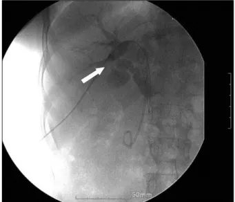
Fig. 1. Preoperative percutaneous transhepatic biliary drainage.
Arrow shows accessory cystic duct connected with right hepatic duct.
한 개의 담낭에서 기시하는 이중 담낭관
고신대학교 의과대학 복음병원 외과 유중재ㆍ신동훈
An Accessory Double Cystic Duct with a Single Gall Bladder
Joongjae Yoo, M.D. and Donghoon Shin, M.D.
Department of Surgery, College of Medicine, Kosin University, Busan, Korea
We report a rare case of a double cystic duct in a 55-year-old woman. The patient complained of upper ab- dominal pain and jaundice. The patient was diagnosed with distal common bile duct (CBD) cancer by endoscopic retro- grade cholangiopancreatography (ERCP). ERCP was unable to reveal the presence of a double cystic duct. However, magnetic resonance imaging (MRI) and a percutaneous transhepatic biliary drainage (PTBD) were performed as im- ages for obstructive jaundice before surgery showed two ducts that looked like cystic ducts-one duct branched from the common bile duct and the other duct branched from the right hepatic duct. A pancreatoduodenectomy was performed to remove the distal CBD cancer and the presence of a dou- ble cystic duct was confirmed with the naked eye. This case suggests that the imaging studies, such as preoperative ERCP and MRI or intraoperative cholangiography are re- quired to avoid complications during hepatobiliary surgery. (J Korean Surg Soc 2008;74:319-321)
Key Words: Double cystic duct, Congenital anomaly
서 론
이중 담낭관은 우리나라에 보고가 거의 없는 매우 드문
담도계의 선천성 기형으로 수술 전 방사선학적인 검사로 발견될 수도 있지만 수술 중 우연히 발견되기도 한다.(1,2) 이중 담낭관과 같은 담도계의 기형이 있을 경우 담도계의 수술 시에 주의를 기울이지 않으면 담관의 손상으로 인한 합병증이 발생할 수 있어 외과적으로 중요한 의미가 있 다.(3,4) 이에 저자들은 총담관의 악성종양으로 췌십이지장 절제술을 시행한 환자에서 이중 담낭관을 발견하였기에 보 고하는 바이다.
증 례
환자는 55세 여자로 황달과 상복부 통증을 주소로 수술 전 약 2개월 전에 타 병원을 방문하였고 내시경적 역행적 담췌관조영술 등 방사선학적 검사에서 원위부 총담관암으 로 진단 받았다. 황달을 완화시키기 위해 경피적 경간 담즙 배액술을 시행하였으며, 배액관을 통한 조직검사에서 원위 부 담관암으로 확진되었다. 이 후 본원 방문하였고 수술 권 유하여 췌십이지장절제술을 시행하였다. 수술 전 내시경적 역행적 담췌관조영술에서 총담관의 폐쇄로 담도계의 형태
320 J Korean Surg Soc. Vol. 74, No. 4
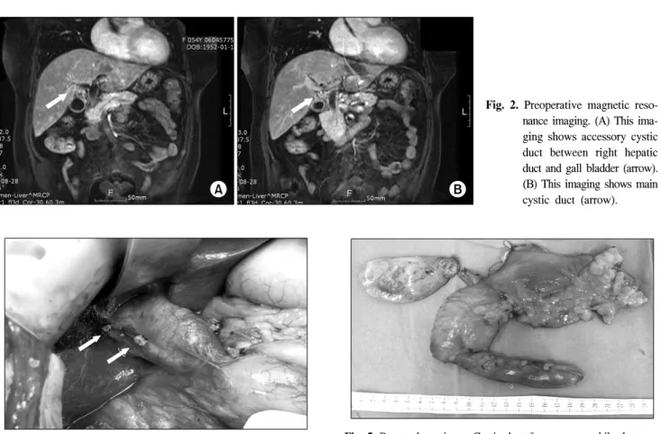
Fig. 2. Preoperative magnetic reso- nance imaging. (A) This ima- ging shows accessory cystic duct between right hepatic duct and gall bladder (arrow).
(B) This imaging shows main cystic duct (arrow).
Fig. 5. Resected specimen. Cystic duct from common bile duct was resected with specimen and the other from right hepatic duct remained.
Fig. 3. Itraoperative finding. Two arrows show two cystic ducts remained on commen bile duct and right hepatic duct.
Fig. 4. Arrows show double cystic duct connected gall bladder.
학적인 이상소견은 알 수 없었으나 경피적 경간 담즙배액 관 조영술에서 이중 담낭관으로 보이는 소견이 발견되었으 며 술전 자기공명영상에서도 이중담낭관이 의심되는 소견 을 보였다(Fig. 1, 2). 수술 당시 담낭절제술을 시행하는 과 정에서 담낭관의 기형을 발견하였고 한 개의 담낭에서 각 각 총담관과 우측간내담관으로 연결된 담낭관이 관찰되었 다. 그 외에 다른 담도계의 기형은 관찰되지 않았으며 타장 기의 기형이나 질환도 관찰되지 않았다. 각 담낭관은 절제
하면서 담즙이 나오는 것을 확인하였고 절제한 담낭에서 두 개의 담낭관이 담낭으로 연결되어 있는 것을 확인하였 다(Fig. 3, 4, 5). 그 외에 다른 담도계의 이상소견은 보이지 않았다. 수술 후 환자는 별다른 합병증 없이 퇴원하였다.
고 찰
담도계의 기형은 매우 흔하고 담낭관과 간외 담관 그리 고 담낭동맥과의 해부학적 이상이 담도계 기형의 33%를 차 지하며 총담관과 담낭관 사이의 기형이 나머지 67%를 차지 하고 있다. 담도계에서 담낭관의 20%정도는 총담관과 평행 하게 주행하여 총담관과 담낭관과의 접합부가 각이 거의 없는 형태이고 담낭관의 5∼8%는 비틀려 있거나 나선형태 를 띠는 기형을 가진다. 그 중에서 이중 담낭관은 담도계의 기형 중 1% 미만으로 매우 드물며 국내에서도 보고가 거의 없다.(1)
Flannery와 Caster(5)는 이중 담낭관을 두 개의 담낭관의 위치에 따라서 세 가지 유형으로 분류하였다. 첫째는 “Y”유 형으로 기존의 담낭관으로부터 하나의 담낭관이 갈라져 나 와 총담관이나 간문부담관에 연결된 형태이고, 두 번째는
“H”유형으로 각각의 담낭관이 담낭으로부터 나와 하나는
Joongjae Yoo and Donghoon Shin:An Accessory Double Cystic Duct with a Single Gall Bladder 321
총담관에 연결되고 다른 하나는 간문부담관이나 총담관에 연결된 형태이며, 마지막은 “지주”유형으로 각각의 총담관 이 하나는 총담관으로 다른 하나는 간실질 내로 직접 연결 되어 들어가는 형태이다. 이들 중 현재까지 가장 많이 보고 된 유형은 본 증례와 같은 “H”유형이다.(6-9)
담관계의 해부학적 기형은 담낭 절제를 전제로 진행하는 수술이나 복강경적 담낭절제술 같은 담관계의 수술 시에 주의를 요하므로 외과적으로 매우 중요한 의미를 가진 다.(3,4)
Tsutsumi 등(8)과 Hirono 등(9)은 담관계의 기형으로 인한 손상을 예방하기 위해 수술 전 내시경적 역행적 담관췌조 영술을 시행하거나 술중 담관조영술을 시행해야 한다고 주 장하기도 하였다.
이중 담낭관이 비록 드문 담도계의 기형이지만 일반적으 로 담도계의 기형은 흔하고 이로 인한 술중 합병증은 환자 에게 치명적인 결과를 초래할 수 있기 때문에 수술 전 검사 를 통해 기형 유무를 확인하거나 술중 검사나 적절한 시야 확보를 통해 기형의 유무를 가려내며 수술하는 것이 중요 하다.
REFERENCES
1) Rabinovitch J, Harandian B, Friedman HP, Arlen M, Rabinov- itch P. Congenital anomalies of the cystic duct. J Int Coll Surg
1964;42:372-8.
2) Benson EA, Page RE. A practical reappraisal of the anatomy of the extrahepatic bile ducts and arteries. Br J Surg 1976;63:
853-60.
3) Lee VS, Chari RS, Cucchiaro G, Meyers WC. Complication of laparoscopic cholecystectomy. Am J Surg 1993;165:527-32.
4) Klotz HP, Schlumpf R, Largiader F. Injury to an accessory bile duct during laparoscopic cholecystectomy. Surg Laparosc Endosc 1992;2:317-20.
5) Flannery MG, Caster MP. Congenital abnormalities of the gallbladder; 101 cases. Int Abstr Surg 1956;103:439-57.
6) Paraskevas G, Papaziogas B, Natsis K, Spanidou S, Kitsoulis P, Atmatzidis K, et al. An accessory double cystic duct with single gallbladder. Chirurgia (Bucur) 2007;102:223-5.
7) Kayahara M, Oyama L, Kitagawa H, Ohta T, Miwa K. Right hepatic duct opening into the cystic duct: the role of pre- and intraoperative cholangiography. Hepatogastroenterology 2005;
52:719-21.
8) Tsutsumi S, Hosouchi Y, Shimura T, Asao T, Kojima T, Takenoshita S, et al. Double cystic duct detected by endo- scopic retrograde cholagiopancreatography and confirmed by intraoperative cholangiography in laparoscopic cholecystec- tomy: a case report. Hepatogastroenterology 2000;47:1266-8.
9) Hirono Y, Takita Y, Nitta N, Hashimoto H. Double cystic duct found by intraoperative cholangiography in laparoscopic chol- ecystectomy. Surg Laparosc Endosc 1997;7:263-5.