131
Open Access
Pulmonary Hypertension in Preterm Infants With Bronchopulmonary Dysplasia
Hyo Soon An, MD, Eun Jung Bae, MD, Gi Beom Kim, MD, Bo Sang Kwon, MD, Jae Suk Beak, MD, Ee Kyung Kim, MD, Han Suk Kim, MD, Jung-Hwan Choi, MD, Chung Il Noh, MD and Yong Soo Yun, MD
Department of Pediatrics, Seoul National University Children’s Hospital, Seoul National University College of Medicine, Seoul, Korea ABSTRACT
Background and Objectives: With the increasing survival of preterm infants, pulmonary hypertension (PH) re- lated to bronchopulmonary dysplasia (BPD) has become an important complication. The aim of this study was to investigate the characteristics and outcome of PH in preterm infants with BPD and to identify the risk factors for PH. Subjects and Methods: We reviewed the records of 116 preterm infants with BPD cared for at a single ter- tiary center between 2004 and 2008. Results: Twenty-nine (25%) infants had PH >2 months after birth. PH oc- curred initially at a median age of 65 days (range, 7-232 days). Severe BPD, a birth weight <800 g, long-term ven- tilator care and oxygen supplementation, a high ventilator setting, infection, and a patent ductus arteriosus (PDA) were related to PH based on univariate analysis (p<0.05). The infants who had longer oxygen supplementation were significantly more likely to have PH (odds ratio, 18.5; 95% confidence interval, 4.1-84.6; p<0.001). PH was improved in 76% of infants after a median of 85 days (range, 20-765 days). Four infants (14%) died. The death of 3 infants was attributed to PH. Conclusion: BPD was frequently complicated by PH. Although PH resolved in the majority of infants, PH in preterm infants with BPD can be fatal. Regular screening for PH and adequate man- agement are required. (Korean Circ J 2010;40:131-136)
KEY WORDS: Hypertension, pulmonary; Infant, premature; Bronchopulmonary dysplasia.
Introduction
Bronchopulmonary dysplasia (BPD) is a chronic lung disease in preterm infants that occurs following mechani- cal ventilation and oxygen therapy for acute respiratory distress after birth. Despite improvement in perinatal care, chronic lung disease after preterm birth remains a major problem. BPD is one of the most significant se- quelae of neonatal intensive care, affecting approxima- tely 10,000 infants in the United States each year.1)
Preterm infants with BPD are at high risk of cardio-
vascular sequelae. Pulmonary hypertension (PH), impair- ed gas exchange due to abnormal vasoregulation, exercise intolerance, systemic hypertension, left ventricular hy- pertrophy, and development of systemic to pulmonary collateral vessels may complicate the course.2)3) PH results in right ventricular hypertrophy (RVH) and cardiome- galy and may lead to right heart failure.4)
Even though severe PH is one of the life-threatening complications in neonates, there are few published stud- ies on the incidence and prognosis of PH in preterm infants with BPD. The aim of this study was to deter- mine the characteristics and outcome of PH in preterm infants and identify the risk factors for PH.
Subjects and Methods
A retrospective study was conducted by reviewing the medical records of all infants <32 weeks gestational age born between June 2004 and April 2008 at our hospital.
The diagnosis of PH was made by echocardiography based on the following criteria: 1) velocity of tricuspid valve regurgitation (TR) ≥3 m/s in the absence of pul-
Received: July 2, 2009
Revision Received: August 13, 2009 Accepted: September 3, 2009
Correspondence: Eun Jung Bae, MD,Department of Pediatrics, Seoul Na- tional University Children’s Hospital, Seoul National University College of Medicine, 101 Daehak-ro, Jongno-gu, Seoul 110-744, Korea Tel: 82-2-2072-3097, Fax: 82-2-743-3455
E-mail: [email protected] ○
○
○ cc This is an Open Access article distributed under the terms of the Creative Commons Attribution Non-Commercial License (http://creativecommons.
org/licenses/by-nc/3.0) which permits unrestricted non-commercial use, distribution, and reproduction in any medium, provided the original work is properly cited.
monary stenosis; and 2) flat or left-deviated interven- tricular septal configuration, and RVH with chamber dilation. Infants with one or both of these findings at
>2 months of age were characterized as having PH. Se- ven BPD cases showed echocardiographic evidence of PH before 1 month of age, and the PH persisted beyond 2 months of age; they were included in this study. How- ever, infants with the typical ‘persistent PH of the new- born’ were excluded. Infants with persistent foramen ovale and patent ductus arteriosus (PDA) were included in the study; however, infants with other congenital heart diseases, congenital diaphragmatic hernias, and meco- nium aspiration syndrome were excluded. Risk factors that contribute to PH were analyzed. Improvement of PH was defined as a TR ≤2.5 m/s, a diminished amount of TR, restoration of interventricular septal configuration, regressed RVH and dilation, and discontinuation of oxy- gen supplementation therapy.
Definition and grading of BPD were based on Jobe- Bancalari criteria.5) The severity of BPD was graded ac- cording to the fraction of inspired oxygen (FiO2) or po- sitive pressure ventilation (PPV), as follows: mild BPD, breathing room air; moderate BPD, requiring oxygen supplementation (FiO2 of <0.30); and severe BPD, requir- ing FiO2 of ≥0.30 or PPV at 36 weeks gestational age.5) Statistical analysis
Data for infant characteristics were expressed as the median and range or mean plus or minus standard de- viation or percentage. Continuous values between the two groups were compared by Student’s t-test. Rate and proportion were tested with the chi-square test. Fac- tors with a p<0.05 by univariate analysis were included in the logistic regression analysis. In all analyses, p<
0.05 were considered significant. Statistical analysis was conducted using the Statistical Package for the Social Sciences (SPSS), version 12.
Results
General characteristics
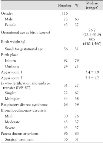
One hundred sixteen preterm infants with BPD met the inclusion criteria for this study. The median gesta- tional age at birth was 26.7 weeks (range, 23.4-31.9 weeks), and the median birth weight was 805 g (range, 430-1,560 g) . Thirty-six patients (31%) were small for gestational age (SGA) at birth, defined as a weight be- low the 10th percentile for the estimated gestational age.
The median age of investigation was 18.5 months (range, 2-48.5 months). The grades of BPD were classified as follows: severe (37%), moderate (37%), and mild (26%).
Sixty-eight (59%) infants had respiratory distress syn- drome (RDS) at birth, and 36 (31%) infants had surgi- cal management for PDA. Seven infants died during the study. The characteristics of the infants studied are
presented in Table 1.
Pulmonary hypertension
Twenty-nine infants with BPD (25%) met the crite- ria of PH beyond 2 months of age. PH was initially diagnosed at a median postnatal age of 65 days (range, 7-232 days). The median gestational age at birth was 26 weeks (range, 23.9-31.0 weeks), and the median birth weight was 700 g (range, 430-1,450 g). RDS was diag- nosed in 21 patients (72%). Severe BPD was diagnosed in 25 infants with PH (86%). Among 116 infants with BPD, PH was diagnosed in 58% of the infants with se- vere BPD, 9.3% of the infants with moderate BPD, and 0% of the infants with mild BPD cases (Fig. 1). The oc- currence of PH was positively correlated with the seve- rity of BPD (p<0.001). The degree of TR on echocar- diography was trivial in 8 (27.6%) infants, mild in 13 (44.8%) infants, and moderate in 8 (27.6%) infants.
Right ventricular dilation or hypertrophy was noted in 20 (68.9%) infants, and 23 infants (79.3%) exhibited a flat interventricular septum on echocardiography.
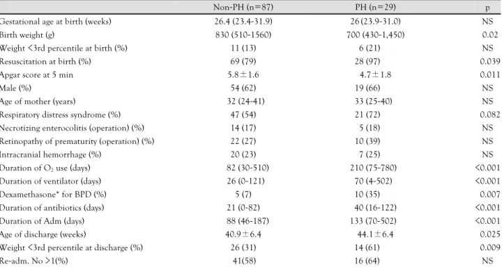
Comparison between the infant groups with and without pulmonary hypertension
The clinical characteristics of the study infants are presented in Table 2. The mean birth weight of the PH group was lower than the non-PH group (p<0.02), al-
Table 1. Characteristics of patients
Number % Median (range)*
Gender 116
Male 073 63
Female 043 37
Gestational age at birth (weeks) 26.7 (23.4-31.9)
Birth weight (g) 805
(430-1,560) Small for gestational age 036 31
Birth place
Inborn 092 79
Outborn 024 21
Apgar score 1 3.4±1.9
Apgar score 5 5.5±1.7
In vitro fertilization and embryo
transfer (IVF-ET) 031 27
Singlet 072 62
Multiplet 044 38
Respiratory distress syndrome 068 59 Bronchopulmonary dysplasia
Mild 030 26
Moderate 043 37
Severe 043 37
Patent ductus arteriosus 096 83 Surgical treatment 036 31
*Median (range) or mean±SD
though the number of infants with a birth weight <3rd percentile was similar at birth. Severe BPD was more prevalent in the PH group (86%) than the non-PH group (20%, p<0.001). The PH group was comprised of more infants who had undergone surgery for PDA than the non-PH group (48% vs. 27%, p=0.025). The Ap- gar score at 5 minutes was lower in the PH group than in the non-PH group (4.7±1.8 vs. 5.8±1.6, respecti- vely, p=0.011). The PH group required more resusci- tation at birth than the non-PH group (97% vs. 79%, respectively, p=0.039).
Except for four of the infants, most of the infants had respiratory support, including mechanical venti- lator and nasal continuous positive airway pressure
(CPAP) therapy during admission. The duration of ven- tilator therapy was longer in the PH group than in the non-PH group {70 (4-502) days vs. 26 (1-121) days, p<
0.001}. The duration of oxygen supplementation was longer in the PH group (median, 210 days; range, 75- 780 days) than in the non-PH group (median, 82 days;
range, 30-510 days, p<0.001). The gestational age at dis- charge was a mean of 44.1±6.4 and 40.9±6.4 days in the PH and non-PH groups, respectively (p=0.025).
The postnatal growth was impaired in both groups from birth to discharge (patients with a weight <3rd percen- tile 13% → 31%, p<0.001 in non-PH; 21% → 61%, p<0.001 in PH group). The proportion of low-body weight infants (<3rd percentile) was higher in the PH group at the time of discharge (31%:61%, p=0.009).
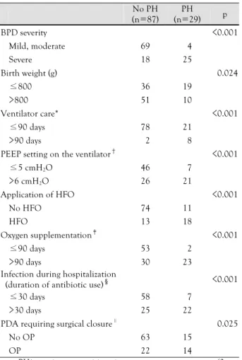
Severe BPD, birth weight <800 g, long-term use of ventilator care and oxygen supplementation, application of high-frequency oscillator ventilator (HFO), high po- sitive end expiratory pressure (PEEP) setting on the ven- tilator, infection during hospitalization (duration of an- tibiotic use), and PDA requiring surgical closure were related to PH based on univariate analysis (p<0.05) (Ta- ble 3). Infants who had longer oxygen supplementa- tion were significantly more likely to have PH than the other patients (odds ratio, 18.5; 95% confidence inter- val, 4.1-84.6; p<0.001). Risk factors that contributed to PH were not identified on multivariate analysis.
Outcome and prognosis
The median duration of follow-up was 484 days (range, 18-999 days) in infants with PH. PH improved
Table 2. Comparison between BPD patients with and without pulmonary hypertension
Non-PH (n=87) PH (n=29) p
Gestational age at birth (weeks) 26.4 (23.4-31.9) 26(23.9-31.0) NS
Birth weight (g) 830 (510-1560) 700 (430-1,450) <0.02
Weight <3rd percentile at birth (%) 11 (13) 6 (21) NS
Resuscitation at birth (%) 69 (79) 28 (97) <0.039
Apgar score at 5 min 5.8±1.6 4.7±1.8 <0.011
Male (%) 54 (62) 19 (66) NS
Age of mother (years) 32 (24-41) 33 (25-40) NS
Respiratory distress syndrome (%) 47 (54) 21 (72) <0.082
Necrotizing enterocolitis (operation) (%) 14 (17) 5 (18) NS
Retinopathy of prematurity (operation) (%) 22 (27) 10 (39) NS
Intracranial hemorrhage (%) 20 (23) 7 (25) NS
Duration of O2 use (days) 82 (30-510) 210 (75-780) <0.001
Duration of ventilator (days) 26 (0-121) 70 (4-502) <0.001
Dexamethasone* for BPD (%) 5 (7) 10 (35) <0.007
Duration of antibiotics (days) 21 (0-82) 40 (16-122) <0.001
Duration of Adm (days) 88 (46-187) 133 (70-502) <0.001
Age of discharge (weeks) 40.9±6.4 44.1±6.4 <0.025
Weight <3rd percentile at discharge (%) 26 (31) 14 (61) <0.009
Re-adm. No >1(%) 41(58) 16 (64) NS
Data are median and range, mean (±SD) or n (%) as appropriate. *dexamethasone rescue therapy was done for severe BPD. BPD: bron- chopulmonary dysplasia, Adm: admission, PH: pulmonary hypertension
Patients (n)
50
40
30
20
10
0
Mild Moderate Severe BPD grade
9.3% 58%
PH No PH
Fig. 1. The occurrence of PH with the degree of BPD severity.
The occurrence of PH positively correlated with the degree of BPD severity (p<0.001). PH: pulmonary hypertension, BPD: br- onchopulmonary dysplasia.
in 22 infants (76%) at a median of 85 (range, 20-765 days) after diagnosis. According to the Kaplan-Meier curve, PH improved in 68% of infants by 6 months of age and 73% of infants by 1 year of age (Fig. 2). Four infants (14%) died during the follow-up period. The deaths of 3 of the infants were attributed to PH. The death of the remaining infant was due to respiratory failure as a result of BPD. PH aggravated the condition in 1 infant. Two other patients have shown improved lung condition and are awaiting follow-up echocardio- graphy. The mortality rate of the PH group was higher than the non-PH group (14%:3.4%, p=0.043). In- fants with PH died by a median age of 162 days (range, 70-274 days), which was a median of 117 days (range, 42-190 days) after being diagnosed with PH. In the non-PH group, 3 infants died. One of them died due to sepsis, and the others died because of respiratory fail- ure aggravated by respiratory infection.
Pulmonary vasodilator therapy was used to treat 20
of the PH infants (69%), and included the following:
inhaled nitric oxide (NO), 13 (44.8%); sildenafil, 18 (62%); bosentan, 3 (10.3%); and iloprost, 4 (13.8%).
Inhaled NO was used for a median of 39 days (range, 2-173 days). Sildenafil was most often administered at a median of 2.6 mg (range, 0.7-5 mg)/(kg·day) for a median of 186 days (range, 32-1,129 days). Oxygen sup- plementation therapy was required for a median of 9.5 months (range, 3-26 months), and PH eventually im- proved 8.5 months (range, 3.2-29.3 months) later.
Discussion
Although the mechanisms responsible for elevated pulmonary vascular resistance and altered reactivity re- main incompletely understood, the development of PH is recognized as a serious complication of BPD that can significantly contribute to the morbidity and mortality of preterm infants.6)7) One study involving 42 preterm infants with BPD complicated by PH (gestational age
<32 weeks) reported that 43% of the infants had se- vere PH and the mortality rate was 38%.6) In the current study, it was estimated that the prevalence of PH among the entire sample of preterm infants with BPD was 25%, and the mortality rate in the PH group was 14%.
We determined the factors related to PH. The fac- tors, including aggressive mechanical ventilation and high inspired oxygen concentrations are known to be related to BPD.8) There are many common factors be- tween BPD and PH; however, we could not identify the specific risk factors for PH in preterm infants with BPD based on multivariate analysis. In BPD, structural ab- normalities of the pulmonary vasculature lead to nar-
Table 3. Factors related to PH
No PH (n=87)
PH (n=29) p
BPD severity <0.001
Mild, moderate 69 04
Severe 18 25
Birth weight (g) <0.024
≤800 36 19
>800 51 10
Ventilator care* <0.001
≤90 days 78 21
>90 days 02 08
PEEP setting on the ventilator† <0.001
≤5 cmH2O 46 07
>6 cmH2O 26 21
Application of HFO <0.001
No HFO 74 11
HFO 13 18
Oxygen supplementation‡ <0.001
≤90 days 53 02
>90 days 30 23
Infection during hospitalization
(duration of antibiotic use)§ <0.001
≤30 days 58 07
>30 days 25 22
PDA requiring surgical closure∥ <0.025
No OP 63 15
OP 22 14
*no PH: continuous positive airway pressure support case (3 pa- tients) excluded; no ventilator care (4), †no PH: continuous posi- tive airway pressure support case (3 patients) excluded; missing data (12), PH: missing data (1), ‡no PH: missing data (4), PH: death case (4 patients) exclueded, §no PH: missing data (4), ∥no PH: missing data (2). PH: pulmonary hypertension, BPD: bronchopulmonary dysplasia, HFO: high-frequency oscillator ventilator, PEEP: positive end expiratory pressure, PDA: patent ductus arteriosus, OP: operation
Percent of persistent PH
1.0
0.8
0.6
0.4
0.2
0.0
0 200 400 600 800 Days from diagnosis of PH
Fig. 2. Kaplan-Meier curve express the resolution of PH. PH im- proved in 68% of patients by 6 months and 73% of patients by 1 year. PH:pulmonary hypertension.
rowing of vessel diameters and decreased vascular com- pliance. Decreased angiogenesis is another consequ- ence of BPD and may contribute to reduce the vascu- lar cross-sectional area. These factors contribute to ele- vated pulmonary vascular resistance.6)9) The pulmonary circulation in these patients abnormally respond to oxy- gen and other pulmonary vasodilators.10)
PH affects the growth of preterm infants. There was no difference in the proportion of infants ≤3rd percen- tile between the PH and the non-PH groups at birth.
However, at the time of discharge, the proportion of in- fants ≤3rd percentile increased significantly in the PH group. Infants with PH had longer periods of oxy- gen supplementation and hospital admission than the other infants. The need for treatment can affect the qu- ality of life and the financial status of the infant’s fa- mily. Although the target systemic arterial oxygen sa- turation in preterm infants is controversial, long-term supplemental oxygen therapy is the standard treatment for PH-associated BPD.5)11) There are some reports that elevations in pulmonary arterial pressure persists beyond infancy and early childhood in preterm infants with
BPD.12)13) The removal of oxygen should be gradual and
completed after evaluation of the infant’s condition.
In the current study, PH was diagnosed at a median of 65 days (range, 7-232 days). The gestational age at birth was 24-26 weeks in 62% of the infants with PH.
Early detection of PH is difficult in children. The diag- nosis of PH in infants and neonates requires a high degree of suspicion because the signs and symptoms of PH may be subtle and non-specific and masked by un- derlying BPD, even in infants with significantly elevat- ed pulmonary artery pressure.14)15) However, there are no optimal methods or screening criteria for preterm infants with BPD. Doppler echocardiography is a non- invasive test commonly used to screen and manage PH.16) Echocardiography also has limitations as a screening st- rategy for PH in this population. Specifically, echocar- diography could fail to detect a measurable TR jet ve- locity in a significant number of high-risk patients; ab- sence of a TR jet velocity does not rule out the presence of severe PH.14) In the current study, more than mode- rate TR on echocardiography was observed in only 28%
of the infants with PH. Because TR is not always meas- urable, qualitative echocardiographic measures of PH, such as right atrial enlargement, RVH, right ventricular dilation, and septal flattening, seem to have good sen- sitivity and a positive predictive value for diagnosing PH in children with chronic lung disease; however, the spe- cificity and negative predictive value are poor.14)
After discharge from the hospital, the diagnosis of PH was confirmed in 4 infants. Although the infants may be well at the time of discharge, symptoms of PH should be monitored with serial echocardiography.
This study was limited by its retrospective design.
Not every infant with BPD in our institution underwent echocardiography during the study period. The time of diagnosis, improvement, and aggravation may not be precise because the evaluation interval was determined based on the infant’s condition.
The number of preterm infants is increasing. Survi- val, as well as a good quality of life are thought to be important in leading a healthy life. The evaluation and management of complications of BPD are imperative.
Preterm infants with BPD must be evaluated periodi- cally for signs and symptoms of PH. More reliable sc- reening methods for PH and severity stratification are needed. For PH treatment guidelines in preterm infants with chronic lung disease, further prospective randomiz- ed controlled studies are warranted in preterm infants with BPD.
REFERENCES
1) Stenmark KR, Abman SH. Lung vascular development: implica- tions for the pathogenesis of bronchopulmonary dysplasia. Annu Rev Physiol 2005;67:623-61.
2) Abman SH, Accurso FJ, Bowman CM. Unsuspected cardiopul- monary abnormalities complicating bronchopulmonary dyspla- sia. Arch Dis Child 1984;59:966-70.
3) Gill AB, Weindling AM. Pulmonary artery pressure changes in the very low birthweight infant developing chronic lung disease.
Arch Dis Child 1993;68(3 Spec No):303-7.
4) Goodman G, Perkin RM, Anas NG, Sperling DR, Hicks DA, Rowen M. Pulmonary hypertension in infants with bronchopul- monary dysplasia. J Pediatr 1988;112:67-72.
5) Jobe AH, Bancalari E. Bronchopulmonary dysplasia. Am J Res- pir Crit Care Med 2001;163:1723-9.
6) Khemani E, McElhinney DB, Rhein L, et al. Pulmonary artery hypertension in formerly premature infants with bronchopulmo- nary dysplasia: clinical features and outcomes in the surfactant era. Pediatrics 2007;120:1260-9.
7) Fouron JC, Le Guennec JC, Villemant D, Perreault G, Davignon A. Value of echocardiography in assessing the outcome of broncho- pulmonary dysplasia of the newborn. Pediatrics 1980;65:529-35.
8) Bancalari E, Claure N. Definitions and diagnostic criteria for br- onchopulmonary dysplasia. Semin Perinatol 2006;30:164-70.
9) Subhedar NV. Recent advances in diagnosis and management of pulmonary hypertension in chronic lung disease. Acta Paediatr Suppl 2004;93:29-32.
10) Abman SH, Wolfe RR, Accurso FJ, Koops BL, Bowman CM, Wiggins JW Jr. Pulmonary vascular response to oxygen in infants with severe bronchopulmonary dysplasia. Pediatrics 1985;75:
80-4.
11) Donti A, Formigari R, Ragni L, Manes A, Galie N, Picchio FM.
Pulmonary arterial hypertension in the pediatric age. J Cardio- vasc Med 2007;8:72-7.
12) Mourani PM, Ivy DD, Gao D, Abman SH. Pulmonary vascular effects of inhaled nitric oxide and oxygen tension in bronchopul- monary dysplasia. Am J Respir Crit Care Med 2004;170:1006-13.
13) Berman W Jr, Yabek SM, Dillon T, Burstein R, Corlew S. Evalu- ation of infants with bronchopulmonary dysplasia using cardiac catheterization. Pediatrics 1982;70:708-12.
14) Mourani PM, Sontag MK, Younoszai A, Ivy DD, Abman SH.
Clinical utility of echocardiography for the diagnosis and mana- gement of pulmonary vascular disease in young children with ch- ronic lung disease. Pediatrics 2008;121:317-25.
15) Kim HW, Kim GB, Je HG, et al. Pulmonary arterial hyperten- sion in children: a single center experience. Korean Circ J 2008;
38:644-50.
16) Bossone E, Bodini BD, Mazza A, Allegra L. Pulmonary arterial hypertension: the key role of echocardiography. Chest 2005;127:
1836-43.