Introduction
Meningioma, a neoplasm derived from meningothelial cells, is one of the most common neoplasms in the central nervous system.1) Although certain types of meningiomas are associated with aggressive clinical behavior, majority of meningiomas appear to be benign.1, 2) Meningiomas are generally slowly growing tumors and produce neurological signs and symptoms either by compression of adjacent structures or by increasing intracranial pressure.1-3) Many meningiomas, however, are asymptomatic and can be found incidentally at imaging study or at
autopsy. Most of meningiomas which encountered in autopsy are incidental findings and are not considered as the chief cause of death.4, 5) Sudden unexpected death caused by meningioma is very rare.6-8) Moreover, the interpretation of meningiomas as a cause of sudden death sometimes can be difficult. In this report, we describe our experience of a sudden, unexpected death caused by olfactory groove meningioma with brain herniation.
Case Report
A 51-year-old woman who was a housewife was found dead in her house with small amount of
208
pISSN 1225-0589 eISSN 2287-2078
ⓒCopyright 2013 by the Korean Society for Legal Medicine This is an Open Access article distributed under the terms of the Creative Commons Attribution Non-Commercial License (http://creativecommons.org/licenses/
by-nc/3.0) which permits unrestricted non-commercial use, distribution, and reproduction in any medium, provided the original work is properly cited.
Korean J Leg Med 2013;37:208-211 http://dx.doi.org/10.7580/kjlm.2013.37.4.208
Sudden Unexpected Death caused by Olfactory Groove Meningioma: A Case Report
Jang-Hee Kim, Min-Hyung Cho, Hantai Kim, Ryun Gil,
Ga-Young Lee, Kyi Beom Lee
Department of Pathology, Ajou University School of Medicine, Suwon-si, Gyeonggi, Korea
Received : August 13, 2013 Revised : August 31, 2013 Accepted : November 25, 2013
Corresponding Author : Kyi Beom Lee
Department of Pathology, Ajou University School of Medicine, San 5, Wonchon-dong, Yeongtong-gu, Suwon-si, Gyeonggi 443-721, Korea.
TEL : +82-31-219-5931 FAX : +82-31-219-5934 E-mail : [email protected]
Meningiomas, one of the most common neoplasms of the central nervous system, may be encountered incidentally during autopsy. Most of these tumors, however, are benign and hence, are not considered as the chief cause of death. Further, sudden unexpected death caused by meningioma is very unusual. Moreover, the diagnosis of an incidental meningioma as the cause of sudden death may sometimes be difficult.
In the present report, we describe an autopsy case of a sudden, unexpected death due to a large olfactory groove meningioma accompanied by severe cerebral edema and tonsillar herniation.
Key Words : Unexpected death, Brain tumor, Meningioma, Cerebral herniation, Cerebral edema
Case Report
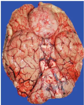
vomitus on her face. She had a history of medicine for depression and hyperlipidemia for two years. On postmortem inspection, there was no evidence of significant external and internal injury except for a focal subcutanous hemorrhage in the occipital area. At autopsy, the brain, weighing 1,500 gm, was soft and edematous with flattening of cerebral gyri (Fig. 1). A
large globular olfactory groove tumor, measuring 5.5
×5.0×4.5 cm, was found and the tumor was also attached to the base of frontal lobe. The medulla was compressed anteriorly and deformed by tonsils, which were consistent with tonsillar herniation (Fig. 2). The tumor was white gray and solid on cut sections and was well demarcated from the brain. A few foci of hemorrhages were in cut sections of the pons and medulla. All other organs were normal and no other abnormality was found. On microscopic examination,
http://dx.doi.org/10.7580/kjlm.2013.37.4.208 http://www.kjlm.or.kr
Pathologic Finding of Sudden Death by Meningioma│Jang-Hee Kim, et al. 209
Fig. 1. Cerebral hemisheres reveal marked edema and congestion.
Fig. 3. Meningotheliomatous meningioma shows brain invasion (H & E, ×20).
Fig. 2. A large meningioma is seen at the base of the frontal lobe. Also present is an anteriorly displacement of medulla with wrap by cerebellar tonsils.
Fig. 4.Mutifocal parenchymal hemorrhages are present in the pons (H & E, ×10).
the tumor was meningotheliomatous meningioma with brain invasion (WHO grade II) (Fig. 3). The brain parenchyme revealed marked edema. The pons and medulla revealed multifocal parenchymal hemorrhages (Fig. 4).
Discussion
Meningiomas are common tumor accounting for 20~30% of primary intracranial tumors.1)Most of the clinical symptoms and signs of meningiomas are caused by local effects of tumor mass. The tumors usually make some pressure on the underlying tissue with resulting damage to cortex or other parts of the brain. Meningiomas sometimes can be large enough or grow rapidly enough to produce generalized increased intracranial pressure.1-3)It may be associated with vascular occlusion, either venous or arterial.7) And it may provoke hemorrhage, edema, or cyst formation in neighboring tissue.3, 8, 9) Many meningiomas, however, are asymptomatic and are incidentally identified at neuro-imaging study or at autopsy.2, 4, 5)Incidence of meningioma found at general autopsy has been reported in the range of 1.4% to 2.4%.4, 5) The majority of incidentally detected meningiomas have been known to be indolent.2, 4)The autopsy cases of meningiomas which had been considered the chief cause of death are uncommon. A previous autopsy study found 172 meningiomas out of a total 11,793 autopies and only 11 of these 172 cases were directly related to patient’s death.4) Moreover, a sudden unexpected death caused by an incidental meningioma is very unusual. About dozen of autopsy cases associated with sudden death due to meningioma have been reported.6-8) Lee et al.7) reported a case of sudden death due to cerebral infarction associated with right frontal meningioma.
Ohaegbulam et al.6) demonstrated that a hemorrhage in sphenoid ridge meningioma could cause a sudden death. Huh et al.8) found pronounced edema in the brain adjacent to the tumor and assumed that peritumoral edema may be related to the sudden
death by intracranial meningioma. In our case, there was also marked cerebral edema producing flattening of gyri. Peritumoral edema has been known to be associated not only with presence of coexisting psychiatric disorders but also with an increase in the morbidity and mortality of patients with meningiomas.1, 3) Nakano et al.3) indicated that severe edema is associated with large tumor size, tumor vasculature, brain invasion, high grade, and the specific variants such as microcystic meningiomas.
Large tumor size and brain invasion could be possible causes of marked cerebral edema in our autopsy case, and the cause of the tonsillar herniation which was found at our autopsy also can be explained by large tumor and profound peritumoral edema. In forensic autopsy practices, it is important to look for evidence of cerebral herniations, especially tonsillar herniation, in cases of sudden death related to brain tumor.
However, interpretation of the presence or degree of tonsillar herniation sometimes may be difficult because of the anatomical variations of the skull base and an artificial impressions occurring after death and brain fixation. Nonetheless, the tonsillar herniations can be considered as a real and significant finding if the tonsils are prominently molded and pushed anteriorly to wrap around the medulla. Hemorrhages and necrosis of parenchyma, brain swelling and other parts of brain herination can support the diagnosis of tonsillar herniation.10) Therefore, in forensic autopsy practices, these findings should be carefully examined in cases of sudden unexpected death with brain tumor.
References
1. Perry A, Louis DN, Scheithauer BW, et al. Menigiomas. In:
Louis DN, Ohgaki H, Wiestler OD, et al, ed. WHO classifi- cation of tumors of the central nervous system. Lyon:
International Agency for Research on Cancer 2007:164-72.
2. Nakamura M, Roser F, Michel J, et al. The natural history of incidental meningiomas. Neurosurgery 2003;53:62-70.
3. Nakano T, Asano K, Miura H, et al. Meningioma with brain edema: radiologic characteristics on MRI and review of the literature. Clin Imaging 2002;26:243-9.
http://www.kjlm.or.kr http://dx.doi.org/10.7580/kjlm.2013.37.4.208
210 Korean Journal of Legal Medicine│2013;37:208-211
http://dx.doi.org/10.7580/kjlm.2013.37.4.208 http://www.kjlm.or.kr Pathologic Finding of Sudden Death by Meningioma│Jang-Hee Kim, et al. 211
4. Rausing A, Ybo W, Stenflo J. Intracranial meningioma-- a population study of ten years. Acta Neurol Scand 1970;46:102-10.
5. Nakasu S, Hirano A, Shimura T, et al. Incidental menin- giomas in autopsy study. Surg Neurol 1987;27:319-22.
6. Ohaegbulam SC. Sudden death from an asymptomatic sphenoid ridge meningioma. J Neurol 1977;215:291-4.
7. Lee BW, Seo JS. Meningioma presenting as cerebral in- farct: case report. Korean J Leg Med 2001;25:53-7.
8. Huh GY, Kim KH, Ahn YW, et al. Sudden death due to un- diagnosed intracranial meningioma: a case report. Korean J Leg Med 2008;32:150-2.
9. Helle TL, Conley FK. Haemorrhage associated with meningioma: a case report and review of the literature. J Neurol Neurosurg Psychiatry 1980;43:725-9.
10. Leestma JE, Kirkpatrick JB. Forensic neuropathology. 1st ed. New York: Raven; 1988. p. 157-83.