© 2017 The Korean Ophthalmological Society
This is an Open Access article distributed under the terms of the Creative Commons Attribution Non-Commercial License (http://creativecommons.org/licenses /by-nc/3.0/) which permits unrestricted non-commercial use, distribution, and reproduction in any medium, provided the original work is properly cited.
Original Article
Diseases such as age-related macular degeneration, dia- betic retinopathy, and branch retinal vein occlusion are se-
rious conditions that can decrease visual acuity and poten- tially lead to blindness [1,2]. An important factor in these diseases is neovascularization, which can be induced by hypoxia, ischemia, and inflammatory reactions. Vascular endothelial growth factor (VEGF), in particular, is thought to be a critical promoter of ophthalmic neovascularization [3-5]. VEGF is essential for neovascularization in the reti- na and choroid. During hypoxia, the expression of VEGF
Effects of Histone Deacetylase Inhibitor (Valproic Acid) on the Expression of Hypoxia-inducible Factor-1 Alpha in Human Retinal
Müller Cells
Young Jun Kim, Sang Jun Park, Na Rae Kim, Hee Seung Chin
Department of Ophthalmology and Inha Vision Science Laboratory, Inha University School of Medicine, Incheon, Korea
Purpose: To evaluate the effects of valproic acid (VPA), a histone deacetylase inhibitor (HDACI), on the expres- sion of hypoxia-inducible factor-1 alpha (HIF-1α) and vascular endothelial growth factor (VEGF) in human reti- nal Müller cells under hypoxic conditions.
Methods: Chemical hypoxia was induced in human retinal Müller cells (MIO-M1) by treatment with increasing concentrations of cobalt(Ⅱ) chloride (CoCl2). Müller cells were also treated with a set concentration of CoCl2, along with various concentrations of VPA. The expression of HIF-1α and VEGF in the treated Müller cells was determined by enzyme-linked immunosorbent assay.
Results: Exposure of human retinal Müller cells to increasing concentrations of CoCl2 produced a dose-depen- dent increase in HIF-1α expression. The addition of increasing concentrations of VPA lead to a dose-depen- dent decrease in expression of HIF-1α and VEGF in Müller cells exposed to a set concentration of CoCl2. Conclusions: HDACI VPA downregulated the expressions of HIF-1α and VEGF in human retinal Müller cells
under hypoxic conditions. Using HDACI to target HIF-1α expression in Müller cells could be a new therapeutic strategy for the treatment of retinal vascular diseases.
Key Words: Histone deacetylase inhibitors, Hypoxia-inducible factor 1 alpha, Retinal Muller cells, Valproic acid, Vascular endothelial growth factor
Received: December 21, 2015 Accepted: March 21, 2016
Corresponding Author: Hee Seung Chin, MD, PhD. Department of Ophthalmology, Inha University Hospital, #27 Inhang-ro, Jung-gu, Incheon 22332, Korea. Tel: 82-32-890-2400, Fax: 82-32-890-2417, E-mail:
is increased to improve vascularization and vascular per- meability [6,7]. Intravitreal injection of anti-VEGF has emerged as a promising treatment for neovasculariza- tion-associated ophthalmic disorders, with the drugs beva- cizumab (Avastin) and ranibizumab (Lucentis) widely used in clinical settings.
In the retina, VEGF is expressed in the Müller cells, pig- ment epithelium, endothelium, astrocytes, and ganglion cells [8-12]. Müller cells are distributed throughout the ret- inal layer, perform a variety of functions, and serve as im- portant mediators of neovascularization [13,14].
To adapt to hypoxic conditions, human cells and organs express a number of genes that affect neovascularization, metabolic processes, cell proliferation, and cell survival.
The primary mediator that controls the expression of these genes is hypoxia-inducible factor-1 alpha (HIF-1α), which regulates the production of VEGF [15,16]. HIF-1α is com- posed of a β and an α subunit, the latter of which reacts to oxygen. In the presence of normal oxygen concentrations, HIF-1α is degraded; however, in a hypoxic state, HIF-1α is not degraded and functions to alter the expression of more than 100 genes. HIF-1α is also involved in the regulation of vascular tone, cell proliferation, apoptosis, and other meta- bolic pathways [17]. Therapeutic agents that target HIF-1α directly could provide widespread control of VEGF-in- duced neovascularization, an important factor in retinal hypoxic diseases, and potentially serve as alternative ther- apeutics to drugs targeting only individual processes in hypoxia-associated disease.
Therapeutics that target HIF-1α have been studied thor- oughly in the field of cancer treatment and many agents that target HIF-1α directly have been developed [18-23].
Among these agents are histone deacetylase inhibitors (HDACIs), which inhibit the histone deacetylase enzyme.
This allows the induction of gene transcription by hyper- acetylation and reduces the activity of HIF-1α [24]. One of the HDAC inhibitors is valproic acid (VPA), which has been widely used for the treatment of epilepsy [25]. Sever- al studies have shown that VPA also inhibits tumor angio- genesis through suppression of angiogenic factors such as VEGF [26,27]. While HDACIs have been widely used in cancer treatment, these agents have yet to be applied to the treatment of ophthalmic disease [28-30].
In this study, the effects of the HDACI, VPA, on the ex- pression of HIF-1α and VEGF in human retinal Müller cells during hypoxia was evaluated.
Materials and Methods
Growth of cell lines
Human retinal Müller cells (MIO-M1) were grown in a humidified incubator at 37°C, 5% CO2, with DMEM + GlutaMAX-I (Gibco BRL, Grand Island, NY, USA) growth media containing 10% FBS (Gibco BRL), 100 U/mL peni- cillin, and 100 µg/mL streptomycin. Cells were provided with fresh media every 2 to 3 days.
Induction of hypoxia
Human retinal Müller cells were seeded at 5 × 10³ cells per well in 96-well cell culture plates. For enzyme-linked immunosorbent assay, the cells were seeded at 1.0 × 10⁵ cells per dish in 100 mm culture dishes. After 24 hours, the growth media was replaced with serum-free media and the cells were starved for 16 hours. Chemical hypoxia was in- duced by incubating cells for 24 hours in serum-free media containing various concentrations of cobalt(Ⅱ) chloride (CoCl2).
Cell viability assay
To investigate the optimal concentration of CoCl2 for in- ducing chemical hypoxia, human retinal Müller cells were exposed to 0, 100, 200, 300, 400, 500, 600, 700, 800, 900, or 1,000 µM CoCl2. The cells were then washed twice with phosphate buffered saline and the cell counting kit-8 (CCK-8; Dojindo Molecular Technologies, Rockville, MD, USA) was used to measure cell viability. CCK-8 measures the amount of live cells by measuring formazan that is produced by dehydrogenase in cells. Ten microliters of CCK-8 and 90 µL of serum-free media were mixed in each well. After 1 hour, optical density was measured by a mi- croplate reader at 450 nm wavelength.
VPA treatment
VPA 1 g/mL (Sigma-Aldrich, Silver Spring, MO, USA) was diluted serially in an aqueous solution of 0.1 N sodium hydroxide (NaOH) to 10 mg/mL and added to wells con- taining hypoxic Müller cells that were treated with 400 µM CoCl2. The wells were exposed to serum-free media for 22 hours with adequate concentrations of VPA (10, 25,
50, 75, 100 µg/mL). The control group (0 µg/mL) was treat- ed with the same amount of 0.1 N NaOH as the experi- mental groups.
Detection of VEGF and HIF-1α
VEGF and HIF-1α proteins were detected using the Quantikine enzyme-linked immunosorbent assay and Sur- veyor IC kits (R&D Systems, Minneapolis, MN, USA), re- spectively, according to the manufacturer’s instructions.
Statistical analyses
The mean ± standard deviation values were reported for the results of three independent experiments and all statis- tics were calculated using the IBM SPSS Statistics ver. 19.0 (IBM Co., Armonk, NY, USA). The Wilcoxon signed-rank test was used to compare differences between the experi- mental and control groups, with a p-value <0.05 consid- ered as statistically significant.
Results
Induction of chemical hypoxia and expression of HIF- 1α in human retinal Müller cells
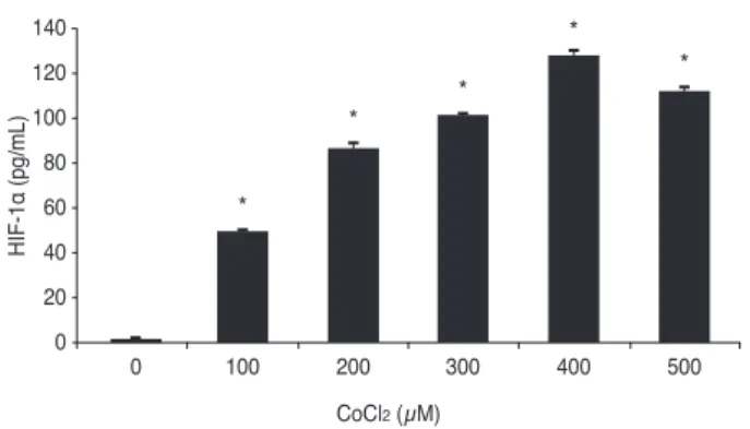
Chemical hypoxia was induced by treating human reti- nal Müller cells with 0, 100, 200, 300, 400, and 500 µM of CoCl2. A dose-dependent increase in the expression of HIF-1α was observed with increasing concentrations of
CoCl2, with maximal HIF-1α expression at 400 μM CoCl2
(Fig. 1).
Viability of human retinal Müller cells during chemical hypoxia
Müller cells were treated with 0, 100, 200, 300, 400, 500, 600, 700, 800, 900, and 1,000 µM CoCl2 and cell viability was assessed using the CCK-8 assay in order to determine the optimal concentration of CoCl2 to induce chemical hy- poxia without reducing cell viability. Cell viability de- creased gradually with increasing concentrations of CoCl2
(Fig. 2). Cell viability decreased even more at 500 µM, but the highest HIF-1α expression was observed with 400 µM of CoCl2. Therefore, the optimal concentration of CoCl2 to induce chemical hypoxia was selected to be 400 µM, based on the results for induction of HIF-1α expression as de- scribed above.
Effect of VPA treatment on the expression of HIF-1α and VEGF by human retinal Müller cells during hypoxia
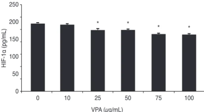
After exposing human retinal Müller cells to 400 μM CoCl2 to induce hypoxia, the cells were treated with 0, 10, 25, 50, 75, and 100 µg/mL of VPA. Expression of HIF-1α decreased significantly with increasing concentrations of VPA (p < 0.05), in a dose-dependent manner (Fig. 3). Sim- ilar results were found for the expression of VEGF (Fig. 4).
CoCl2 (µM)
HIF-1α (pg/mL)
140 120 100 80 60 40 20
0 0 100
⁎
200
⁎
300
⁎
400
⁎
500
⁎
Fig. 1. Effects of cobalt(Ⅱ) chloride (CoCl2) on the expression of hypoxia-inducible factor-1 alpha (HIF-1α) in human retinal Müller cells. *p-value < 0.05.
CCK-8 (%)
100110 9080 7060 5040 3020 100
CoCl2 (µM)
0 100 200 300 400 500 600 700 800 900
Fig. 2. Effects of cobalt(Ⅱ) chloride (CoCl2) on the survival of human retinal Müller cells. CCK-8 = cell counting kit-8.
Discussion
Neovascularization in the retina leads to vision loss and blindness in age-related macular degeneration, diabetic retinopathy, branch retinal vein occlusion, and other oph- thalmic disorders. The primary cause of this neovascular- ization is the physiological response to hypoxia. HIF-1α controls many of the reactions to hypoxia, as well as the expression of other genes, including placental growth factor, stromal-derived factor 1, angiopoietin 2, platelet- derived growth factor B, erythropoietin, and VEGF [31-33].
In ophthalmic diseases associated with neovasculariza- tion, improvements in vision have been demonstrated us- ing therapeutics that target VEGF. However, these im- provements appeared in only half of the patients and the effects were not long-lasting, requiring repeated, expensive treatments [34,35].
As a result, a number of studies have attempted to de- sign therapeutics targeting mediators other than VEGF.
HIF-1α, in particular, has been one of the more promising targets. As noted, many potential mechanisms are avail-
able to suppress HIF-1α. Studies of the pro-apoptotic, an- ti-angiogenic effects of HDACIs as anti-cancer therapeu- tics have demonstrated that HDACI-mediated suppression of HIF-1α has a significant anti-angiogenic effect [36-38].
The HDACI VPA has been used widely in the treatment of epilepsy and bipolar disorder and this compound has been found to have neuroprotective or neuroregenerative effects on retinal ganglion cells in these patients [39-41].
This study determined that VPA suppresses HIF-1α ex- pression by human retinal Müller cells during hypoxia (Fig. 3), suggesting that VPA could possibly be used to tar- get HIF-1α in humans. However, further studies are need- ed to determine the precise mechanism by which HDACIs suppress HIF-1α [42-44].
This study found that VPA suppresses VEGF expression in human retinal Müller cells during hypoxia (Fig. 4), like- ly due to suppression of HIF-1α, which normally activates transcription of the VEGF gene [45].
The HIFs are heterodimeric nuclear proteins consisting of α and β subunits. There are three types of oxygen-reac- tive α subunits: HIF-1α, HIF-2α, and HIF-3α. HIF-3α had not been studied extensively and it is currently unknown whether HIF-1α or HIF-2α has a greater affect in the retina [46,47]. This study analyzed HIF-1α, but further studies are needed to determine whether HIF-1α or HIF-2α is more important in human retinal Müller cells.
One of the limitations of this study is that targeting HIF- 1α is more non-specific than treatments targeting VEGF, because it controls the upper regulatory pathway. Treat- ments for specific targets would be better pharmacological- ly, but current widely used treatments that directly target VEGF have limitations, as mentioned above. Thus, con- trolling various hypoxia-induced pathways could be advan- tageous. This study identified some of the regulatory inter- actions between HIF-1α and VEGF, but further studies on other factors that are controlled by hypoxia are needed.
This was an in vitro study, so further in vivo studies should also be performed before making conclusions about the ac- tual effects of VPA on human retinal Müller cells.
In this study, CoCl2 was used to simulate the hypoxic state. One drawback of the cobalt model is that it rep- resents only the chronic activation state of HIF-1, although it is widely established and thought to be an appropriate model for hypoxic states. With this concern in mind, ex- periments using CoCl2 may not actually reflect a hypoxic state with respect to the upper regulatory level of HIF-1.
VPA (µg/mL)
VEGF (pg/mL)
600 500 400 300 200 100
0 0 10 25
⁎
50
⁎
75
⁎
100
⁎ ⁎
Fig. 4. Effects of increasing concentrations of valproic acid (VPA) on vascular endothelial growth factor (VEGF) expression by hu- man retinal Müller cells during hypoxia. *p-value < 0.05.
VPA (µg/mL)
HIF-1α (pg/mL)
250 200 150 100 50
0 0 10 25
⁎
50
⁎
75
⁎
100
⁎
Fig. 3. Effects of increasing concentrations of valproic acid (VPA) on hypoxia-inducible factor-1 alpha (HIF-1α) expression by human retinal Müller cells during hypoxia. *p-value < 0.05.
In summary, VPA was found to decrease the expression of HIF-1α and VEGF in human retinal Müller cells during hypoxia. Using VPA or other HDACIs to target HIF-1α in retinal Müller cells could be a potential therapeutic strate- gy for the treatment of retinal vascular diseases.
Conflict of Interest
No potential conflict of interest relevant to this article was reported.
Acknowledgements
Human retinal Müller cells (Moorfields/Institute of Oph- thalmology-Müller 1, MIO-M1) were provided generously by professor G. Astrid Limb (Institute of Ophthalmology, University College London, London, UK).
References
1. Congdon N, O’Colmain B, Klaver CC, et al. Causes and prevalence of visual impairment among adults in the Unit- ed States. Arch Ophthalmol 2004;122:477-85.
2. Congdon NG, Friedman DS, Lietman T. Important causes of vi- sual impairment in the world today. JAMA 2003;290:2057-60.
3. Stalmans I, Ng YS, Rohan R, et al. Arteriolar and venular patterning in retinas of mice selectively expressing VEGF isoforms. J Clin Invest 2002;109:327-36.
4. Haigh JJ, Morelli PI, Gerhardt H, et al. Cortical and retinal defects caused by dosage-dependent reductions in VEGF-A paracrine signaling. Dev Biol 2003;262:225-41.
5. Marneros AG, Fan J, Yokoyama Y, et al. Vascular endothe- lial growth factor expression in the retinal pigment epithe- lium is essential for choriocapillaris development and visu- al function. Am J Pathol 2005;167:1451-9.
6. Aiello LP, Avery RL, Arrigg PG, et al. Vascular endothelial growth factor in ocular fluid of patients with diabetic reti- nopathy and other retinal disorders. N Engl J Med 1994;331:1480-7.
7. Adamis AP, Miller JW, Bernal MT, et al. Increased vascu- lar endothelial growth factor levels in the vitreous of eyes with proliferative diabetic retinopathy. Am J Ophthalmol 1994;118:445-50.
8. Pierce EA, Avery RL, Foley ED, et al. Vascular endothelial growth factor/vascular permeability factor expression in a mouse model of retinal neovascularization. Proc Natl Acad Sci U S A 1995;92:905-9.
9. Miller JW, Adamis AP, Aiello LP. Vascular endothelial growth factor in ocular neovascularization and prolifera- tive diabetic retinopathy. Diabetes Metab Rev 1997;13:37- 50.
10. Aiello LP, Northrup JM, Keyt BA, et al. Hypoxic regula- tion of vascular endothelial growth factor in retinal cells.
Arch Ophthalmol 1995;113:1538-44.
11. Stone J, Itin A, Alon T, et al. Development of retinal vascu- lature is mediated by hypoxia-induced vascular endothelial growth factor (VEGF) expression by neuroglia. J Neurosci 1995;15(7 Pt 1):4738-47.
12. Stone J, Chan-Ling T, Pe’er J, et al. Roles of vascular endo- thelial growth factor and astrocyte degeneration in the gen- esis of retinopathy of prematurity. Invest Ophthalmol Vis Sci 1996;37:290-9.
13. Bai Y, Ma JX, Guo J, et al. Muller cell-derived VEGF is a significant contributor to retinal neovascularization. J Pathol 2009;219:446-54.
14. Wang J, Xu X, Elliott MH, et al. Muller cell-derived VEGF is essential for diabetes-induced retinal inflammation and vascular leakage. Diabetes 2010;59:2297-305.
15. Kaelin WG Jr. The von Hippel-Lindau tumor suppressor protein and clear cell renal carcinoma. Clin Cancer Res 2007;13:680s-684s.
16. Liao D, Johnson RS. Hypoxia: a key regulator of angiogen- esis in cancer. Cancer Metastasis Rev 2007;26:281-90.
17. Ke Q, Costa M. Hypoxia-inducible factor-1 (HIF-1). Mol Pharmacol 2006;70:1469-80.
18. Pili R, Donehower RC. Is HIF-1 alpha a valid therapeutic target? J Natl Cancer Inst 2003;95:498-9.
19. Welsh SJ, Powis G. Hypoxia inducible factor as a cancer drug target. Curr Cancer Drug Targets 2003;3:391-405.
20. Semenza GL. Targeting HIF-1 for cancer therapy. Nat Rev Cancer 2003;3:721-32.
21. Giaccia A, Siim BG, Johnson RS. HIF-1 as a target for drug development. Nat Rev Drug Discov 2003;2:803-11.
22. Powis G, Kirkpatrick L. Hypoxia inducible factor-1alpha as a cancer drug target. Mol Cancer Ther 2004;3:647-54.
23. Brown JM, Wilson WR. Exploiting tumour hypoxia in cancer treatment. Nat Rev Cancer 2004;4:437-47.
24. Poon E, Harris AL, Ashcroft M. Targeting the hypoxia-in- ducible factor (HIF) pathway in cancer. Expert Rev Mol
Med 2009;11:e26.
25. Gottlicher M, Minucci S, Zhu P, et al. Valproic acid defines a novel class of HDAC inhibitors inducing differentiation of transformed cells. EMBO J 2001;20:6969-78.
26. Zhang ZH, Hao CL, Liu P, et al. Valproic acid inhibits tu- mor angiogenesis in mice transplanted with Kasumi1 leu- kemia cells. Mol Med Rep 2014;9:443-9.
27. Shan Z, Feng-Nian R, Jie G, Ting Z. Effects of valproic acid on proliferation, apoptosis, angiogenesis and metasta- sis of ovarian cancer in vitro and in vivo. Asian Pac J Can- cer Prev 2012;13:3977-82.
28. Marks PA, Richon VM, Breslow R, Rifkind RA. Histone deacetylase inhibitors as new cancer drugs. Curr Opin On- col 2001;13:477-83.
29. Drummond DC, Noble CO, Kirpotin DB, et al. Clinical de- velopment of histone deacetylase inhibitors as anticancer agents. Annu Rev Pharmacol Toxicol 2005;45:495-528.
30. Johnstone RW, Licht JD. Histone deacetylase inhibitors in cancer therapy: is transcription the primary target? Cancer Cell 2003;4:13-8.
31. DeNiro M, Al-Halafi A, Al-Mohanna FH, et al. Pleiotropic effects of YC-1 selectively inhibit pathological retinal neo- vascularization and promote physiological revasculariza- tion in a mouse model of oxygen-induced retinopathy. Mol Pharmacol 2010;77:348-67.
32. Adams JM, Difazio LT, Rolandelli RH, et al. HIF-1: a key mediator in hypoxia. Acta Physiol Hung 2009;96:19-28.
33. Aiello LP, Pierce EA, Foley ED, et al. Suppression of reti- nal neovascularization in vivo by inhibition of vascular en- dothelial growth factor (VEGF) using soluble VEGF-re- ceptor chimeric proteins. Proc Natl Acad Sci U S A 1995;92:10457-61.
34. Rey S, Semenza GL. Hypoxia-inducible factor-1-dependent mechanisms of vascularization and vascular remodelling.
Cardiovasc Res 2010;86:236-42.
35. Brown DM, Kaiser PK, Michels M, et al. Ranibizumab versus verteporfin for neovascular age-related macular de- generation. N Engl J Med 2006;355:1432-44.
36. Yu F, White SB, Zhao Q, Lee FS. HIF-1alpha binding to VHL is regulated by stimulus-sensitive proline hydroxyl-
ation. Proc Natl Acad Sci U S A 2001;98:9630-5.
37. Mie Lee Y, Kim SH, Kim HS, et al. Inhibition of hypox- ia-induced angiogenesis by FK228, a specific histone deacetylase inhibitor, via suppression of HIF-1alpha activi- ty. Biochem Biophys Res Commun 2003;300:241-6.
38. Williams RJ. Trichostatin A, an inhibitor of histone deacetylase, inhibits hypoxia-induced angiogenesis. Expert Opin Investig Drugs 2001;10:1571-3.
39. Biermann J, Grieshaber P, Goebel U, et al. Valproic ac- id-mediated neuroprotection and regeneration in injured retinal ganglion cells. Invest Ophthalmol Vis Sci 2010;51:526-34.
40. Zhang Z, Qin X, Zhao X, et al. Valproic acid regulates an- tioxidant enzymes and prevents ischemia/reperfusion inju- ry in the rat retina. Curr Eye Res 2012;37:429-37.
41. Zhang Z, Qin X, Tong N, et al. Valproic acid-mediated neuroprotection in retinal ischemia injury via histone deacetylase inhibition and transcriptional activation. Exp Eye Res 2012;94:98-108.
42. Jeong JW, Bae MK, Ahn MY, et al. Regulation and desta- bilization of HIF-1alpha by ARD1-mediated acetylation.
Cell 2002;111:709-20.
43. Kim MS, Kwon HJ, Lee YM, et al. Histone deacetylases induce angiogenesis by negative regulation of tumor sup- pressor genes. Nat Med 2001;7:437-43.
44. Kong X, Lin Z, Liang D, et al. Histone deacetylase inhibi- tors induce VHL and ubiquitin-independent proteasomal degradation of hypoxia-inducible factor 1alpha. Mol Cell Biol 2006;26:2019-28.
45. Qian DZ, Kachhap SK, Collis SJ, et al. Class II histone deacetylases are associated with VHL-independent regula- tion of hypoxia-inducible factor 1 alpha. Cancer Res 2006;66:8814-21.
46. Lin M, Chen Y, Jin J, et al. Ischaemia-induced retinal neo- vascularisation and diabetic retinopathy in mice with con- ditional knockout of hypoxia-inducible factor-1 in retinal Muller cells. Diabetologia 2011;54:1554-66.
47. Mowat FM, Luhmann UF, Smith AJ, et al. HIF-1alpha and HIF-2alpha are differentially activated in distinct cell pop- ulations in retinal ischaemia. PLoS One 2010;5:e11103.