Veterinary Science
Simultaneous detection of Lawsonia intracellularis, Brachyspira hyodysenteriae and Salmonella spp. in swine intestinal specimens by multiplex polymerase chain reaction
Dong Kyun Suh
1, Jae Chan Song
2,*
1
Research Institute of Health and Environment, Daegu 706-841, Korea
2
College of Veterinary Medicine, Kyungpook National University, Daegu 702-701, Korea
A multiplex PCR assay was developed for the simultaneous detection of the etiologic agents associated with porcine proliferative enteropathies (PPE), swine dysentery (SD) and porcine salmonellosis (PS) in a single reaction using DNA from swine intestinal samples. Single and multiplex PCR amplification of DNA from Lawsonia intracellularis, Salmonella typhimurium and Brachyspira hyodysenteriae with each primer set produced fragments of the predicted size without any nonspecific amplification, 210-bp, 298-bp and 403-bp bands, respectively. The single PCR assay could detect as little as 100 pg of purified DNA of S. typhimurium and L. intracellularis, and 50 pg of B. hyodysenteriae, respectively. However, multiplex PCR turned out to be 10 times lower sensitivity with S. typhimurium compared with single PCR. With 23 swine intestinal specimens suspected of having PPE, SD and/or PS, the multiplex PCR assay showed identical results with conventional methods except one. In conclusion, this multiplex PCR is a feasible alternative to standard diagnostic methods for detection of L.
intracellularis, B. hyodysenteriae and Salmonella spp. from swine intestinal specimens.
Key words: Brachyspira hyodysenteriae , Lawsonia intracel- lularis , Multiplex PCR, Salmonella spp.
Introduction
Porcine proliferative enteropathies (PPE) caused by Lawsonia intracellularis, swine dysentery (SD) by Brachyspira hyodysenteriae and porcine salmonellosis (PS) are acknowledged as important diseases of suboptimal performance and mortality in grower-finisher pigs, causing tremendous financial loss due to death of pigs, decreased rate of growth and poor feed conversion [22]. PPE, also
known as ileitis, intestinal adenomatosis, or necrotic enteritis is a naturally occurring disease that can affect pigs from their weaning to young adult stage. The disease is of economic importance due to death loss, increased medication costs, poor weight gain and decreased feed conversion, etc.
Estimates of the reduction in the weight gain and feed conversion efficiency were 20 to 30% [14,21]. Salmonellosis is a worldwide problem and causes zoonotic disease. PS manifests itself in postweaning pigs of all ages, and is most often attributed to S. choleraesuis var. kunzendorf and S.
typhimurium . Infection in swine typically results in diarrhea with septicemia and pneumonia more common in older swine. Prevalence of Salmonella infection is widespread from 3 to 21% depending in part on which tissues were examined [24]. Diarrhea is the most consistent sign of SD.
As the diarrhea progresses, watery stools containing blood, mucus and shreds of white mucofibrinous exudate are seen, with concurrent staining of rear quarters [12]. SD also causes a tremendous financial loss due to the expenses for therapy because it seems to occur in a cyclic manner with 3 to 4 weeks intervals.
Diagnosis of PPE had been done by the observation of typical histopathological lesions characterized by the marked proliferation of immature enterocytes within crypts of intestine; affected cells were demonstrated by Warthin- Starry (silver) stains. However, the culture and isolation of this organism require specialized cell culture techniques [16,17,19]. Culture was also the most widely used tool for Salmonella detection. However, the current standard laboratory procedure to culture and identify Salmonella spp . takes approximately 4 to 7 days [8]. In addition, Salmonella serovars are not detectable in certain clinical samples that contain small numbers of organism. The diagnosis of SD is based on herd history, clinical signs, observation of characteristic intestinal lesions and isolation of B.
hyodysenteriae from feces or the intestine. Isolating B.
hyodysenteriae from other intestinal bacteria becomes more difficult when attempting to recover the organism from swine infected with B. hyodysenteriae but having chronic
*Corresponding author
Tel: +82-53-950-5958; Fax: +82-53-950-5955
E-mail: [email protected]
diarrhea or no diarrhea. Laboratory confirmation of B.
hyodysenteriae by culture is based upon colony morphology, pattern and intensity of hemolysis and other growth characteristics, all of which are very similar for nonpathogenic B. innocens . As a result, a definitive diagnosis of swine dysentery can be very challenging [13].
The advent of molecular techniques has allowed for the development of more rapid diagnostic test of pathogenic organism. Use of polymerase chain reaction (PCR) has been reported for the definitive identification of several pathogenic organisms mentioned above [4-7,15]. We have already developed one step PCR for detection of L. intracellularis from diagnostic sample [28]. This study further developed a multiplex PCR assay for simultaneous detection of the etiologic agents associated with PPE, SD and PS to reduce the preparation and analysis time required to identify multiple target sites in 1 assay. Also, we tested whether this assay can be a useful alternative to single PCR and used as a complement test for standard diagnostic method.
Materials and Methods
Bacterial strains and DNA
L. intracellularis genomic DNA, 11 Salmonella spp., 10 Brachyspira spp. including 8 field isolates and 3 other enteric bacteria were used in this study (Table 1). All bacterial strains were identified biochemically and serologically [3]. Chromosomal DNA of Salmonella spp . and other bacterial strains listed in Table 1 were isolated as previously described [1,23].
Preparation of DNA from intestinal specimens
A total of 23 porcine intestinal specimens consisting of feces and mucosal scrapings were obtained from the slaughter pigs and field cases of Youngnam province during the periods between 1997 and 2000. Culture and identification of Salmonella spp. and B. hyodysenteriae , and histopathological examinations of tissue specimens for L.
intracellularis were performed by standard techniques [3,18]. DNA from mucosal scrapings of swine intestinal specimens diagnosed as PPE was extracted by the method described by Jones et al. [2,17]. The ileal mucosa from pigs with PPE was scraped from the ileum and homogenized in tissue grinder. The homogenate was centrifuged, and the supernatant was filtered sequentially through 5 µ m, 1.2 µ m and 0.8 µ m filters. A 20% diatomaceous earth suspension (50 µ l) in 0.17 M HCl was vortexed with infected mucosal filtrate (50 µ l) in a sterile microcentrifuge tube containing 950 ml of lysis buffer consisting of 5 M guanidine thiocyanate (GuSCN), 22 mM EDTA, 0.05 M Tris-Cl (pH 6.4) and 0.65% Triton X-100. The lysis buffer was drawn off with a pipette, dried at 56
oC for 15 min and dissolved in TE buffer.
After centrifugation at 12,000 × g for 2 min, the supernatant was stored at − 20
oC. Fecal specimen (0.2 g) was suspended in lysis buffer, vortexed and was then centrifuged at 14,000 × g for 20 sec after standing for 1 hr at room temperature. The supernatant was placed in a tube containing 50 µ l of DE suspension. Further processing needed was the same as described above for the extraction of DNA from mucosal filtrate.
Oligonucleotides and PCR reaction
The primers for specific amplification of L. intracellularis , B. hyodysenteriae and Salmonella spp. by multiplex PCR assays were designed from Bioneer Co. (Korea) (Table 2).
The 50 µ l of PCR mixture contained 5 µ l of 10 × PCR buffer, 3 µ l of 25mM MgCl
2, 4 µ l of 10mM deoxynucleotide triphosphate mixture, 20 pmol of each primers, 1 µ l of each DNA template and 0.5 unit of Taq DNA polymerase (Takara, Japan). PCR amplification was conducted on a DNA thermocycler (Robocycler; Stratagene, USA). The initial mixture was heated to 94
oC for 5 min. This step was followed by 45 cycles, each consisting of denaturation at 95
oC for 30 sec, annealing at 56
oC for 30 sec and polymerization at 72
oC for 1 min, followed by additional polymerization at 72
oC for 5 min. The presence of PCR
Table 1. List of bacterial strains (DNA) and sources
Bacterial strains Sources Bacterial strains Sources
L.intracellularis DNA
S.enteritidis (D1) S.reading (B) S.durazzo (A) S.typhimurium (B) S. typhi (D) S.newport (C2) S.derby (B) S.muenchen (C2) S.montevideo (C1) S.choleraesuis (C1) S.meleagridis (E1)
NVRQS*
ATCC13076 ATCC11511 ATCC6967 ATCC29946 NVRQS NVRQS NVRQS
NVRQS (chicken) NVRQS (chicken) NVRQS (swine) NVRQS (chicken)
B.innocens
B.hyodysenteriae (B204) B.hyodysenteriae 1
B.hyodysenteriae 2
B.hyodysenteriae 3
B.hyodysenteriae 4
B.hyodysenteriae 5
B.hyodysenteriae 6
B.hyodysenteriae 7
B.hyodysenteriae 8
Escherichia coli (ML1410) Campylobacter jejuni Listeria monocytogens
ATCC29796 ATCC31287 swine swine swine swine swine swine swine swine NVRQS ATCC33560 ATCC15313
*NVRQS: National Veterinary Research and Quarantine Services, Ministry of Agriculture and Forestry, Korea
products was determined by electrophoresis of 5 ml of the amplified products in a 1.8% metaphore agarose gel with tris boric acid electrophoresis buffer (0.45 M Tris, 0.45 M boric acid, 0.01 M EDTA, pH 7.8) and visualized using the Eagle Eye II (Stratagene, USA) according to manufacturer's manual.
Sensitivity and specificity of multiplex PCR
To assess the minimal amount of DNA detectable by multiplex PCR, genomic DNA from L. intracellularis, B.
hyodysenteriae B204 and S. typhimurium ATCC 29946 were prepared by 10-fold serial dilutions from 100 ng to 10 pg and subjected to PCR reaction. For specificity determination, DNA from all strains of bacteria listed in Table 1 was used in DNA amplification. Negative control for clinical samples were collected from slaughter pigs of the farm from which there were no previous symptoms or history of PPE, SD and PS for last years since 1998. They were determined whether the 3 organisms were detected by the bacteriological cultures for Salmonella spp. and B.
hyodysenteriae , and by the histological examination for L.
intracellularis .
Cloning and sequencing of PCR product
To confirm the identity of the PCR products, they were purified by using GeneClean II kit (Invitrogen, USA) after agarose gel electrophoresis and then cloned into p Bluescript KS plasmid in Eco RV site. The 8 cloned each PCR products were sequenced by PCR sequencing method with TopTM DNA sequencing kit (Injae, Korea). The identities of the products were confirmed by comparison of the sequence with previous report obtained from the GenBank [9-11].
Results
PCR reaction
Single PCR amplification of purified DNA from L.
intracellularis, S. typhimurium and B. hyodysenteriae with each primer set resulted in a fragment of the predicted size:
210-bp, 298-bp and 403-bp bands, respectively (Fig. 1).
Also, multiplex PCR amplification of purified DNA from each species yielded products corresponding to the same molecular weight of DNA as single PCR. To determine the
minimal detectable concentration of each template DNA, PCR was conducted on serial dilutions of each purified DNA from 100 ng to 10 pg. The single PCR assay could detect as little as 100 pg of purified DNA for S. typhimurium and L. intracellularis , and 50 pg for B. hyodysenteriae, respectively. However, multiplex PCR resulted in a sensitivity 10 times lower with S. typhimurium (Fig. 2).
Subcloning of the amplified product from L. intracellularis, S. typhimurium ATCC 29946 and B. hyodenteriae B204 into pBluescript KS strain and sequencing of the product indicated that it had the identical sequences with previous report obtained from the GenBank [9-11].
Specificity and sensitivity of multiplex PCR
In order to analyze the specificity of the PCR assay, DNA isolated from each bacterial strains as well as DNA from several other bacterial strains including E. coli, Campylobacter spp. and Listeria monocytogenes which cause intestinal diseases in swine, was used as a template DNA in each PCR reactions. The PCR assay produced the expected DNA fragment with template DNA purified each bacterial strains, and did not produce any nonspecific amplified DNA fragments derived from other untargeted organisms listed in Table 1 (Figs. 3 and 4).
Table 2. Primers for multiplex PCR amplification of L. intracellularis, B. hyodysenteriae and Salmonella spp. from porcine intestinal specimens
Organism Name Sequences (forward; reverse) Size
L. intracellularis LIR LIR 5'-GCAGCACTTGCAAACAATAAACT-3' 5'-TTCTCCTTTCTCATGTCCCATAA-3' 210 bp B. hyodysenteriae BHR BHF 5'-GCTGGAGATGATGCTTCTGG-3' 5'-GTCCAAGAGCTTGGCTGTTC-3' 403 bp Salmonella spp. SAR SAF 5'-TTGGTGTTTATGGGGTCGTT-3' 5'-GGGCATACCATCCAGAGAAA-3' 298 bp
Fig. 1. Single PCR amplified DNA pattern of L. intracellularis, S. typhimurium and B. hyodysenteriae with 45 cycles at different annealing temperature. M; øX174 digested by Hae III, Lane 1~4;
L. intracellularis template DNA at 52
oC, 54
oC, 56
oC and 58
oC,
respectively, Lane 5~8; S. typhimurium template DNA at
different DNA at 52
oC, 54
oC, 56
oC and 58
oC, respectively, Lane
9~12; B. hyodysenteriae template DNA at 52
oC, 54
oC, 56
oC and
58
oC, respectively.
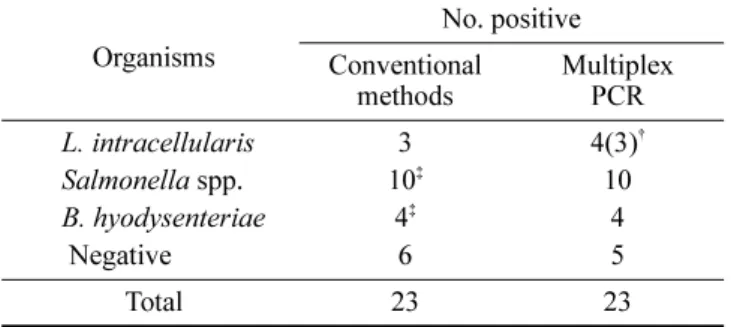
Evaluation of clinical samples by multiplex PCR A total of 23 swine intestinal samples suspected of having PPE, SD and/or PS alone or in combinations were screened by cultivation and/or histopathological examination and multiplex PCR. Multiplex PCR assay yielded PCR products alone or in combination corresponding to the expected molecular weight of DNA from L. intracellularis, Salmonella spp. and B. hyodysenteriae : 210-bp, 298-bp and 403-bp bands, respectively (Fig. 5). Out of the 23 specimens, all specimens had multiplex PCR assay results that corresponded to the results of conventional method except one sample. It was negative with L. intracellularis by histopathological examination of tissues, but positive with multiplex PCR (Table 3).
Discussion
Recently, DNA sequences unique to each of the bacterial agents associated with PPE, SD and PS were independently identified. Moreover, DNA analysis techniques have been shown to be more sensitive than standard diagnostic methods for L. intracellularis, B. hyodysenteriae and Salmonella spp.
[6,17,26]. In terms of each single PCR, PCR/Southern hybridization and PCR assay for the detection of L.
intracellularis specific DNA have proven to be more sensitive than other conventional methods [7,17]. However, the PCR methods particularly focused on nested PCR to confirm the amplified PCR products which was laborious and time consuming. To minimize this problem, the present
Fig. 2. Sensitivity of the amplified DNA from L. intracellularis, B. hyodesenteriae, and S. typhimurium by multiplex PCR. Each template DNA was serially diluted. M; øX174 digested by Hae III, Lane 1; 100 ng of template DNA, Lane 2; 10 ng of DNA, Lane 3; 1 ng of DNA, Lane 4; 100 pg of DNA, Lane 5; 50 pg of DNA.
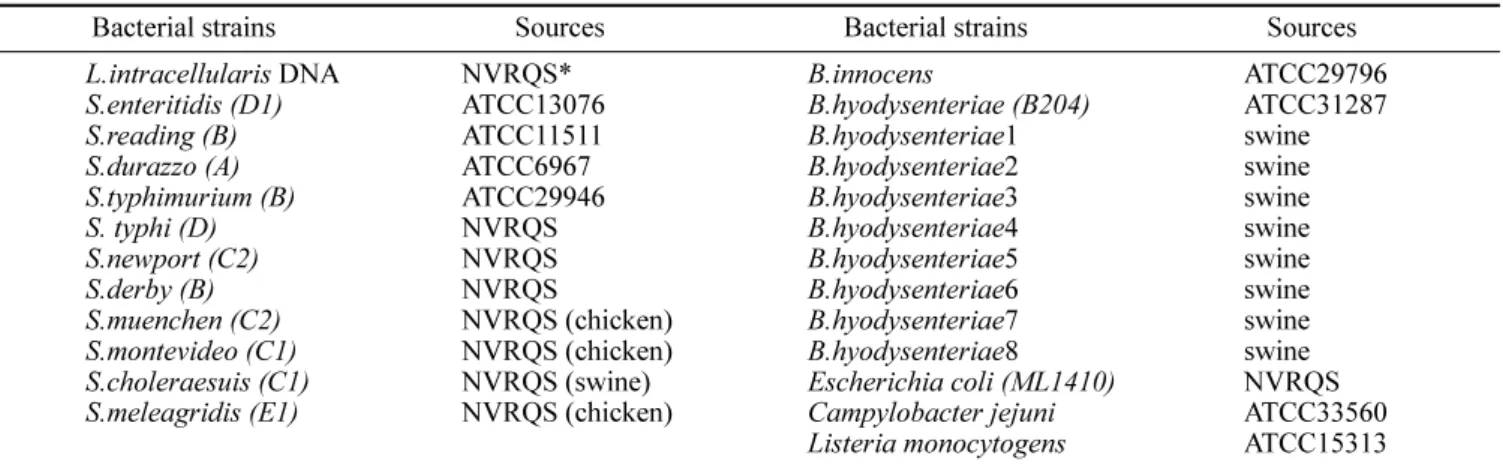
Fig. 3. Specificity of the multiplex PCR for detection of B.
hyodysenteriae with 45 cycles. M; øX174 digested by Hae III, Lane 1; B. hyodysenteriae ATCC31287, Lane 2~9; B. hyodysenteriae isolates, Lane 10; B. innocens, Lane 11; Campylobacter jejuni, Lane 12; Escherichia coli , Lane 13; Listeria monocytogens .
Fig. 4. Specificity of multiplex PCR for detection of L.
intracellularis and Salmonella spp . M; øX174 digested by Hae III, Lane 1; L. intracellularis, Lane 2~12; Salmonella spp . listed in Table 1., Lane 13; Listeria monocytogenes, LaLane 14;
Campylobacter jejuni, Lane 15; E. coli.
Fig. 5. Results of the multiplex PCR for detection of individual or combinations of bacterial agents from porcine intestinal feces.
M; øX174 digested by Hae III, Lane 1; L. intracellularis DNA, Lane 2; S, typhimurium, Lane 3; B. hyodysenteriae , Lane 4; L.
intracellularis & B. hyodysenteriae, Lane 5; L. intracellularis &
S. typhimurium, Lane 6; S, typhimurium & B. hyodysenteriae,
Lane 7; L. intracellularis & S. typhimurium & B. hyodysenteriae
(sample from herd No. 6).
study reconstructed the previously reported PCR analysis system, which included synthesis of DNA primers, annealing temperature and the number of reaction cycles.
The one step PCR assay could detect a predicted 210-bp PCR product, which was identical to the source DNA sequences without the reamplification step of PCR product.
Jones et al. [17] detect as few as 10
1and 10
3L.
intracellularis from 1 cm
2of intestinal mucosa and one g of feces, respectively. However, a nonspecific band was detected from the PCR product amplified with fecal DNA.
The numbers of amplification cycles are one of the important factors for increasing the sensitivity in PCR and might produce nonspecific DNA. However, nonspecific DNA was not detected in this study despite an increase in the amplification cycles from over 45 cycles. The increased sensitivity of this PCR protocol was about 10 times over that previously reported and likely due to the increase of the number of PCR cycles [20].
Sensitivity of the PCR for detection of Salmonella spp.
was up to 100 pg of DNA. This was comparable to an earlier report in which 27 pg of purified chromosomal DNA were needed for detection of Salmonella spp. by PCR [25]. This sensitivity was lower than the detection limit of 330 fg by Nguyen et al. [23]. Earlier studies have described PCR- based probes for detection of Salmonella spp. [4,25,29].
There seems to a limitation of this PCR that it does not differentiate Salmonella spp. at the level of species because we used Salmonella common primer set from invA gene, which enable Salmonella spp. to invade the cell. Only a limited number of serotypes have been associated with PS and swine sources, such as serotypes choleraesuis , typhimurium and heidelberg though the genus Salmonella comprises more than 2,400 serotypes [27]. Due to its rapidity and sensitivity, however, this PCR can be useful in a Salmonella spp. reduction program of swine production.
Previous study reported a PCR assay for B. hyodysenteriae on the basis of sequence analysis of a recombinant clone designated pRED3C6, with a sensitivity between 1 and 10 organisms per 0.1 g of feces [6]. Sensitivity of the PCR in this study was 50 pg of DNA: a little lower than that of Elder
et al [6]. Further studies are required to increase the sensitivity, and examine the specificity with others such as B. pilosicoli, B. intermedia and B. murdochii.
Elder et al. [7] also developed a multiplex PCR for detection of L. intracellularis, B. hyodysenteriae and Salmonella spp. based on the results of a single PCR of each organism [6,17,26]. The sensitivity and specificity of multiplex PCR results compared with the results of standard culture/histopathology for detection of the 3 bacterial agents from a total of 79 porcine intestinal specimens were 100%.
The detection limit of its single PCR was higher than that in this study. However, that of the multiplex PCR could not be compared to each other because it was not tested in the earlier report. The present study developed a multiplex PCR assay to detect L. intracellularis, B. hyodysenteriae and Salmonella spp . simultaneously from porcine intestinal specimens based on the results of a single PCR of each organism. The major advantages of multiplex PCR over conventional PCR are the conservation of reagents (such as polymerase) and template, and the reduction in preparation and analysis time required to identify multiple target sites in 1 assay as opposed to running separate analysis for each target. Attempts to detect each species by multiplex PCR had some difficulties in setting the PCR condition such as annealing temperature and the numbers of PCR cycles. We first set 52
oC as the annealing temperature when designing each single PCR primer set, and tested the broad range between 50~60
oC in each PCR. The results showed best condition between 54~56
oC of annealing temperature, but unclear band patterns with 50
oC and 60
oC depending on the single PCR (data not shown). The most specific and clear band was detected at 56
oC when the multiplex PCR was tested between 52~58
oC. Results of a single PCR assay could detect as little as 100 pg, 50 pg and 10 pg of purified chromosomal DNA from L. intracellularis , B. hyodysenteriae and S. typhimurium , respectively. However, the multiplex PCR resulted in a sensitivity of 10 times lower with S.
typhimurium and same range with L. intracellularis and B.
hyodysenteriae. This result might be explained by resulting from the combination of 3 primer sequences with a template from certain other organisms or by interference from PCR inhibitors.
There has been little recent information on the prevalence of 3 major enteric organisms affecting finishing pigs in Korea, specially no data for B. hyodysenteriae . We have developed this multiplex PCR to determine the prevalence of those organisms mentioned above with clinical field samples, which will be published later. The present study applied this method to clinical samples suspected of having PPE, SD and PS alone or in combination whether the multiplex PCR was available for screening the prevalence of L. intracellularis, B. hyodysenteriae and Salmonella spp. on pig farms. The accuracy for the detection of each organism using multiplex PCR results was increased compared with
Table 3. Results of conventional methods and multiplex PCR for detection of 3 organisms from porcine intestinal specimens
Organisms No. positive
Conventional
methods Multiplex PCR
L. intracellularis 3 4(3)
†Salmonella spp. 10
‡10
B. hyodysenteriae 4
‡4
Negative 6 5
Total 23 23
†
No. positive in feces
‡