DOI 10.3349/ymj.2008.49.6.931
Purpose: To assess the estimate prevalence and risk factors for age-related maculopathy (ARM) in Seoul, Korea. Patients and Methods: We examined 9,530 subjects with, 40 years of age or older between January 2006 and December 2006 in Seoul, Korea. Subjects underwent fundus photography, clinical examinations (including blood analyses), and completed detailed questionnaires. Fundus images were graded according to definitions from the Wisconsin Age-Related Maculopathy Grading System. Results: ARM was present in 235 subjects, corresponding to an estimate prevalence of 2.46%. Hepatitis B infection (positive status for HBsAg and HBcAb), serum triglyceride levels and high density lipoprotein levels remained as significant risk factors after age-adjustment. Multivariate analyses showed that the prevalence of ARM was significantly higher in older subjects [odds ratio (OR) 1.134; 95% CI 1.114- 1.154] and those who were seropositive for hepatitis B surface antigen (OR 2.566; 95% CI 1.519-4.335). Conclusion: The estimated prevalence of ARM was 2.46%. Age and hepatitis B infection may increase the risk of ARM.
Key Word: Age-related maculopathy, hepatitis B, triglyceride, high-density lipoprotein
INTRODUCTION
Age-related maculopathy (ARM) is the main cause of adult blindness in developed countries.1 Although there is considerable information on the nature and prevalence of visual impairment and blindness in Western countries, only a few popula- tion-based articles have appeared, describing the prevalence of ARM and the association of ARM
with various patient characteristics in different racial and ethnic groups in Western countries.2-5 Only limited population-based data on the pre- valence, characteristics, and risk factors for ARM in Asian populations are available.6-13
Studies from Western countries implicate lifestyle choices, nutritional parameters, and genetic risk factors in the pathogenesis of ARM.14Known risk factors for developing ARM are age,14-18 smoking,
14-17,19-21and hypertension.22-24 Hyperlipidemia and fat intake are related to the presence of exudative ARM,24 and obesity25-28 and atherosclerosis24 con- tribute to ARM development in some studies.
Asian lifestyles and nutritional choices are differ- ent, however, from those of Western populations.
The purpose of this report is to provide an estimate of the prevalence of ARM in a large survey sample, and to evaluate possible risk factors for ARM in the South Korean population.
PATIENTS AND METHODS
The Yonsei Eye Study was carried out in Seoul, which is the largest city in South Korea. The Medical Ethics Committee of the Yonsei Uni- versity medical center approved the study protocol, and all participants were well-briefed on the pur- poses of the study before giving written informed consent in accordance with the tenets of the Declaration of Helsinki. Baseline interviews and ophthalmic examinations, with concurrent general medical examinations, took place between January 2006 and December 2006. Of 13,016 participants who came in for a health check-up during that period, 9,530 subjects (73.2%) were 40 years old.
All examinations were carried out at the Yonsei
Estimated Prevalence and Risk Factor for Age-related Maculopathy
Mi In Roh, Ji Hyun Kim, Suk Ho Byeon, Hyoung Jun Koh, Sung Chul Lee, and Oh Woong Kwon Department of Ophthalmology, The Institute of Vision Research, Yonsei University College of Medicine, Seoul, Korea.
Received February 22, 2008 Accepted May 21, 2008
Reprint address: requests to Dr. Oh Woong Kwon, Department of Ophthalmology, Yonsei University College of Medicine, 250 Seongsanno, Seodaemun-gu, Seoul 120-752, Korea. Tel: 82-2-2228- 3573, Fax: 82-2-312-0541, E-mail: [email protected]
Medical Examination Center in Seoul, Korea. After measuring visual acuities and intraocular pressures (using a Type WT-200 instrument; Canon Inc, Tokyo, Japan), digital photographs of the maculae and optic discs were taken using a digital fundus camera (model TRC.NW 100; Topcon, Tokyo, Japan). Medical examinations were performed, which included blood analyses, urine analyses, and electrocardiograms. Additionally, the educa- tion level, income status, smoking habits, and illness history of each subject were assessed by questionnaire. Subjects were asked to report exist- ing or previous diagnoses of diabetes mellitus, hypertension, cardiac disease, or hepatic disease.
Ophthalmic examination
For the assessment of ARM, the Wisconsin Age- Related Maculopathy Grading system was used, as described previously in detail.29,30 Fundus photo- graphs were examined by ophthalmologists with special training in retinal diseases (MIR, JHK).
Intergrader and intragrader agreement was assess- ed using the quadratic weighted kappa statistic on a random subset of 30 subjects. There was excellent inter-and intraobserver agreement (κ = 0.82) for the grading of fundus photographs. Senior investiga- tors also reviewed all photographs graded as late ARM (ARM 4). Grading of the fundus photographs was based on the staging systems developed by the investigators of the Rotterdam Eye Study31: ARM 0, no feature characteristic of early ARM, or hard drusen only; ARM 1, either soft drusen or pigmentary irregularities only; ARM 2, either soft, indistinct ( 125 m) or reticular drusen only, orμ soft, distinct, drusen with pigmentary irregul- arities; ARM 3, soft, indistinct, or reticular drusen with pigmentary irregularities; and ARM 4, neo- vascular ARM or geographic atrophy. Neovascular ARM was considered to be present if any of the following lesions were found: serous or hemor- rhagic retinal pigment epithelial detachment, subretinal neovascular membrane, or subretinal hemorrhage. Geographic atrophy was considered to be present if preretinal fibrous scarring or retinal pigment atrophy with visible choroidal vessels was found. We defined ARM stage 2 and 3 as early ARM and stage 4 as late ARM. Subjects were ex- cluded from the study whose fundi could not be
graded because of poor photograph quality or dense ocular media.
Data analysis
For smoking analysis, subjects were categorized as non-smokers (those who had never smoked), current smokers, and prior smokers (those who had smoked earlier but were not smoking for 6 months at the time of the study). Education level was graded on the basis of final education level attained, as follows: 0, no school education; 1, graduated from elementary school; 2, graduated from middle school; 3, graduated from high school;
4, bachelors degree from college and, 5; masters degree from college. Monthly income was divided into 6 categories: 1, monthly income less than 1.5 million won; 2, monthly income between 1.5 million and 2.5 million won; 3, monthly income between 2.5 million and 3.5 million won; 4, month- ly income between 3.5 million and 4.5 million won;
5, monthly income between 5.5 million won and 6.5 million won, and; 6, monthly income more than 6.5 million won. Hypertension and hepatitis were considered to be present when subjects self- reported these conditions, and diabetes was con- sidered to be present when subjects self-reported the condition and the self-report was confirmed by a measurement of fasting blood glucose level > 126 mg/dL.
Statistical analyses
Student t-test was used for analyses of con- tinuous variables, and the Pearson 2test was also employed for nominal variables. Age-adjusted logistic regression analysis was performed to determine risk factors for early and late ARM, using odds ratio calculations, and estimations of the 95% confidence intervals. We then developed multivariable models using the following pro- cedure. The initial specification included indepen- dent variables consistently identified in previous research as risk factors for early and late ARM, or independent variables identified in univariate analyses as related to outcomes (p > 0.10). We then sequentially removed non-significant (p > 0.15) covariates, making it certain that these eliminations did not change estimated coefficients of the ex-
posure variable by more than 10%.32 Age and gender were adjusted regardless of association.
Because our models might have been overspeci- fied, given the limited number of ARM in the study group, we also constructed reduced form models by selecting covariates, based on their statistical significance in the adjusted models. SPSS version 12 (SPSS Inc, Chicago, IL, USA) was used to per- form statistical analyses. A two sided p value <
0.05 was considered to be statistically significant.
RESULTS Study population
A total of 13,016 potential participants (7,109 men and 5,770 women), who underwent examina- tions at the Yonsei Medical Examination Center between January 1, 2006 and December 31, 2006, were identified. Of the participants, 9,530 (73.2%) were aged 40 years, and underwent full clinical examinations. Of these, 9,468 (99.3%) completed all study procedures, including dilated fundus photo-
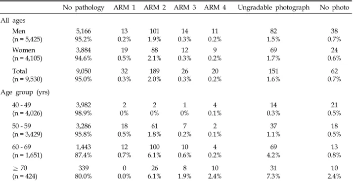
graphy. Fundus images were not available for 62 (0.7%) participants because subjects refused to be photographed, and poor-quality, unusable photo- graphs were obtained from 151 (1.6%) subjects because of corneal opacities, phthisis bulbi, or small pupils. Table 1 shows the prevalence of ARM by gender and age. Percentages of the population in various age groups were 42.3% in the group aged 40 - 49 years, 36.0% in the group aged 50 - 59 years, 17.3% in the group aged 60 - 69 years, and 4.5% in the group aged 70 years. ARM was seen in 235 (2.46%) subjects. Estimated prevalence of early ARM (ARM 2 and ARM 3) and late ARM (ARM 4) increased from 0% and 0.1%, respectively, in the 40 - 49 year age group, to 2.0% and 0.1% in the group aged 50 - 59 years, to 6.7% and 0.2% in the group aged 60 - 69 years, and to 8.0% and 2.4%
in subjects aged 70 years.
Mean age of the subjects with ARM was 63.17
± 7.45 years[mean ± standard deviation (SD)]. One hundred and twenty-six (53.6%) ARM patients were male and 109 (46.4%) were female, thus males showing a slight predominance. Early ARM and late ARM were present in 2.3% (215 subjects) and
Table 1. Numbers of Subjects over 40 Years of Age with ARM, and ARM Prevalence Rates (%)
No pathology ARM 1 ARM 2 ARM 3 ARM 4 Ungradable photograph No photo All ages
Men
(n = 5,425) 5,166
95.2% 13
0.2% 101
1.9% 14
0.3% 11
0.2% 82
1.5% 38
0.7%
Women
(n = 4,105) 3,884
94.6% 19
0.5% 88
2.1% 12
0.3% 9
0.2% 69
1.7% 24
0.6%
Total
(n = 9,530) 9,050
95.0% 32
0.3% 189
2.0% 26
0.3% 20
0.2% 151
1.6% 62
0.7%
Age group (yrs) 40 - 49
(n = 4,026) 3,982
98.9% 2
0% 2
0% 1
0% 4
0.1% 14
0.3% 21
0.5%
50 - 59
(n = 3,429) 3,286
95.8% 18
0.5% 61
1.8% 7
0.2% 2
0.1% 37
1.1% 18
0.5%
60 - 69
(n = 1,651) 1,443
87.4% 12
0.7% 100
6.1% 10
0.6% 4
0.2% 69
4.2% 13
0.8%
70
(n = 424) 339
80.0% 0
0.0% 26
6.1% 8
1.9% 10
2.4% 31
7.3% 10
2.4%
ARM, age-related maculopathy.
0, no features of early ARM, or hard drusen only; 1, either soft drusen or pigmentary irregularities only; 2, either soft, indistinct ( 125 m), or reticular drusen only, or soft, distinct, drusen with pigmentary irregularities; 3, soft, indistinct, or reticular drusen,μ with pigmentary irregularities; 4, neovascular ARM or geographic atrophy.
0.2% (20 subjects) of participants, respectively.
Among these, ARM was bilateral in 123 subjects (52.3%) and unilateral in 112 subjects (47.7%).
Among bilateral subjects, geographic atrophy was found in both eyes in a single subject, and 2 subjects each showed geographic atrophy in 1 eye and multiple drusens in the other eye. The remain- ing subjects showed multiple drusens in both eyes with or without pigment abnormalities. Among the unilateral ARM subjects, 17 (15.2%) eyes showed late ARM (neovascular AMD in 5 subjects and geographic atrophy in 12 subjects), and remaining 95 eyes showed early ARM. Among ARM subjects, there were no differences in dis- tributions of ARM features between males and females (ARM 2, ARM 3, and ARM 4) (Pearson chi-square test, p = 0.991), or late ARM features (Pearson chi-square test, p = 0.897). Bilateral ARM was more prevalent in females (Pearson chi-square test, p = 0.011).
Neovascular AMD was found in 5 subjects.
Among these, 2 patients showed subretinal hemor- rhages with exudates, a single subject showed serous pigment epithelial detachment with hyper- pigmentation, and 2 subjects had large subretinal hemorrhages with orange nodules.
Risk factors for ARM
When Student t-test and Pearson 2 test were used for analyses of variables, the following risk factors for early and late ARM (ARM 2, 3 and 4) were subjected to age-adjusted univariate analysis:
gender, socioeconomic status (monthly income and education level), smoking history, self-reported diagnosis of hypertension, self-reported diagnosis of cerebral vascular accident, self-reported diagno- sis of hepatitis, and body mass index. Serum levels of cholesterol, triglycerides (TG), high-density lipoprotein (HDL), low-density lipoprotein (LDL), lactate dehydrogenase (LDH) and C-reactive protein (CRP) were measured. In addition, serum hepatitis B surface antigen status (HBsAg), hepa- titis B core antibody level (HBcAb), hepatitis B surface antibody level (HBsAb), and hepatitis C antibody level (HCVAb) were also assessed.
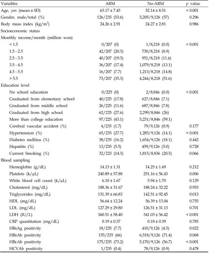
Subjects with ARM were significantly older (p
< 0.001), and monthly income and final education level were lower than subjects without ARM. Also,
while 27% of the ARM subjects had hypertension, only 14.1% of the subjects without ARM had hyper- tension history (p < 0.001). As for blood sample analysis, significantly different platelet, LDH, and TG levels were found with ARM subjects (Table 2).
Therefore, estimated prevalence of ARM was strongly age-related (logistic regression; p < 0.001;
OR 1.134; 95% CI; 1.114 - 1.154), as expected. The results of age-adjusted logistic regression analyses of risk factors for ARM are listed in Table 3. After adjusting for age, hepatitis B infection (positive status for HBsAg and HBcAb) and serum trigly- ceride levels and HDL levels remained as signi- ficant risk factors for ARM. Multivariate regres- sion analysis indicated that serum HBsAg-posi- tive status was significant and associated with a
> 2-fold increase in the risk of ARM, relative to normal macular subjects, in both the adjusted (OR 2.55 ; 95% CI 1.476 - 4.421) and reduced (OR 2.736;
95% CI 1.588 - 4.714) analyses (Table 4).
DISCUSSION
We estimated the prevalence and risk factors for ARM in Seoul, Korea. In our study population, 2.46% of participants had signs of early or late ARM in one or both eyes, suggesting that ARM is relatively rare among adult South Koreans in Seoul. A total of 2.26% of the study subjects showed early ARM, defined as soft indistinct drusen with or without pigmentation abnor- malities. Late ARM (defined as neovascular AMD or geographic atrophy) was seen in 0.2% of the study population. Among those with late ARM, neovascular AMD in at least one eye was seen in 5 subjects and geographic atrophy in 15 subjects.
Although direct comparison is difficult due to the fact that our studies showed estimate pre- valence of ARM, these estimates of early and late ARM are lower than the incidence rates observed in Western countries. In the Beaver Dam study,33 the prevalence of indistinct soft drusen was 8.4%
and patients with either pure geographic atrophy or exudative macular degeneration constituted 0.6% of the study population. The Framingham Eye Study34showed an incidence of 1.5% of exuda- tive macular degeneration in people 52 years of age. In a population-based prevalence study (the
Table 2. Statistical Data on Risk Factors for Age-related Maculopathy
Variables ARM No-ARM p value
Age, yrs (mean ± SD) 63.17 ± 7.45 52.14 ± 8.51 < 0.001
Gender, male/total (%) 126/235 (53.6) 5,205/9,126 (57) 0.296
Body mass index (kg/m2) 24.26 ± 2.91 24.27 ± 2.81 0.986
Socioeconomic status
Monthly income/month (million won)
< 1.5 0/207 (0) 1/8,218 (0.0) < 0.001
1.5 - 2.5 42/207 (20.3) 730/8,218 (8.9)
2.5 - 3.5 40/207 (19.3) 951/8,218 (11.6)
3.5 - 4.5 36/207 (17.4) 1,079/8,218 (13.1)
4.5 - 5.5 16/207 (7.7) 1,213/8,218 (14.8)
> 5.5 73/207 (35.3) 4,244/8,218 (51.6)
Education level
No school education 0/225 (0) 2/8,846 (0.0) < 0.001
Graduated from elementary school 40/225 (17.8) 627/8,846 (7.1) Graduated from middle school 26/225 (11.6) 687/8,846 (7.8) Graduated from high school 62/225 (27.6) 2,299/8,846 (26) More than college education 97/225 (43.1) 5,231/8,846 (59.1)
Cerebral vascular accident (%) 4/235 (1.7) 79/9,126 (0.9) 0.177
Hypertension (%) 65/235 (27.7) 1,285/9,126 (14.1) < 0.001
Diabetes mellitus (%) 38/235 (16.2) 1,654/9,126 (18.1) 0.442
Hepatitis (%) 13/235 (5.5) 459/9,126 (5.0) 0.728
Current Smoking (%) 32/223 (14.3) 1,815/8,836 (20.5) 0.066
Blood sampling
Hemoglobin (g/dL) 14.13 ± 1.31 14.25 ± 1.49 0.212
Platelets (k/µL) 240.89 ± 57.88 251.16 ± 56.43 0.006
White blood cell count (k/µL) 6.10 ± 1.67 5.94 ± 1.70 0.129
Cholesterol (mg/dL) 188.36 ± 31.67 188.24 ± 32.22 0.953
Triglycerides (mg/dL) 131.39 ± 66.83 142.51 ± 92.45 0.013
HDL (mg/dL) 56.64 ± 12.24 56.39 ± 13.04 0.755
LDL (mg/dL) 127.29 ± 29.80 126.51 ± 31.13 0.701
LDH (IU/L) 360.51 ± 58.40 341.03 ± 56.42 < 0.001
CRP quantitation (mg/dL) 0.19 ± 0.37 0.18 ± 0.39 0.785
HBsAg positivity 18/235 (7.7) 410/9,126 (4.5) 0.022
HBsAb positivity 155/235 (66) 6,518/9,126 (71.4) 0.068
HBcAb positivity 175/235 (73.2) 5,170/9,126 (56.7) < 0.001
HCVAb positivity 1/235 (0.4) 78/9,126 (0.9) 0.478
ARM, age related maculopathy = ARM2 + ARM3 + ARM4; SD, standard deviation; HDL, high-density lipoprotein; LDL, low- density lipoprotein; LDH, lactate dehydrogenase; CRP, C-reactive protein; HBsAg, hepatitis B surface antigen; HBsAb, hepatitis B surface antibody; HBcAb, hepatitis B core antibody; HCVAb, hepatitis C antibody.
Chi-square test was used for nominal variables.
Student's t test was used for continuous variables.
Beijing Eye Study), Li et al.13 found that the overall prevalence rates of early and late ARM were 1.4%
and 0.2%, respectively, and these data are similar to our results. The prevalence of late ARM in the black population was 0.59% in a Barbados study,4
and was 0.5% in Hispanic individuals aged 50 years in Arizona.35
The prevalence of early ARM obtained in the present study on South Koreans living in the Seoul area was slightly higher (2.46% vs. 1.4%) than in Table 3. Age-adjusted Univariate Logistic Analyses to Find Associations of Prevalence Rates of ARM with Other Parameters
Variables
ARM Age-adjusted
p value OR (95% CI)
Sex 0.396 1.123 (0.859 - 1.468)
Body mass index (kg/m2) 0.779 0.993 (0.947 - 1.042)
Socioeconomic status
Monthly income/month 0.910 0.995 (0.917 - 1.081)
Education level 0.582 0.970 (0.868 - 1.083)
Self-reported diagnosis
Cerebral vascular accident 0.277 1.803 (0.622 - 5.220)
Hypertension 0.437 1.131 (0.829 - 1.542)
Diabetes mellitus 0.978 1.005 (0.699 - 1.445)
Hepatitis 0.162 2.514 (0.691 - 9.148)
Current Smoking 0.866 1.034 (0.700 - 1.528)
Blood sampling
Hemoglobin (g/dL) 0.709 1.019 (0.924 - 1.123)
Platelets (k/µL) 0.261 0.999 (0.996 - 1.001)
White blood cell count (k/µL) 0.678 1.013 (0.952 - 1.078)
Cholesterol (mg/dL) 0.459 0.998 (0.994 - 1.003)
Triglycerides (mg/dL) 0.017 0.998 (0.996 - 1.000)
HDL (mg/dL) 0.040 1.011 (1.000 - 1.021)
LDL (mg/dL) 0.570 0.999 (0.995 - 1.003)
LDH (IU/L) 0.820 1.000 (0.998 - 1.003)
CRP quantitation (mg/dL) 0.303 0.813 (0.548 - 1.205)
HBsAg positivity < 0.001 2.653 (1.591 - 4.425)
HBsAb positivity 0.547 0.916 (0.690 - 1.218)
HBcAb positivity 0.011 1.475 (1.092 - 1.992)
HCVAb positivity 0.103 0.187 (0.025 - 1.405)
ARM, age related maculopathy = ARM2 + ARM3 + ARM4; HDL, high-density lipoprotein; LDL, low-density lipoprotein; LDH, lactate dehydrogenase; CRP, C-reactive protein; HBsAg, hepatitis B surface antigen; HBsAb, hepatitis B surface antibody; HBcAb, hepatitis B core antibody; HCVAb, hepatitis C antibody; OR, odds ratio; 95% CI, 95% confidence interval.
the Chinese population group of the Beijing Eye Study,13 nevertheless, the two studies yielded similar estimates of the prevalence of late ARM (0.2% vs. 0.2%). The prevalence rates of early ARM in the age groups of 40 - 49 years, 50 - 59 years, 60 - 69 years, and 70 years were 1.54%, 2.74%, 3.41%, and 6.31%, respectively, in the Beijing study.
In the same study, prevalence rates of late ARM were 0.14%, 0.52%, 0.14% and 1.11%, respectively, in these age groups. These figures are similar to our data (Table 1). The slight between-study difference in prevalence of early ARM might have been due to a population difference, geographic difference (rural vs. urban), ethnic differences between Chinese and Koreans, differences in dietary or nutritional habits, environmental parameters, and difference in the study design.
Several studies14-19,22,23 have identified risk factors for ARM, and both population-based studies and case control studies have been carried out. Re- fractive error,36-38 iris color,39,40 hypertension and
cardiovascular disease,23,24,28,41 smoking,20,41-44 and alcohol consumption,44-46are known risk factors for ARM. In our study, age was significantly associ- ated with estimated early and late ARM pre- valence, as expected. When adjustments were made for subject age in univariate analysis, seropositivity for HBsAg and HBcAb, indicating (past or present) infections with hepatitis B, serum levels of HDL and TG all remained significantly associated with ARM (Table 3).
Two previous studies47,48 have demonstrated statistically insignificant, but inverse associations between education levels attained and ARM.
Although crude analysis showed significant rela- tionships between education level and monthly income and ARM (Table 2), no significant inverse association was noted after age adjustment (p = 0.582, OR 0.970, 95% CI 0.868 - 1.083 for education, p= 0.910, OR 0.995, 95% CI 0.917 - 1.081 for monthly income).
Also, although smoking has been reported to be Table 4. Multivariate Logistic Analysis of Risk Factors for ARM
Variables
ARM Unadjusted model
OR (95% CI)
Adjusted model*
OR (95% CI)
Reduced form model OR (95% CI) Primary independent variable
HBsAg positivity 1.763 (1.079 - 2.881) 2.555 (1.476 - 4.421) 2.736 (1.588 - 4.714) Adjustment variable
Age (yrs) - 1.135 (1.115 - 1.156) 1.134 (1.114 - 1.154)
Sex - 1.019 (0.716 - 1.449) 1.090 (0.772 - 1.538)
Monthly income/month - 1.020 (0.925 - 1.125) 1.023 (0.928 - 1.128)
Education level - 0.929 (0.800 - 1.078) 0.942 (0.812 - 1.092)
Body mass index (kg/m2) - 0.997 (0.942 - 1.055)
Hypertension - 1.305 (0.920 - 1.852) 1.239 (0.880 - 1.745)
Current smoking - 1.172 (0.752 - 1.827) 1.104 (0.712 - 1.713)
CRP quantitation (mg/dL) - 0.579 (0.302 - 1.113) -
Triglycerides (mg/dL) - 0.998 (0.996 - 1.001) -
HDL (mg/dL) - 1.005 (0.991 - 1.018) -
ARM, age related maculopathy = ARM2 + ARM3 + ARM4; BMI, body mass index; HDL, high-density lipoprotein; CRP, C-reactive protein; OR, odds ratio; 95% CI, 95% confidence interval.
*Hosmer and Lemeshow Test: p = 0.239.
Hosmer and Lemeshow Test: p = 0.260.
one of risk factors for ARM, our study showed no definite association between smoking and ARM (p
= 0.866, OR 1.034, 95% CI 0.700 - 1.528). This might have been due to relatively small number of late ARM in our study, which is more likely related with smoking.20,42
One of the most distinct risk factors for ARM found in the present study is the association between hepatitis B and ARM in South Korea.
Even after adjusted multivariate analysis for socioeconomic factors such as education level and monthly income in this study, HBsAg (p < 0.001;
OR 2.736; 95% CI 1.588 - 4.714) and anti HBc antibody (p < 0.05; OR 1.475; 95% CI 1.092 - 1.992) were significantly associated with ARM, (Tables 3 and 4). The statistical significance of both HBs antigen and anti HBc antibody with ARM suggest that there is less opportunity than that this asso- ciation is by chance alone (1/20 × 1/20 = 1/400).
The reason why there has been no epidemiologic study about Hepatitis B and ARM was that Hepa- titis viral marker has not been included as covaria- tes in previous epidemiologic studies. Since Hepa- titis B infection is highly prevalent in South Korea, viral markers were checked in most examinees.
Although the prevalence of hepatitis B surface antigenemia has declined because of an effective vaccination program, HBV remains endemic in South Korea. Seropositivity for HBsAg was esti- mated at 7 - 9% in the 1980s.49It was also reported in 1998 Korean National Health and Nutrition survey that prevalence of HBsAg was 5.1% in males and 4.1% in females, with low prevalence rates in individuals under 20 years old.50 There is a biologic plausibility underlying this association.
First, HBs Ag is found in subretinal fluids with increased detection rate of antibodies to S-antigen in healthy virus carriers, increasing risk for uveore- tinal pathology.51,52This inflammatory process may induce drusen formation. Secondly, molecular mimicry between retinal S-antigen and Hepatits S antigen53,54 can induce cross reactivity which can predispose to uveoretinal inflammation. Further- more, as viral hepatitis is associated with de- creased level of complement C3, C4 and comple- ment factor H related protein 1,55this may activate the alternative complement pathway, thereby increasing the risk of drusen formation.56
Associations between ARM and hypertension,
cardiovascular disease,23,24,28 serum lipid level,14,41 obesity,24,27 cardiovascular biomarkers,28,57-59 and BMI26 have previously been documented. In our subjects, no significant associations between ARM and hypertension, serum cholesterol level, or cardiovascular biomarker CRP were found after age-adjustment. The serum level of TG was inve- rsely associated with the prevalence of ARM, whereas the serum level of HDL showed a signifi- cant positive association. However, the association between low TG level, high HDL level and ARM adjusted for age was not maintained in the adjusted model (Table 4), which was adjusted for age, HBs Ab positivity, gender and hypertension. This could be explained by the positive confounding by one or more of the above risk factors for ARM, although there are some reports to support the association between low TG, high HLD and ARM.28,57-59
Strength of this study is the relatively large number of subjects examined. The protocol was standardized, thereby providing an accurate estimate of the prevalence, and risk factors for ARM in Seoul, Korea. Also, the strength of this study is that socioeconomic factors, cardiovascular risk factors, and hepatitis viral markers were considered for analysis. A limiting factor of the present study is that this is a cross-sectional design which cannot assess the causality. Therefore, pro- spective data are needed to confirm the issue of ARM causality. Although this data may not repre- sent the general population of South Korea, sub- jects were those who visited the health examina- tion center for regular check-ups, but not for the treatment of confirmed disease, which may lower the selection bias compared with previous hos- pital-based studies. Moreover, since there was only small number of subjects with ARM, the power of the study to identify all significant factors may be decreased. Therefore, the results of the present study may be taken as practical evidence for those associations that were statistically signi- ficant, but not as a proof for those associations which did not show statistical significance. Also, subjects, in whom a dense cataract or hazy cornea media were considered to be reasons for visual impairment, might additionally have had ARM.
This fact might artificially reduce the prevalence figures for ARM.
In conclusion, the single most important risk
factor for ARM in our study, besides age, is seropositivity for HBsAg. However, factors such as current smoking, history of previous smoking, and the level of smoking, in addition to self-reported diagnoses of systemic diseases such as diabetes mellitus and cerebrovascular condition which had previously been reported as risk factors for ARM, did not show statistically significant influences on the prevalence of ARM in this study.
REFERENCES
1. Thylefors B, Negrel AD, Pararajasegaram R, Dadzie KY. Global data on blindness. Bull World Health Organ 1995;73:115-21.
2. Klein BE, Klein R. Cataracts and macular degeneration in older Americans. Arch Ophthalmol 1982;100:571-3.
3. Klein R, Rowland ML, Harris MI. Racial/ethnic differences in age-related maculopathy. Third National Health and Nutrition Examination Survey. Ophthal- mology 1995;102:371-81.
4. Schachat AP, Hyman L, Leske MC, Connell AM, Wu SY. Features of age-related macular degeneration in a black population. The Barbados Eye Study Group. Arch Ophthalmol 1995;113:728-35.
5. Cruickshanks KJ, Hamman RF, Klein R, Nondahl DM, Shetterly SM. The prevalence of age-related maculo- pathy by geographic region and ethnicity. The Colorado- Wisconsin Study of Age-Related Maculopathy. Arch Ophthalmol 1997;115:242-50.
6. Lim JI, Kwok A, Wilson DK. Symptomatic age-related macular degeneration in Asian patients. Retina 1998;
18:435-8.
7. Chang TS, Hay D, Courtright P. Age-related macular degeneration in Chinese-Canadians. Can J Ophthalmol 1999;34:266-71.
8. Tsai SY, Hsu WM, Cheng CY, Liu JH, Chou P. Epide- miologic study of age-related cataracts among an eld- erly Chinese population in Shih-Pai, Taiwan. Ophthal- mology 2003;110:1089-95.
9. Saw SM, Foster PJ, Gazzard G, Seah S. Causes of blindness, low vision, and questionnaire-assessed poor visual function in Singaporean Chinese adults: The Tanjong Pagar Survey. Ophthalmology 2004;111:1161-8.
10. Hsu WM, Cheng CY, Liu JH, Tsai SY, Chou P.
Prevalence and causes of visual impairment in an eld- erly Chinese population in Taiwan: the Shihpai Eye Study. Ophthalmology 2004;111:62-9.
11. Tsai CY, Woung LC, Chou P, Yang CS, Sheu MM, Wu JR, et al. The current status of visual disability in the elderly population of Taiwan. Jpn J Ophthalmol 2005;
49:166-72.
12. Sivaprasad S, Membrey WL, Sivagnanavel V, Gonzalez JG, Liu DT, Chan WM, et al. Second eye of patients with unilateral neovascular age-related macular dege-
neration: Caucasians vs Chinese. Eye 2006;20:923-6.
13. Li Y, Xu L, Jonas JB, Yang H, Ma Y, Li J. Prevalence of age-related maculopathy in the adult population in China: the Beijing eye study. Am J Ophthalmol 2006;
142:788-93.
14. Smith W, Assink J, Klein R, Mitchell P, Klaver CC, Klein BE, et al. Risk factors for age-related macular degeneration: Pooled findings from three continents.
Ophthalmology 2001;108:697-704.
15. Age-Related Eye Disease Study Research Group. Risk factors associated with age-related macular degenera- tion. A case-control study in the age-related eye disease study: Age-Related Eye Disease Study Report Number 3. Ophthalmology 2000;107:2224-32.
16. Miyazaki M, Kiyohara Y, Yoshida A, Iida M, Nose Y, Ishibashi T. The 5-year incidence and risk factors for age-related maculopathy in a general Japanese popula- tion: the Hisayama study. Invest Ophthalmol Vis Sci 2005;46:1907-10.
17. McCarty CA, Mukesh BN, Fu CL, Mitchell P, Wang JJ, Taylor HR. Risk factors for age-related maculopathy:
the Visual Impairment Project. Arch Ophthalmol 2001;
119:1455-62.
18. Buch H, Vinding T, la Cour M, Jensen GB, Prause JU, Nielsen NV. Risk factors for age-related maculopathy in a 14-year follow-up study: the Copenhagen City Eye Study. Acta Ophthalmol Scand 2005;83:409-18.
19. Clemons TE, Milton RC, Klein R, Seddon JM, Ferris FL 3rd. Risk factors for the incidence of Advanced Age- Related Macular Degeneration in the Age-Related Eye Disease Study (AREDS) AREDS report no. 19. Ophthal- mology 2005;112:533-9.
20. Evans JR, Fletcher AE, Wormald RP. 28,000 Cases of age related macular degeneration causing visual loss in people aged 75 years and above in the United Kingdom may be attributable to smoking. Br J Ophthalmol 2005;
89:550-3.
21. Tomany SC, Wang JJ, Van Leeuwen R, Klein R, Mitchell P, Vingerling JR, et al. Risk factors for incident age-related macular degeneration: pooled findings from 3 continents. Ophthalmology 2004;111:1280-7.
22. Chaine G, Hullo A, Sahel J, Soubrane G, Espinasse- Berrod MA, Schutz D, et al. Case-control study of the risk factors for age related macular degeneration.
France-DMLA Study Group. Br J Ophthalmol 1998;82:
996-1002.
23. Hyman L, Schachat AP, He Q, Leske MC. Hyperten- sion, cardiovascular disease, and age-related macular degeneration. Age-Related Macular Degeneration Risk Factors Study Group. Arch Ophthalmol 2000;118:351-8.
24. van Leeuwen R, Ikram MK, Vingerling JR, Witteman JC, Hofman A, de Jong PT. Blood pressure, athero- sclerosis, and the incidence of age-related maculopathy:
the Rotterdam Study. Invest Ophthalmol Vis Sci 2003;
44:3771-7.
25. Schaumberg DA, Christen WG, Hankinson SE, Glynn RJ. Body mass index and the incidence of visually significant age-related maculopathy in men. Arch
Ophthalmol 2001;119:1259-65.
26. Klein BE, Klein R, Lee KE, Jensen SC. Measures of obesity and age-related eye diseases. Ophthalmic Epidemiol 2001;8:251-62.
27. Moeini HA, Masoudpour H, Ghanbari H. A study of the relation between body mass index and the incidence of age related macular degeneration. Br J Ophthalmol 2005;89:964-6.
28. Delcourt C, Michel F, Colvez A, Lacroux A, Delage M, Vernet MH. Associations of cardiovascular disease and its risk factors with age-related macular degeneration:
the POLA study. Ophthalmic Epidemiol 2001;8:237-49.
29. Klein R, Davis MD, Magli YL, Segal P, Klein BE, Hubbard L. The Wisconsin age-related maculopathy grading system. Ophthalmology 1991;98:1128-34.
30. Klein R, Klein BE, Knudtson MD, Wong TY, Cotch MF, Liu K, et al. Prevalence of age-related macular degene- ration in 4 racial/ethnic groups in the multi-ethnic study of atherosclerosis. Ophthalmology 2006;113:373- 80.
31. Klaver CC, Assink JJ, van Leeuwen R, Wolfs RC, Vingerling JR, Stijnen T, et al. Incidence and progression rates of age-related maculopathy: the Rotterdam Study.
Invest Ophthalmol Vis Sci 2001;42:2237-41.
32. Greenland S. Modeling and variable selection in epide- miologic analysis. Am J Public Health 1989;79:340-9.
33. Klein R, Klein BE, Linton KL. Prevalence of age-related maculopathy. The Beaver Dam Eye Study. Ophthal- mology 1992;99:933-43.
34. Leibowitz HM, Krueger DE, Maunder LR, Milton RC, Kini MM, Kahn HA, et al. The Framingham Eye Study monograph: An ophthalmological and epidemiological study of cataract, glaucoma, diabetic retinopathy, macular degeneration, and visual acuity in a general population of 2631 adults, 1973-1975. Surv Ophthalmol 1980;24(Suppl):335-610.
35. Muñoz B, West SK, Rubin GS, Schein OD, Quigley HA, Bressler SB, et al. Causes of blindness and visual impairment in a population of older Americans: The Salisbury Eye Evaluation Study. Arch Ophthalmol 2000;118:819-25.
36. Sandberg MA, Tolentino MJ, Miller S, Berson EL, Gaudio AR. Hyperopia and neovascularization in age- related macular degeneration. Ophthalmology 1993;100:
1009-13.
37. Wang JJ, Mitchell P, Smith W. Refractive error and age-related maculopathy: the Blue Mountains Eye Study. Invest Ophthalmol Vis Sci 1998;39:2167-71.
38. Xu L, Li Y, Zheng Y, Jonas JB. Associated factors for age related maculopathy in the adult population in China: the Beijing eye study. Br J Ophthalmol 2006;
90:1087-90.
39. Khan JC, Shahid H, Thurlby DA, Bradley M, Clayton DG, Moore AT, et al. Age related macular degeneration and sun exposure, iris colour, and skin sensitivity to sunlight. Br J Ophthalmol 2006;90:29-32.
40. Nicolas CM, Robman LD, Tikellis G, Dimitrov PN, Dowrick A, Guymer RH, et al. Iris colour, ethnic origin
and progression of age-related macular degeneration.
Clin Experiment Ophthalmol 2003;31:465-9.
41. Tan JS, Mitchell P, Smith W, Wang JJ. Cardiovascular risk factors and the long-term incidence of age-related macular degeneration: the Blue Mountains Eye Study.
Ophthalmology 2007;114:1143-50.
42. Chakravarthy U, Augood C, Bentham GC, de Jong PT, Rahu M, Seland J, et al. Cigarette smoking and age- related macular degeneration in the EUREYE Study.
Ophthalmology 2007;114:1157-63.
43. Khan JC, Thurlby DA, Shahid H, Clayton DG, Yates JR, Bradley M, et al. Smoking and age related macular degeneration: the number of pack years of cigarette smoking is a major determinant of risk for both geo- graphic atrophy and choroidal neovascularisation. Br J Ophthalmol 2006;90:75-80.
44. Arnarsson A, Sverrisson T, Stefánsson E, Sigurdsson H, Sasaki H, Sasaki K, et al. Risk factors for five-year incident age-related macular degeneration: the Reykjavik Eye Study. Am J Ophthalmol 2006;142:419-28.
45. Liu S, Lee IM, Linson P, Ajani U, Buring JE, Hennekens CH. A prospective study of physical activity and risk of prostate cancer in US physicians. Int J Epidemiol 2000;29:29-35.
46. Moss SE, Klein R, Klein BE, Jensen SC, Meuer SM.
Alcohol consumption and the 5-year incidence of age- related maculopathy: the Beaver Dam eye study.
Ophthalmology 1998;105:789-94.
47. Goldberg J, Flowerdew G, Smith E, Brody JA, Tso MO.
Factors associated with age-related macular degenera- tion. An analysis of data from the first National Health and Nutrition Examination Survey. Am J Epidemiol 1988;128:700-10.
48. The Eye Disease Case-Control Study Group. Risk factors for neovascular age-related macular degeneration. Arch Ophthalmol 1992;110:1701-8.
49. Jang MK, Lee JY, Lee JH, Kim YB, Kim HY, Lee MS, et al. Seroepidemiology of HBV infection in South Korea, 1995 through 1999. Korean J Intern Med 2001;16:
153-9.
50. Lee DH, Kim JH, Nam JJ, Kim HR, Shin HR. Epide- miological findings of hepatitis B infection based on 1998 National Health and Nutrition Survey in Korea.
J Korean Med Sci 2002;17:457-62.
51. Friberg TR, Williamson D. Hepatitis B surface antigen in human subretinal fluid. Am J Ophthalmol 1983;95:
712-3.
52. Slepova OS, Kushnir VN, Zayseva NS, Titarenko ZD, Dumbrava VA. Clinical and immunological signs of retinal lesions and possibilities of their correction by drugs in patients with chronic diffuse liver diseases of viral etiology and carriers of Australia antigen. Vestn Oftalmol 1994;110:27-9.
53. Singh VK, Kalra HK, Yamaki K, Abe T, Donoso LA, Shinohara T. Molecular mimicry between a uveito- pathogenic site of S-antigen and viral peptides. Induc- tion of experimental autoimmune uveitis in Lewis rats.
J Immunol 1990;15;144:1282-7.
54. Shinohara T, Singh VK, Tsuda M, Yamaki K, Abe T, Suzuki S. S-antigen: from gene to autoimmune uveitis.
Exp Eye Res 1990;50:751-7.
55. Gangadharan B, Antrobus R, Dwek RA, Zitzmann N.
Novel serum biomarker candidates for liver fibrosis in hepatitis C patients. Clin Chem 2007;53:1792-9.
56. Yates JR, Sepp T, Matharu BK, Khan JC, Thurlby DA, Shahid H, et al. Complement C3 variant and the risk of age-related macular degeneration. N Engl J Med 2007;9;357:553-61.
57. Vine AK, Stader J, Branham K, Musch DC, Swaroop A.
Biomarkers of cardiovascular disease as risk factors for
age-related macular degeneration. Ophthalmology 2005;
11:2076-80.
58. Klein R, Klein BE, Jensen SC, Mares-Perlman JA, Cruickshanks KJ, Palta M. Age-related maculopathy in a multiracial United States population: the National Health and Nutrition Examination Survey III. Ophthal- mology 1999;106:1056-65.
59. van Leeuwen R, Klaver CC, Vingerling JR, Hofman A, van Duijn CM, Stricker BH, et al. Cholesterol and age-related macular degeneration: is there a link? Am J Ophthalmol 2004;137:750-2.