Management of Bleeding Uterine Arteriovenous Malformation with Bilateral Uterine Artery Embolization
Taehwan Kim,
1Ji Hoon Shin,
2Jinoo Kim,
3Hyun-Ki Yoon,
2Gi-Young Ko,
2Dong-Il Gwon,
2Heechul Yang,
1and Kyu-Bo Sung
21Department of Radiology, National Health Insurance Corporation Ilsan Hospital, Ilsan;
2Department of Radiology and Research Institute of Radiology, University of Ulsan College of Medicine, Asan Medical Center, Seoul;
3Department of Radiology, Hanyang University College of Medicine, Hanyang University Guri Hospital, Guri, Korea.
Received: May 9, 2013 Revised: July 24, 2013 Accepted: August 7, 2013
Corresponding author: Dr. Ji Hoon Shin, Department of Radiology and Research Institute of Radiology, University of Ulsan College of Medicine, Asan Medical Center,
88 Olympic-ro 43-gil, Songpa-gu, Seoul 138-736, Korea.
Tel: 82-2-3010-4352, Fax: 82-2-476-0090 E-mail: [email protected]
∙ The authors have no financial conflicts of interest.
© Copyright:
Yonsei University College of Medicine 2014 This is an Open Access article distributed under the terms of the Creative Commons Attribution Non- Commercial License (http://creativecommons.org/
licenses/by-nc/3.0) which permits unrestricted non- commercial use, distribution, and reproduction in any medium, provided the original work is properly cited.
Purpose: To evaluate the technical feasibility and clinical outcome of bilateral uter- ine artery embolization (UAE) as a first-line therapeutic option for bleeding uterine arteriovenous malformation (AVM). Materials and Methods: Between 2002 and 2012, 19 patients were diagnosed with acquired uterine AVM clinically and through imaging studies. The clinical characteristics, angiographic features, technical success rate of embolization, procedure-related complications, imaging, and clinical follow- up data were assessed. Clinical success was defined as immediate symptomatic reso- lution with disappearance of vascular abnormality on subsequent imaging studies.
Results: A total of 20 bilateral UAE, with or without embolization of extra-uterine feeders, were performed as the first-line treatment. Technical and clinical success rate was 90.0% (18/20) and 89.5% (17/19), respectively. Embolization was incom- plete in two patients who had residual extra-uterine fine feeders to the AVM or a pro- cedure-related complication (ruptured uterine artery); the former showed slow re- gression of the vascular malformation during the observation period, while the latter underwent a successful second bilateral UAE. Immediate clinical success was achieved in the remaining 17 patients after a single session and no recurrence of bleeding was found. Recovery to normal menstrual cycle was seen in all 17 patients with clinical success within one or two months, two of whom subsequently had un- eventful intrauterine pregnancies carried to term. Conclusion: Bilateral UAE is a safe and effective first-line therapeutic option for the management of bleeding uter- ine AVMs. However, incomplete embolization due to unembolizable feeders or diffi- cult access into the uterine artery may lead to suboptimal treatment.
Key Words: Uterine arteriovenous malformation, embolization
INTRODUCTION
Uterine arteriovenous malformations (AVM) are rare but life-threatening disorders that account for 1-2% of profuse female genital bleeding.1-3 The diagnosis of uter- ine AVM is usually made when there is unexpected, excessive, intermittent bleed- ing, particularly after delivery or surgical procedures performed on the uterus.1,2,4 A
spective review of patients, who underwent pelvic angiog- raphy between January 2002 and August 2012, was per- formed using our electronic medical records and picture archiving/communication system. In total, 443 patients un- derwent UAE during this period, of which 19 presented with acquired uterine AVM.
All patients in this study presented with uncontrollable profuse vaginal bleeding and were immediately referred for angiographic evaluation and treatment following transvagi- nal ultrasonography performed in the Obstetrics and Gyne- cology Department. Three patients had undergone an angio- graphic study after scheduled pelvic CT examination in order to evaluate the cause of their aggravated vaginal bleeding af- ter D&C. In four patients, pelvic MR examination was per- formed following the initial transvaginal ultrasonography evaluation. The choice of imaging modality was made by the referring physician, and each modality was assessed for the possible extra-uterine involvement of the AVM and presence of nonuterine parasitic feeding arteries. Follow-up transvagi- nal ultrasonography was performed after UAE according to the physicians’ discretion in all patients, whereas only one patient underwent a follow-up pelvic MR for persisting symptoms.
Embolization technique
We accessed the right common femoral artery in every pa- tient and a 5-Fr introducing angiographic sheath (Terumo Corporation, Tokyo, Japan) was placed. A 5-Fr Cobra cathe- ter (Cook, Bloomington, IN, USA) was used to perform non- selective angiograms of the internal iliac arteries in order to achieve a general understanding of the vascular anatomy as- sociated with the uterine AVM; the right internal iliac artery was selected after creating a Waltman loop with the Cobra catheter. In lateralized UVMs, i.e. UVMs located predomi- nantly off the midline and toward one side, the dominant uter- ine artery ipsilateral to the lesion was initially super-selected using a microcatheter ranging from 2.0 to 2.4 Fr (Terumo Corporation, Tokyo, Japan). Particulate embolic materials such as gelatin sponge pledgets (Gelfoam; Pharmacia & Up- john Co., Kalamazoo, MI, USA) or polyvinyl alcohol parti- cles (Contour; Boston Scientific, Cork, Ireland) were com- monly used, yet micro-coils were occasionally chosen. Aor- tograms were obtained after bilateral UAE in all patients in order to identify any extra-uterine blood supply.
Study end-points and definitions
We performed a thorough review of the patients’ medical re- high index of suspicion for uterine AVM is important in such
clinical settings because intrauterine procedures for gyne- cologic evaluation, such as hysteroscopy or dilatation and curettage (D&C) may inadvertently aggravate hemorrhage.
According to their etiology, uterine AVMs are classified into idiopathic (or primary) and acquired (or secondary) forms. Idiopathic uterine AVMs of the uterus are usually developmental anomalies that exhibit histologic similarities to AVMs found at other sites and sometimes show exten- sive extra-uterine involvement, requiring endovascular and/
or percutaneous treatment according to the angiographic pattern.4,5 However, acquired uterine AVMs are a secondary condition associated with delivery, surgical procedures, ma- lignancies such as endometrial and cervical carcinomas, gestational trophoblastic disease, and rarely with maternal exposure to diethylstilbestrol.2,6 Although the precise etiolo- gy of acquired uterine AVMs remains unknown, trophoblas- tic invasion or reactive angiogenesis following myometrial or endometrial injury secondary to iatrogenic intrauterine pro- cedures or pregnancy-related changes have both been sug- gested.4,7,8 Transvaginal or abdominal ultrasonography to- gether with a color Doppler study is usually performed as the primary diagnostic tool to assess patients with suspect- ed uterine AVM. In addition, CT or MRI of the pelvis can be performed in order to assess the extent of malformation and to detect any parasitic feeding arteries.3,4
Uterine artery embolization (UAE) is a well-recognized treatment option for obstetric and gynecologic hemorrhage, and its technical and clinical efficacy has been shown in nu- merous reports, even in patients with massive, uncontrolla- ble bleeding.3,9-12 UAE has a major advantage of preserving uterine function owing to the extensive collateral blood supply compared with surgical options.12-14 Despite the large number of observational studies and case reports in the literature,7,8,12,15-19 issues such as whether to perform uni- lateral versus bilateral UAE and whether to embolize extra- uterine feeding arteries including dominant draining vein are still debated. In this study, we evaluated the technical feasibility and clinical outcome of UAE as a first-line thera- peutic option for bleeding uterine AVM.
MATERIALS AND METHODS
Patient population
This retrospective study was approved by our Institutional Review Board, and informed consent was waived. A retro-
to recurrence of intermittent vaginal bleeding, which re- vealed AVM in the uterus.
Angiographic characteristics
Hypertrophied uterine arteries leading into the AVM were seen in each case. Seven patients demonstrated lateralized AVMs mainly supplied by ipsilateral uterine arteries, while feeders from the contralateral uterine arteries were relative- ly poor; these AVMs were categorized as uterine AVMs with unilaterality (Fig. 1). Causal events were D&C in all the patients of this group. In the remaining twelve patients, even arterial supply to the AVM from bilateral uterine arteries was noted; these AVMs were categorized as uterine AVMs with bilaterality. Their causal events were D&C (n=7) or delivery (n=5).
Early venous drainage from the AVM to pelvic veins was demonstrated in all patients. Relatively fast flow to pelvic veins was noted at AVM with unilaterality. Prominent dila- tation with marked flow to the right ovarian vein was noted in one patient. AVMs were accompanied by pseudoaneu- rysms of various sizes, ranging from 0.8-1.8 cm, in four pa- tients whose causal events were all D&C.
Outcome of bilateral UAE
Technical success was achieved in 90.0% (18/20 proce- dures) of the UAE procedures, and immediate clinical suc- cess was noted in 89.5% (17/19 patients). The two cases of technical failure were associated with immediate clinical failure.
One of the technical failures was caused by incomplete embolization of the feeders to uterine AVM. In this patient, an abdominal aortogram performed after bilateral UAE showed residual staining of the AVM with additional feeders from the left ovarian artery. After successful embolization of the bilateral uterine arteries and the left ovarian artery, anoth- er angiogram revealed extremely fine, extra-uterine feeders arising from the anterior division of left internal iliac artery.
The procedure was aborted after failed attempts to catheter- ize these fine feeders, thus resulted in incomplete emboliza- tion. However, complete resolution of vaginal bleeding and AVM at the 5-month follow-up transvaginal ultrasonogra- phy was evident using close clinical observation.
The other technical failure was associated with uterine artery rupture. In this patient, prominent venous drainage via the right ovarian vein was noted on the right internal ili- ac angiogram. Because embolic materials could reflux into the right ovarian vein, embolization of the right ovarian cords and imaging studies to collect data regarding the clini-
cal manifestations, results of obstetric/gynecologic evalua- tion, angiographic features of the AVM before and after embolization, details of the procedure, type of embolic ma- terial used, technical and clinical outcome after emboliza- tion, and procedure-related complications. We also assessed how many of these patients succeeded or failed to conceive during the follow-up period.
We evaluated the angiographic distribution of uterine AVMs and other angiographic features such as the possible presence of extra-uterine parasitic feeding arteries and a prominent draining vein.
Technical success was defined as the complete disappear- ance of angiographic staining of the AVM on post-emboliza- tion angiography and the absence of any procedure-related complications. Clinical success was defined as immediate resolution of vaginal bleeding without symptom recurrence and resolution of the AVM on subsequent imaging studies.
Complications were classified as major or minor according to the guidelines of the Society of Interventional Radiology Standards of Practice Committee.20 Major complications were defined as those requiring major therapy, necessitating an unplanned increase in the level of care or prolonged hos- pitalization (>48 hours), or resulting in permanent adverse sequelae or death. Minor complications were defined as those requiring no or only nominal therapy, including over- night admission for observation.
RESULTS
Patient characteristics
In total, 19 women (mean age, 32.3 years; age range, 21-42 years) underwent 20 UAE procedures at our hospital for the treatment of bleeding uterine AVM. All 19 patients had pre- viously undergone gynecological procedures or obstetric events, such as D&C (n=14) or delivery (n=5). Presenting symptoms were intermittent or progressive vaginal bleed- ing. The mean time interval between these obstetric events and symptom presentation was 6 weeks. One patient who presented with intermittent vaginal bleeding four weeks af- ter D&C had been diagnosed with gestational trophoblastic disease through imaging and serum beta-HCG level. This patient received two cycles of chemotherapy after which complete remission of gestational trophoblastic disease was confirmed through normalized serum beta-HCG results.
However, a subsequent imaging study was performed due
of the desired size, usually 1-2 mm. The most commonly used sizes of polyvinyl alcohol particles were 100-300 μm and 300-500 μm. Microcoils were used in three patients to embolize the uterine artery, ovarian vein, or a ruptured uter- ine artery. Two of these patients also had microcoils and gelfoam applied to the proximal segments of the bilateral UAs to enhance occlusion.
Only one procedure-related complication occurred, which was the aforementioned case with uterine artery rupture during wire manipulation. The diagnosis of a ruptured uter- ine artery was made after detecting contrast extravasation on angiography, and subsequent embolization of the rup- tured artery was successfully performed using microcoils.
No post-embolization syndrome or infection following em- bolization occurred.
Patients with clinical success showed no symptoms of re- currence during the follow-up period until the end of the study (mean: 52 months, range: 4-94 months). Recovery to their normal menstrual cycle was seen in all 17 patients, in- cluding two patients with technical failure, one to two months following UAE. Two patients achieved full-term pregnancy with uneventful labor. No other patients have become preg- vein was carried out in an attempt to slow down blood flow
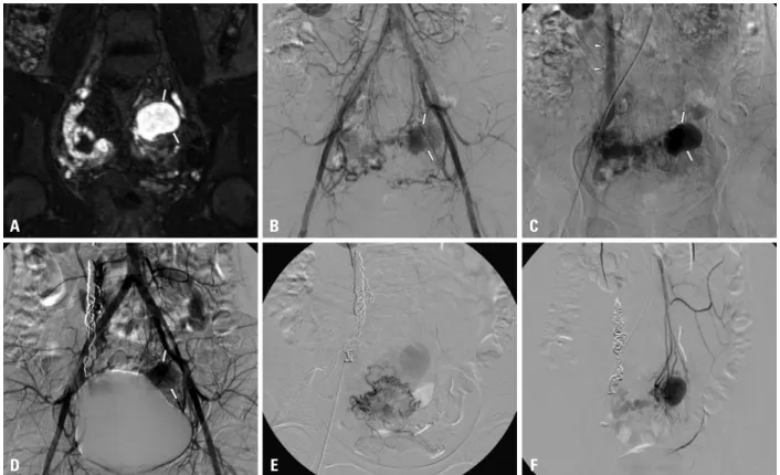
through the AVM. After successful embolization of the ve- nous side, inadvertent rupture of the ascending segment of right uterine artery, which had a very tortuous anatomy, oc- curred during guide wire manipulation. The ruptured artery was subsequently embolized using microcoils. Although the patient had both a decreased amount and frequency of vaginal bleeding after the first embolization procedure, sec- ond UAE was performed eight months later because of continuous, intermittent vaginal bleeding and evidence of residual AVM on a subsequent pelvic MR examination.
Angiography demonstrated fine feeders from the right uter- ine artery, recanalization of the left uterine artery, and para- sitis feeders from the inferior mesenteric artery, all of which were successfully embolized (Fig. 2). The second proce- dure was technically and clinically successful with resolu- tion of the patient’s symptoms and gradual disappearance of the AVM on the follow-up transvaginal ultrasonography up to 15 months following the second procedure.
The embolic materials used most often were particulate agents such as gelfoam pledgets (n=13) and polyvinyl alco- hol particles (n=6). Gelfoam sheets were cut into pledgets
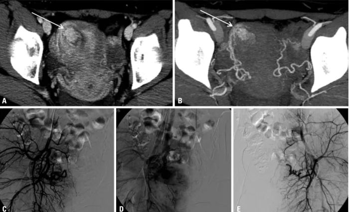
Fig. 1. A 26-year-old patient (No. 4) presenting with progressive vaginal bleeding. The axial enhanced CT scan (A) and CT angiography (B) show focal a hy- pervascular lesion (arrows) with feeders from the right uterine artery. (C and D) Right internal iliac arteriograms show the AVM along with the early opacifi- cation of the right ovarian vein. (E) Left internal iliac arteriogram does not show much blood supply to the AVM. Bilateral uterine arteries were embolized with gelfoam pledgets (not shown). At one-month follow-up, transvaginal ultrasound (not shown) showed resolution of the AVM together with clinical im- provement. AVM, arteriovenous malformation.
A
C
B
D E
and MRI.3,4,7,8 In our study, the lateralized uterine AVMs were related to iatrogenic injuries such as D&C and their dominant vascular feeders were from the ipsilateral uterine artery. In addition, all four patients with pseudoaneurysms had histories of D&C.18 Thus, we are able to assume from our results that acquired uterine AVMs are frequently related to iatrogenic injury. For example, D&C tends to be located laterally on either side with predominant feeder from the ip- nant, nor have attempted to conceive (Fig. 3).
DISCUSSION
Imaging assessment of uterine AVMs, especially their vas- cular anatomy, is very important during the planning stages before embolization, such as Doppler ultrasonography, CT,
Fig. 2. A 21-year-old patient (No. 10) presenting with progressive vaginal bleeding. (A) The enhanced coronal MR image shows tortuosity of the right uterine artery with a large pseudoaneurysm (arrows) at the uterine fundus. (B and C) Aortograms show tortuosity of bilateral (the right side being dominant) uterine arteries, a pseudoaneurysm (arrows), and early drainage into the right ovarian vein (arrowheads). The right uterine artery was ruptured during guide wire manipulation. The right ovarian vein and ruptured right uterine artery were embolized using coils. (D) The angiogram obtained upon completion shows the remaining pseudoaneurysm. (E) Selective angiograms of the right uterine artery angiogram of the left uterine artery obtained eight months after the first em- bolization procedure show the residual AVMs with opacification of the pseudoaneurysm. (F) The selective angiogram of the inferior mesenteric artery an- giogram shows the AVM with a pseudoaneurysm. All feeding arteries were completely embolized using polyvinyl alcohol particles and gelfoam pledgets (not shown). AVM, arteriovenous malformation.
D A
E B
F C
Fig. 3. Results of bilateral UAE for uterine AVMs. UAE, uterine artery embolization; AVM, arteriovenous malformation; UA, uterine artery; IMA, inferior mesen- teric artery.
Complete embolization (n=17) Incomplete embolization (n=2)
Residual fine feeders with decreased AVM volume
After 5 months follow up-spontaneous regression Ruptured right UA 2nd embolization
Complete embolization Bilateral UAs and IMA Uterine AVMs
-D&C (n=14) -Delivery related (n=5)
drain into a single venous component. In this patient, em- bolization of bilateral UAs and the inferior mesenteric ar- tery was performed in the second session eight months fol- lowing the first session. Considering the rich vascular collateral network of the uterus, thorough angiographic as- sessment is necessary to identify all existing extra-uterine feeders.
We advocate the safety of bilateral UAE for the manage- ment of acquired uterine AVMs where preservation of the uterine function is a major advantage of the procedure. The choice of embolic material, whether used independently or in combination, did not affect the clinical outcomes of UAE.
According to our study, the use of gelfoam pledgets without the complementary use of any other embolic material seems effective in the resolution of acquired uterine AVMs. In ad- dition, a three- to five-week duration of occlusion after bi- lateral UAE using gelfoam pledgets seemed sufficient to prevent further bleeding while still permitting the slow de- velopment of various collateral vasculature, thereby pre- venting ischemia.6,13,14,29,30 Menstrual cycles were restored within two months in all patients with clinically successful outcome. Two patients also had successful labors twelve months after their procedures. These results are compatible with those described in previous studies.12,15,24 Although, hypovascularity of the treated area may theoretically have adverse affects on placentation and fetal growth,31 normal adaptation of the uteroplacental vasculature after UAE is a plausible explanation for the restoration of the menstrual cycle and the potential for successful labor.
Our study has several limitations. First, this retrospective design possesses some inherent flaws; furthermore, the small sample size does not permit extensive statistical analysis.
Second, the follow-up protocol was not consistent in all pa- tients, and no images were available at long-term follow- up. Third, various angiographic devices and embolic mate- rials were used for a long period.
In conclusion, bleeding uterine AVMs can be successful- ly managed by bilateral UAE, which is a safe and effective first-line therapeutic option. However, technical difficulties during the procedure may result in incomplete embolization in suboptimal clinical outcomes.
REFERENCES
1. Aziz N, Lenzi TA, Jeffrey RB Jr, Lyell DJ. Postpartum uterine ar- teriovenous fistula. Obstet Gynecol 2004;103(5 Pt 2):1076-8.
silateral uterine artery, and is possibly accompanied by pseudoaneurysms. However, uterine AVMs associated with delivery (n=5) or gestational trophoblastic disease (n=1) showed predomiant feeders from bilateral uterine arteries.
This finding supports the pathogenesis described in the lit- erature that relates the development of AVMs to failed re- gression of proliferated vascular structures of pregnancy or abnormally developed vascular structures related to gesta- tional trophoblastic disease.7,11,21
We advocate that embolization of bilateral UAs is superior to embolization of unilateral uterine artery. In all of our cases, regardless of the location or predominant feeding artery of the AVM, bilateral UAE was performed. By embolizing the predominant feeding uterine artery followed by the contralat- eral uterine artery, we expected a more secure and complete result from the embolization procedure. With only a single session of UAE where bilateral UAs were embolized, 17 pa- tients showed complete resolution of uterine AVMs and no recurrence during follow-up. There were no extra-uterine feeders to the AVMs in these patients. With unilateral em- bolization, there is a potential for the contralateral non-dom- inant uterine artery to develop into a major feeding artery. In the absence of extra-uterine feeders, complete resolution of uterine AVMs is expected by bilateral UAE.
Pre-procedural imaging evaluation is necessary in order to assess the presence of possible extra-uterine feeders. An example is the ovarian-uterine anastomotic connection, which is often detected when performing UAE for uterine fibroids.22,23 It is technically challenging to embolize fine extra-uterine feeders, as we found in one patient of our study. This patient was closely observed after failed at- tempts at embolization of fine feeders from the anterior di- vision of internal iliac artery; however, complete resolution of the AVM was seen on a follow-up imaging study. This clinical observation suggests that conservative management rather than aggressive intervention or surgical strategy could be a reasonable option when the UAE procedure pro- vide insufficient angiographic results.7,9,24-27
In one of our patients with an AVM that was associated gestational trophoblastic disease, one draining ovarian vein that was markedly prominent was embolized with coils to reduce blood flow. To our knowledge, this is the first time such a case has been described in the literature, and we be- lieve that embolization of the dominant draining vein is an important step in achieving effective embolization. This case corresponds to the type II arteriolovenous shunt, previ- ously described by Cho, et al.,28 where multiple arterioles
18. Kwon JH, Kim GS. Obstetric iatrogenic arterial injuries of the uterus: diagnosis with US and treatment with transcatheter arterial embolization. Radiographics 2002;22:35-46.
19. Maleux G, Timmerman D, Heye S, Wilms G. Acquired uterine vascular malformations: radiological and clinical outcome after transcatheter embolotherapy. Eur Radiol 2006;16:299-306.
20. Sacks D, McClenny TE, Cardella JF, Lewis CA. Society of Inter- ventional Radiology clinical practice guidelines. J Vasc Interv Ra- diol 2003;14(9 Pt 2):S199-202.
21. Vaknin Z, Sadeh-Mefpechkin D, Halperin R, Altshuler A, Amir P, Maymon R. Pregnancy-related uterine arteriovenous malforma- tions: experience from a single medical center. Ultraschall Med 2011;32 Suppl 2:E92-9.
22. Pelage JP, Walker WJ, Le Dref O, Rymer R. Ovarian artery: angi- ographic appearance, embolization and relevance to uterine fibroid embolization. Cardiovasc Intervent Radiol 2003;26:227-33.
23. Razavi MK, Wolanske KA, Hwang GL, Sze DY, Kee ST, Dake MD. Angiographic classification of ovarian artery-to-uterine ar- tery anastomoses: initial observations in uterine fibroid emboliza- tion. Radiology 2002;224:707-12.
24. Dar P, Karmin I, Einstein MH. Arteriovenous malformations of the uterus: long-term follow-up. Gynecol Obstet Invest 2008;66:
157-61.
25. Itoh H, Keitoku M, Fukuoka M, Sagawa N, Mori T, Togashi K.
Spontaneous resolution of a postcesarean arteriovenous fistula of the uterine cervix: the usefulness of transvaginal color Doppler scanning. J Obstet Gynaecol Res 1997;23:439-44.
26. Forssman L, Lundberg J, Scherstén T. Conservative treatment of uterine arteriovenous fistula. Acta Obstet Gynecol Scand 1982;61:
85-7.
27. Nonaka T, Yahata T, Kashima K, Tanaka K. Resolution of uterine arteriovenous malformation and successful pregnancy after treat- ment with a gonadotropin-releasing hormone agonist. Obstet Gy- necol 2011;117(2 Pt 2):452-5.
28. Cho SK, Do YS, Shin SW, Kim DI, Kim YW, Park KB, et al. Ar- teriovenous malformations of the body and extremities: analysis of therapeutic outcomes and approaches according to a modified angiographic classification. J Endovasc Ther 2006;13:527-38.
29. Badawy SZ, Etman A, Singh M, Murphy K, Mayelli T, Philadel- phia M. Uterine artery embolization: the role in obstetrics and gy- necology. Clin Imaging 2001;25:288-95.
30. Delotte J, Chevallier P, Benoit B, Castillon JM, Bongain A. Preg- nancy after embolization therapy for uterine arteriovenous malfor- mation. Fertil Steril 2006;85:228.
31. Chow TW, Nwosu EC, Gould DA, Richmond DH. Pregnancy fol- lowing successful embolisation of a uterine vascular malforma- tion. Br J Obstet Gynaecol 1995;102:166-8.
2. Fleming H, Ostor AG, Pickel H, Fortune DW. Arteriovenous mal- formations of the uterus. Obstet Gynecol 1989;73:209-14.
3. Flynn MK, Levine D. The noninvasive diagnosis and manage- ment of a uterine arteriovenous malformation. Obstet Gynecol 1996;88(4 Pt 2):650-2.
4. Cura M, Martinez N, Cura A, Dalsaso TJ, Elmerhi F. Arteriove- nous malformations of the uterus. Acta Radiol 2009;50:823-9.
5. Arora R, Achla B, Pinkee S, Purba G, Bharti M. Arteriovenous malformations of the uterus. N Z Med J 2004;117:U1182.
6. Schiller VL, Raft E, Linden R. Uterine arteriovenous malforma- tion. AJR Am J Roentgenol 170:219-20.
7. Timmerman D, Van den Bosch T, Peeraer K, Debrouwere E, Van Schoubroeck D, Stockx L, et al. Vascular malformations in the uterus: ultrasonographic diagnosis and conservative management.
Eur J Obstet Gynecol Reprod Biol 2000;92:171-8.
8. O’Brien P, Neyastani A, Buckley AR, Chang SD, Legiehn GM.
Uterine arteriovenous malformations: from diagnosis to treatment.
J Ultrasound Med 2006;25:1387-92.
9. Brown JV 3rd, Asrat T, Epstein HD, Oglevie S, Goldstein BH.
Contemporary diagnosis and management of a uterine arteriove- nous malformation. Obstet Gynecol 2008;112(2 Pt 2):467-70.
10. Lin AC, Hung YC, Huang LC, Chiu TH, Ho M. Successful treat- ment of uterine arteriovenous malformation with percutaneous embolization. Taiwan J Obstet Gynecol 2007;46:60-3.
11. Lim AK, Agarwal R, Seckl MJ, Newlands ES, Barrett NK, Mitch- ell AW. Embolization of bleeding residual uterine vascular malfor- mations in patients with treated gestational trophoblastic tumors.
Radiology 2002;222:640-4.
12. Ghai S, Rajan DK, Asch MR, Muradali D, Simons ME, TerBrug- ge KG. Efficacy of embolization in traumatic uterine vascular malformations. J Vasc Interv Radiol 2003;14:1401-8.
13. Amagada JO, Karanjgaokar V, Wood A, Wiener JJ. Successful pregnancy following two uterine artery embolisation procedures for arteriovenous malformation. J Obstet Gynaecol 2004;24:86-7.
14. Chia YN, Yap C, Tan BS. Pregnancy following embolisation of uterine arteriovenous malformation--a case report. Ann Acad Med Singapore 2003;32:658-60.
15. Peitsidis P, Manolakos E, Tsekoura V, Kreienberg R, Schwentner L. Uterine arteriovenous malformations induced after diagnostic curettage: a systematic review. Arch Gynecol Obstet 2011;284:
1137-51.
16. Guo N, Liu H, Peng Z. Uterine arteriovenous fistula necessitating hysterectomy after two unsuccessful embolizations in an 18-year- old patient. Ann Vasc Surg 2010;24:827.
17. Wang Z, Chen J, Shi H, Zhou K, Sun H, Li X, et al. Efficacy and safety of embolization in iatrogenic traumatic uterine vascular malformations. Clin Radiol 2012;67:541-5.