황반부 부종을 동반한 분지망막정맥폐쇄의 치료에 있어서 유리체강내 베바시주맙의 반응을 예측하는 인자
Predictive Factors for a Favorable Response to Intravitreal Bevacizumab for Macular Edema Associated with Branch Retinal Vein Occlusion
강현구1, 서유리2, 최은영1, 이성철2, 김민1
Hyun Goo Kang1, Yuri Seo2, Eun Young Choi1, Sung Chul Lee2, Min Kim1
1연세대학교 의과대학 강남세브란스병원 안과학교실 시기능개발연구소, 2연세대학교 의과대학 세브란스병원 안과학교실 시기능개발연구소
1Department of Ophthalmology, Institute of Vision Research, Gangnam Severance Hospital, Yonsei University College of Medicine, Seoul, Korea
2Department of Ophthalmology, Institute of Vision Research, Severance Hospital, Yonsei University College of Medicine, Seoul, Korea
Purpose: To determine the factors for predicting a favorable response to intravitreal bevacizumab (IVB) for macular edema (ME) associ- ated with branch retinal vein occlusion (BRVO).
Methods: Thirty-seven eyes of patients first diagnosed with BRVO-associated ME and treated with IVB more than twice were included in this retrospective case series. Baseline characteristics, initial best-corrected visual acuity (BCVA), initial central macular thickness (CMT), and change in CMT after 2 consecutive monthly IVB injections were reviewed. Patients were classified into 2 groups according to their IVB response: non-responders were defined as those in whom CMT was not decreased by greater than 10% of the initial value after 2 con- secutive monthly injections. The types of observed macular edema were further subdivided into the cystoid macular edema (CME) only, serous retinal detachment (SRD) only, and combined CME and SRD groups for analysis.
Results: Thirty-three patients were classified as responders and 4 patients were classified as non-responders. The responder group was comprised of significantly older patients than the non-responder group (63.8 ± 11.7 vs. 54.5 ± 1.0, p = 0.034). The initial BCVA of the non-responder group was significantly higher than that of the responder group (logMAR 0.08 ± 1.04 vs. 0.37 ± 0.60, p = 0.003). The an- atomical type of ME did not significantly influence the response to IVB. There were no differences in the histories of diabetes mellitus or hypertension between the groups, and the existence of an epiretinal membrane did not appear to affect treatment response.
Conclusions: Patients with better initial BCVA and those who were older appeared to have a more favorable response to IVB treatment in ME due to BRVO.
Keywords: Branch retinal vein occlusion; Intravitreal bevacizumab; Macular edema; Prognostic factors
Address reprint requests to Min Kim, MD
Department of Ophthalmology, Institute of Vision Research, Gangnam Severance Hospital, Yonsei University College of Medicine, #211 Eonju-ro, Gangnam-gu, Seoul 06273, Korea
Tel: 82-2-2019-3440; Fax: 82-2-3463-1049 E-mail: [email protected]
Received: 2017. 8. 30 Revised: 2017. 11. 7 Accepted: 2017. 11. 11
Introduction
Branch retinal vein occlusion (BRVO) is an occlusion in the small veins of the retina resulting in reduced circulation that leads to ischemia, hemorrhages, and fluid leakage [1,2].
The severity of symptoms depends upon the location of the occlusion and, in general, the more proximal the occlusion, the more severe the area affected and the greater the degree of edema [3]. In severe cases involving the macula, this can result in secondary macular edema (ME) and may present as a sudden painless decrease in the vision of the affected eye [4]. Disease prevalence ranges from 0.3% to 1.1% and it is the second most frequent disease of the retinal vasculature after diabetic retinopathy [5,6]. Several known major risk factors for BRVO include increasing age, a history of hyper- tension or other cardiovascular diseases, and precursor signs of chronic hypertensive damage on fundoscopy [7,8].
Although a range of new treatments has been introduced in recent decades, the effectiveness and safety of many of these are debated [9,10]. As such, the current main goals of clinical management are to stabilize vision and reduce the secondary complications associated with BRVO, either with intravitreal anti-vascular endothelial growth factor (VEGF) injections or corticosteroids introduced via intravitreal im- plants or sub-Tenon’s injection [11-13].
Intravitreal bevacizumab (IVB), a monoclonal antibody of VEGF, has been shown to be a cost-effective method to improve visual acuity and reduce central macular thickness (CMT) [14,15]. Although the introduction of newer an- ti-VEGF agents, such as aflibercept (Eylea, Bayer Pharma- ceuticals, Berlin, Germany) and ranibizumab (Lucentis, Ge- nentech Inc., South San Francisco, CA, USA), has provided alternative modalities with significant therapeutic benefits, the off-label use of Avastin (Genentech Inc., South San Fran- cisco, CA, USA) has allowed for a relatively less expensive alternative with evidence-based effectiveness [16,17]. IVB has therefore remained an important mainstay therapeutic option, especially for patients with chronic and/or recurring secondary ME due to BRVO [18-20].
Therefore, the objective of the present study was to in- vestigate factors predicting a favorable response to IVB for BRVO-associated ME. The results of the present study will allow ophthalmologists to provide proper recommendations to patients, especially in cases where a favorable response to IVB monotherapy can be predicted.
Materials and Methods
This retrospective, comparative case study was performed at the Department of Ophthalmology of Yonsei University at Severance Hospital and Gangnam Severance Hospital. The study adhered to the tenets of the Declaration of Helsinki and was approved by the Institutional Review Board/Ethics Committee of Yonsei University. Written informed consent was obtained from all subjects.
The medical charts and imaging studies of patients di- agnosed with secondary ME associated with BRVO from January 2012 to December 2014 were reviewed. BRVO was diagnosed using a wide-field scanning laser ophthalmoscope (Optomap, Optos PLC., Dunfermline, United Kingdom) and included characteristic flame-shaped retinal hemorrhages in the area of the occluded vein, and hypo-perfusion noted in the affected vessels as revealed by fluorescein angiography with a confocal scanning system (HRA-2; Heidelberg En- gineering Inc., Heidelberg, Germany). ME was defined as a CMT greater than 300 μm on optical coherence tomography (OCT) (Heidelberg Spectralis, Heidelberg Engineering Inc., Heidelberg, Germany). After pupil dilation, well-trained examiners performed OCT examinations according to a pre-approved protocol, and an average CMT was calculated using the 3D macular volumetric scan protocol (30 × 20 de- grees, 25 sections with 235 micrometers spacing) using the included software.
Inclusion criteria consisted of patients treated with IVB at least twice consecutively with the same protocol at month- ly intervals. Briefly, 1.25 mg/0.1 mL of bevacizumab was injected through the pars plana under sterile conditions in a designated facility. Subsequent re-treatment after a period of at least 4 weeks post-injection was considered at the discre- tion of the primary retina specialist based on the improve- ment in best-corrected visual acuity (BCVA) and in ME as revealed by OCT. Patients with a history of ocular surgery, intravitreal corticosteroid implants, sub-Tenon’s triamcino- lone injection, and laser photocoagulation were excluded.
The patients were grouped according to the quality of response to IVB therapy: non-responders were defined as patients whose CMT was not decreased by more than 10% of the initial after 2 injections, which was a standard definition used in a previously published study by Dabir et al. [21]. All patients were assessed for response 1 month after the second injection, which is a routine follow-up interval at our retina
center. At this follow-up visit, all patients underwent BCVA measurement, OCT imaging, and fundus photography. Pa- tients were also subdivided based on the initial type of mac- ular edema: cystoid macular edema (CME) only, serous reti- nal detachment (SRD) only, and combined CME and SRD.
Data from each outpatient visit consisting of BCVA, intraocular pressure, and imaging examination by fundus photography and OCT were compiled. Baseline data in- cluding initial BCVA and initial CMT, as well as additional demographic data consisting of age, sex, significant medical history, types of observed ME, and presence of an epiretinal membrane were collected as possible prognostic factors in- fluencing the response of treatment outcome.
Statistical analyses were performed using the Statistical Package for the Social Sciences (SPSS version 22.0, IBM Corp., Armonk, NY, USA). Values are expressed as the mean ± standard deviation or percentage. BCVA measured by Snellen charts were converted to the logarithm of the minimal angle of resolution (logMAR) for statistical purpos- es. Data distribution and homogeneity of variance were ana- lyzed. Fisher's exact test and independent T-test were used to compare the two groups. Multivariable analysis using binary logistic regression was used to investigate the relationships
between the possible prognostic factors.
Results
A total of 37 eyes from 37 patients were included in this study and treatment response was assessed: 33 patients were classified as responders and 4 were classified as non-re- sponders to IVB therapy. The baseline characteristics of the two groups are summarized in Table 1. There were no significant differences in the sex ratio, history of diabetes mellitus or hypertension, duration of symptoms, time to first injection, presence of an epiretinal membrane, or initial CMT height. However, the initial BCVA was significantly different between the two groups (p = 0.03): responders had a mean initial BCVA of 20/47 (logMAR 0.37 ± 0.6) while in non-responders the value was 20/24 (0.08 ± 1.0). The mean percentage in CMT reduction was -39.5 ± 15.9% for respond- ers while +6.95 ± 16.6% for non-responders, and was signifi- cant (p = 0.010).
The anatomical type of ME observed on OCT did not ap- pear to significantly influence the response to IVB (Table 2).
There were no cases of ME with serous retinal detachment
Table 1. Baseline characteristics of responders and non-responders to intravitreal bevacizumab monotherapy for secondary macular edema associated with branch retinal vein occlusion
IVB responders IVB non-responders p-value
Total (n, %) 33 (89.2) 4 (10.8)
Age (years) 63.8 ± 11.7 54.5 ± 1.0 0.034*
Gender (n, %)
Female 23 (69.7) 2 (50.0) 0.583†
Male 10 (30.3) 2 (50.0) 0.518†
Diabetes mellitus (n, %) 3 (8.1) 1 (25.0) 0.380†
Hypertension (n, %) 12 (36.4) 2 (50.0) 0.625†
Initial BCVA (logMAR [Snellen]) 0.37 ± 0.6 (20/47) 0.08 ± 1.0 (20/24) 0.003*
Initial CMT (μm) 474.8 ± 129.8 497.3 ± 150.8 0.749*
Duration of symptoms (days) 41.2 ± 50.2 74.7 ± 54.6 0.279*
Time to first injection (days) 4.7 ± 4.6 6.0 ± 1.0 0.641*
Presence of epiretinal membrane (n, %) 4 (12.1) 1 (25.0) 0.456†
CMT reduction rate (%) -39.5 ± 15.9 +6.95 ± 16.6 0.010*
Values are presented as mean ± SD or n (%) unless otherwise indicated.
IVB = intravitreal bevacizumab; BCVA = best corrected visual acuity; logMAR = logarithm of minimal angle of resolution; CMT = central macular thickness.
*Independent student t-test; †Fisher's exact test.
only in either group, and no significant differences in the ratios of CME only compared to combined CME- and SRD- type ME with regards to favorable treatment response.
Representative imaging of a case from each treatment re- sponse group can be seen in Fig. 1 and 2.
Discussion
Bevacizumab is a monoclonal antibody that inhibits angio- genesis by targeting and inhibiting VEGF, which effectively inhibits new blood vessel formation. Intravitreal sampling of BRVO patients revealed increased levels of VEGF, which is a potent inductor of vascular permeability and intraocular neovascularization [22]. Increased VEGF in the vitreous cavity has been correlated with the severity of ME and neo- vascularization [23]. Several long-term, large-sample studies have already shown the effectiveness of IVB as a therapy for secondary ME due to BRVO [13,24]; however, in our study we sought to examine in which cases IVB may prove a cost-effective alternative, especially with regard to patient financial burden and the chronic, recurrent nature of the dis- ease.
There have been few studies that have sought to determine predictive factors for the effectiveness of IVB therapy. In 2010, Ach et al. [25] evaluated retinal vein occlusion patients for such predictive factors and found that the CMT and age of patients had prognostic value in cases of central retinal vein occlusion, but that there were no observed predictive factors in cases of BRVO. In contrast, Jaissle et al. [26] conducted a study in 2011 that observed that baseline BCVA, patient’s age, and duration of BRVO were relevant prognostic factors for visual improvement. The results of this second study cor- relate with our findings based on Korean patients, which also found significance in baseline BCVA and patient age.
In our study, the anatomical type of observed ME that the patient presented with did not appear to influence the favora- bility of response. Several studies have included an addition- al classification called diffuse retinal thickening, defining it as edema without subretinal fluid or cystic lesions. However, we have found that in our cases high definition OCT revealed the presence of microcysts which allowed us to classify the anatomical type as CME, which was similar to findings from Catier et al. [27] and Trichonas and Kaiser et al. [2]. Further- more, the presence of systemic comorbidities such as diabe- tes mellitus and hypertension, and existence of an epiretinal membrane did not appear to affect treatment response.
As can be concluded from our results based on Korean pa- tients, IVB could be more effective in treating older patients diagnosed with BRVO-associated ME who present with low vision. For younger patients who show poor initial BCVA, alternative treatment agents such as ranibizumab or dexa- methasone implants may be used for more effective manage- ment [28,29].
This study has several limitations. Due to its small sam- ple size, the data analyzed may not be representative of the entire population and as such, conclusions must be drawn carefully. However, as our results correlate with previous large sample-sized studies, we believe that the results are significant and merit consideration. Additionally, we did not exclude patients with diabetes mellitus as we sought to determine whether such an underlying systemic disease may affect the treatment response; however, we applied rigorous diagnostic criteria to isolate BRVO as the cause of the ob- served ME. Our results may allow for proper recommenda- tions to be made to patients with BRVO associated ME. In conclusion, patients with better initial BCVA and those who were older appeared to have a more favorable response to IVB treatment in ME due to BRVO.
Table 2. Comparison of responders to intravitreal bevacizumab monotherapy according to the anatomic type of macular edema observed at the time of initial diagnosis
IVB responders IVB non-responders p-value
Anatomical type
CME only 20 (60.6) 2 (50.0) 0.990*
CME and SRD 13 (39.4) 2 (50.0) 0.867*
Values are presented as n (%) unless otherwise indicated.
IVB = intravitreal bevacizumab; CME = cystoid macular edema; SRD = serous retinal detachment.
*Fisher's exact test.
Conflicts of interest
No conflicting relationship exists for any author.
References
1. Finkelstein D. Ischemic macular edema. Recognition and favor- able natural history in branch vein occlusion. Arch Ophthalmol 1992;110:1427-34.
2. Trichonas G, Kaiser PK. Optical coherence tomography imaging of macular oedema. Br J Ophthalmol 2014;98 Suppl 2:ii24-9.
3. Opremcak EM, Bruce RA. Surgical decompression of branch
retinal vein occlusion via arteriovenous crossing sheathotomy: a prospective review of 15 cases. Retina 1999;19:1-5.
4. Klein R, Klein BE, Moss SE, Meuer SM. The epidemiology of ret- inal vein occlusion: the Beaver Dam Eye Study. Trans Am Oph- thalmol Soc 2000;98:133-41; discussion 141-3.
5. Lim LL, Cheung N, Wang JJ, et al. Prevalence and risk factors of retinal vein occlusion in an Asian population. Br J Ophthalmol 2008;92:1316-9.
6. Rogers SL, McIntosh RL, Lim L, et al. Natural history of branch retinal vein occlusion: an evidence-based systematic review.
Ophthalmology 2010;117:1094-101.e5.
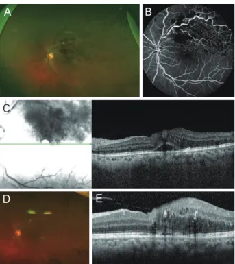
7. Mitchell P, Smith W, Chang A. Prevalence and associations of Figure 1. A representative case from the responder group. A man
in his early sixties presented with vision loss in his left eye. He had no prior medical, ocular surgery or trauma history. His best corrected visual acuity (BCVA) at presentation was 20/100 in the affected eye.
Fundus photography revealed a characteristic flame-shaped retinal hemorrhage in a cone-shape around the superior arcade (A). Corre- sponding areas of hypo-fluorescence due to retinal hypoperfusion were observed on fluorescein angiography (B). A central macular thickness of 454 μm on optical coherence tomography (OCT) con- firmed the diagnosis of secondary macular edema due to branch ret- inal vein occlusion (C). The anatomical type was classified as cystoid macular edema only. After 2 injections of intravitreal bevacizumab, there was improvement of the hemorrhage on fundus imaging and a dramatic reduction in macular edema on OCT (D, E). The final BCVA at the most recent follow-up was 20/25 for his affected eye.
A
C
D E
B
Figure 2. A representative case from the non-responder group. A fifty-year-old man presented with visual discomfort in his left eye.
He had a history of hypertension and diabetes mellitus, and was a known carrier of hepatitis B. He had no known history of ocular sur- gery, trauma, or treatment. His initial best corrected visual acuity was 20/29 in his affected eye. A characteristic flame-shaped retinal hem- orrhage on fundus photography (A) and hypoperfusion noted on fluorescein angiography (B) confirmed the diagnosis of branch retinal vein occlusion. Macular edema with a central macular thickness (CMT) of 659 μm was seen on optical coherence tomography (C). The anatomical type was classified as combined cystoid macular edema with serous retinal detachment. After 2 injections with intravitreal bevacizumab, though the retinal hemorrhage had resolved signifi- cantly, there were hard exudates remaining in the parafoveal area (D).
Furthermore, CMT was nearly unchanged at 612 μm, which was only a 7.1% reduction (E) post-injection.
A
C
D E
B
retinal vein occlusion in Australia: the Blue Mountains Eye Study.
Arch Ophthalmol 1996;114:1243-7.
8. Wong TY, Larsen EKM, Klein R, et al. Cardiovascular risk factors for retinal vein occlusion and arteriolar emboli: the atherosclero- sis risk in communities & cardiovascular health studies. Ophthal- mology 2005;112:540-7.
9. McIntosh RL, Mohamed Q, Saw SM, Wong TY. Interventions for branch retinal vein occlusion: an evidence-based systematic review. Ophthalmology 2007;114:835-54.
10. Shahid H, Hossain P, Amoaku W. The management of retinal vein occlusion: is interventional ophthalmology the way for- ward? Br J Ophthalmol 2006;90:627-39.
11. Hayreh SS, Zimmerman MB. Branch retinal vein occlusion: natural history of visual outcome. JAMA ophthalmology 2014;132:13-22.
12. Klein R, Moss SE, Meuer SM, Klein BE. The 15-year cumulative incidence of retinal vein occlusion: the Beaver Dam Eye Study.
Arch Ophthalmol 2008;126:513-8.
13. Papadia M, Misteli M, Jeannin B, Herbort CP. The influence of anti-VEGF therapy on present day management of macular edema due to BRVO and CRVO: a longitudinal analysis on visual function, injection time interval and complications. Int Ophthal- mol 2014;34:1193-201.
14. Rabena MD, Pieramici DJ, Castellarin AA, et al. Intravitreal bevaci- zumab (Avastin) in the treatment of macular edema secondary to branch retinal vein occlusion. Retina 2007;27:419-25.
15. Abegg M, Tappeiner C, Wolf-Schnurrbusch U, et al. Treatment of branch retinal vein occlusion induced macular edema with bevacizumab. BMC Ophthalmol 2008;8:18.
16. Wu L, Arevalo JF, Roca JA, et al. Comparison of two doses of intravitreal bevacizumab (Avastin) for treatment of macular ede- ma secondary to branch retinal vein occlusion: results from the Pan-American Collaborative Retina Study Group at 6 months of follow-up. Retina 2008;28:212-9.
17. Kreutzer TC, Alge CS, Wolf AH, et al. Intravitreal bevacizumab for the treatment of macular oedema secondary to branch retinal vein occlusion. Br J Ophthalmol 2008;92:351-5.
18. Campochiaro PA, Sophie R, Pearlman J, et al. Long-term out- comes in patients with retinal vein occlusion treated with ranibi- zumab: the RETAIN study. Ophthalmology 2014;121:209-19.
19. Heier JS, Campochiaro PA, Yau L, et al. Ranibizumab for macular
edema due to retinal vein occlusions: long-term follow-up in the HORIZON trial. Ophthalmology 2012;119:802-9.
20. Brown DM, Heier JS, Clark WL, et al. Intravitreal aflibercept injec- tion for macular edema secondary to central retinal vein occlu- sion: 1-year results from the phase 3 COPERNICUS study. Am J Ophthalmol 2013;155:429-37.e7.
21. Dabir SS, Das D, Nallathambi J, et al. Differential systemic gene expression profile in patients with diabetic macular edema: re- sponders versus nonresponders to standard treatment. Indian J Ophthalmol 2014;62:66-73.
22. Noma H, Funatsu H, Yamasaki M, et al. Pathogenesis of macular edema with branch retinal vein occlusion and intraocular levels of vascular endothelial growth factor and interleukin-6. Am J Ophthalmol 2005;140:256-61.
23. Pe'er J, Shweiki D, Itin A, et al. Hypoxia-induced expression of vascular endothelial growth factor by retinal cells is a com- mon factor in neovascularizing ocular diseases. Lab Invest 1995;72:638-45.
24. Jaulim A, Ahmed B, Khanam T, Chatziralli IP. Branch retinal vein occlusion: epidemiology, pathogenesis, risk factors, clinical fea- tures, diagnosis, and complications. an update of the literature.
Retina 2013;33:901-10.
25. Ach T, Hoeh AE, Schaal KB, et al. Predictive factors for changes in macular edema in intravitreal bevacizumab therapy of retinal vein occlusion. Graefes Arch Clin Exp Ophthalmol 2010;248:155-9.
26. Jaissle GB, Szurman P, Feltgen N, et al. Predictive factors for func- tional improvement after intravitreal bevacizumab therapy for macular edema due to branch retinal vein occlusion. Graefes Arch Clin Exp Ophthalmol 2011;249:183-92.
27. Catier A, Tadayoni R, Paques M, et al. Characterization of macular edema from various etiologies by optical coherence tomogra- phy. Am J Ophthalmol 2005;140:200-6.
28. Lee KH, Kang EC, Koh HJ. Dexamethasone intravitreal implant rescue treatment for bevacizumab refractory macular edema secondary to branch retinal vein occlusion. Korean J Ophthal- mol 2017;31:108-14.
29. Son BK, Kwak HW, Kim ES, Yu SY. Comparison of ranibizumab and bevacizumab for macular edema associated with branch retinal vein occlusion. Korean J Ophthalmol 2017;31:209-16.